Introduction
Nasopharyngeal carcinoma is common in Southern China and Southeast Asia. Orbital involvement of nasopharyngeal carcinoma is classified as tumour stage (T) four disease. Involvement of the lacrimal apparatus is very rare due to the distance from the primary tumour.
We report three cases of nasolacrimal relapse in patients with previously treated nasopharyngeal carcinoma. There was no local nasopharyngeal recurrence in any of these cases. At the initial stage, all cases were either T1 or T2 without orbital involvement. The mode of tumour spread, and the computed tomography (CT) findings, are discussed.
Case reports
Case one
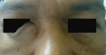
A 48-year-old man presented with a two-month history of a small, hard nodule at the right medial canthus (Figure 1). He had previously been diagnosed with nasopharyngeal carcinoma, of tumour-node-metastasis (TNM) stage T2b N2 M0. He had completed concomitant chemoradiotherapy three years ago. For the previous two months, he had had persistent epiphora with occasional blood-stained discharge from his right eye. His vision was unimpaired.
Clinical photograph of case one, showing the small, hard, fixed nodule at the right medial canthus. The medial half of the right lower eyelid is elevated by underlying tumour.

Nasal endoscopy found no local recurrence of nasopharyngeal carcinoma.
Fine needle aspiration of the right lacrimal sac nodule confirmed lacrimal relapse of nasopharyngeal carcinoma.
A CT scan showed localised tumour in the lacrimal fossa, with nasolacrimal canal invasion and soft tissue thickening at the osteomeatal complex.
The patient received further chemoradiotherapy.
After a year of follow up, he was well with no evidence of tumour recurrence.
Case two
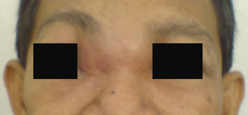
A 39-year-old man presented to us with a three-week history of right medial canthal swelling, epiphora and mild rhinorrhoea. Two years previously, he had been diagnosed with nasopharyngeal carcinoma, staged as T2a N1 M0. He had subsequently completed concomitant chemoradiotherapy. His vision was otherwise unaffected. The right medial canthal swelling had gradually extended across the midline (Figure 2).
Clinical photograph of case two, in which the tumour in the right lacrimal sac extended across the midline and involved the nasal bone and bilateral fronto-ethmoidal sinuses.

On nasal endoscopy, there was mild mucosal oedema at the osteomeatal complex, with no signs of local nasopharyngeal recurrence.
Fine needle aspiration of the medial canthal swelling revealed metastatic carcinoma, in keeping with the previously diagnosed nasopharyngeal carcinoma.
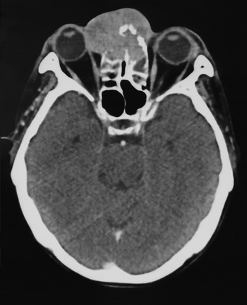
A CT scan showed an enhancing lacrimal sac lesion in the medial canthus, associated with erosion of the underlying nasal bone and extension into the frontal and ethmoid sinuses. There was mucosal thickening over the right maxillary sinus (Figure 3).
Axial computed tomography scan of case two, showing an enhancing lacrimal sac lesion in the medial canthus, associated with erosion of the underlying nasal bone and extension into the frontal and ethmoid sinuses. There is mucosal thickening over the right maxillary sinus. The posterior two-thirds of the right orbit is spared from tumour infiltration.

The initial response to chemotherapy was satisfactory. However, the tumour recurred six months after treatment. The patient succumbed a year later with multi-organ distant metastases.
Case three
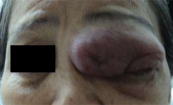
A 56-year-old woman presented to us with a two-month history of left eye proptosis, epiphora, diplopia and blurred vision (Figure 4). Two years previously, she had been diagnosed with nasopharyngeal carcinoma, staged as T1 N3 M0. She had completed radical radiotherapy with concomitant chemotherapy. However, she had defaulted from follow up after treatment. Prior to the current presentation she had developed blood-stained epiphora of her left eye, followed by medial canthal swelling, rhinorrhoea and other orbital symptoms.
Clinical photograph of case three, in which there was late presentation of a left lacrimal sac relapse with extensive tumour infiltration of both the upper and lower eyelids.

Nasal endoscopy showed no signs of local nasopharyngeal recurrence, but the mucosa of the left lateral nasal wall was oedematous.
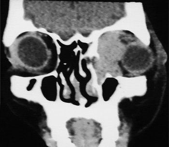
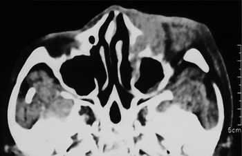
A CT scan showed an enhancing lesion in the medial part of the left orbit. There was widening of the left nasolacrimal canal and erosion of the lower medial orbital wall (Figures 5 and 6). The tumour involved the anterior one-third of the medial rectus muscle and displaced the globe anterolaterally. It extended posteriorly to the anterior half of the extraconal space. The globe, optic nerve and remaining recti muscles were preserved.
Coronal computed tomography scan of case three, showing the tumour involving the anterior half of the left extraconal space. The scan also shows the inferior meatal soft tissue mass, lacrimal sac infiltration and globe displacement.

Axial computed tomography scan of case three, showing that the left nasolacrimal duct has been widened and obliterated by tumour.

Histopathological analysis of a biopsy of the left medial canthal mass revealed a metastatic, undifferentiated carcinoma consistent with the previous nasopharyngeal carcinoma.
Further investigation revealed distant metastases to the lungs and liver. Palliative chemotherapy and supportive care were delivered. Unfortunately, the patient died six months later.
Discussion
Nasopharyngeal carcinoma is a common head and neck cancer in Southern China and Southeast Asia. The incidence of nasopharyngeal carcinoma is 30 times greater among Chinese individuals compared with Caucasians.Reference Amrith 1
Nasopharyngeal carcinoma with orbital involvement is not common. In a series reported by Heng et al., only 2.6 per cent of non-disseminated nasopharyngeal carcinoma cases had orbital involvement, and this was reported mainly in cases of recurrent nasopharyngeal carcinoma.Reference Heng, Wee and Fong 2 Kelvin et al. described nine cases of nasopharyngeal carcinoma with orbital involvement; eight of these nine cases had previously been treated for nasopharyngeal carcinoma, with subsequent new orbital relapse.Reference Lee, Seah, Tow, Cullen and Fong 3
Advanced nasopharyngeal carcinoma tends to invade into the posterior part of the orbit by infiltrating the inferior orbital fissure via the pterygopalatine fossa. The tumour may also extend anterosuperiorly to the cavernous sinus and infiltrate the superior orbital fissure to reach the posterior orbit. Both pathways contribute to compressive optic neuropathy, resulting in blurring of vision, diplopia and proptosis. Our three cases demonstrated different orbital presentations, suggesting different routes of tumour spread. In all cases, the tumour only involved the anterior half of the orbit, and especially the lacrimal apparatus; the posterior half of the orbit was spared from tumour invasion, as evidenced by the CT findings.
The route of tumour dissemination to the lacrimal system in locally treated nasopharyngeal carcinoma is especially relevant to our three cases. There is only limited literature describing nasolacrimal relapse in the anteromedial orbit, because of the rarity of this event.Reference Tang, Periyannan, Prepageran, Shashinder, Singh and Bhagubhai 4 , Reference Ching, Chong and Khoo 5 Lymphatic dissemination to the lacrimal system appears unlikely, due to the lack of lymphatics in the lacrimal apparatus. Ching et al. have proposed a route of tumour spread to the anteromedial orbit, after analysing the CT findings in five cases.Reference Ching, Chong and Khoo 5 The CT scan of all these patients showed nasolacrimal duct invasion and osteomeatal complex obliteration, while four cases demonstrated a medial canthal mass or pre-septal thickening, ethmoidal bulla opacification, and inferior meatus involvement. Nasolacrimal relapse of nasopharyngeal carcinoma is believed to spread along the lateral nasal wall. It infiltrates the nasolacrimal duct in retrograde fashion and extends further into the lacrimal sac and ethmoidal and sphenoidal sinuses.Reference Kallman, Foster, Wulc, Yousem and Kennedy 6
The emergence of nasolacrimal relapse may suggest failure of previous radical treatment. Standard external beam radiotherapy with an eye shield may deliver a lower dose of irradiation to the lateral nasal wall, compromising the effectiveness of radical radiotherapy in nasopharyngeal carcinoma cases. The occurrence of nasolacrimal relapse is classified as stage T4 disease, which carries a poor prognosis, with an overall five-year survival rate of only 28 to 35 per cent.Reference Heng, Wee and Fong 2
-
• Epiphora and medial canthal swelling are common in nasolacrimal relapse of nasopharyngeal carcinoma
-
• Retrograde invasion through the lateral nasal wall is the route of spread
-
• Computed tomography shows nasolacrimal duct widening and osteomeatal complex obliteration
-
• Irradiation of nasopharyngeal carcinoma should be more targeted
-
• Eye preservation remains a challenge in such cases
Radiotherapy with concomitant chemotherapy remains the chief treatment for nasolacrimal relapse. In order to safeguard the eyes from post-radiation complications, targeted intracavitary brachytherapy may be the preferred option in this condition. The roles of neo-adjuvant chemotherapy and surgery in nasolacrimal relapse are still controversial.
Conclusion
Although nasolacrimal relapse is rare, epiphora and medial canthal swelling are alarming developments which should not be overlooked. Relevant CT findings should be actively sought to ensure early detection of such relapse. In order to prevent under-treatment of nasopharyngeal carcinoma, more targeted radiotherapy should be delivered. The prevention of eye loss due to post-radiation complications remains a challenge in such cases.