Introduction
Cyanobacteria, algae and plants carry out oxygenic photosynthesis. They produce oxygen (O2) from water and take up carbon dioxide (CO2) to yield biomass. Cell respiration reverses this process. Oxygenic photosynthesis has formed the oxygen we breathe and most of the biomass we use as food, feed, fibre and fuel. Solar driven oxygen evolution started very early in evolution. A dramatic rise of the atmospheric oxygen level (by more than four orders of magnitude), the Great Oxygenation Event (GOE), dates 2.4 billion years back from now (Bekker et al., Reference Bekker, Holland, Wang, Rumble, Stein, Hannah, Coetzee and Beukes2004; Kump, Reference Kump2008). Both geological (Planavsky et al., Reference Planavsky, Asael, Hofmann, Reinhard, Lalonde, Knudsen, Wang, Ossa, Pecoits, Smith, Beukes, Bekker, Johnson, Konhauser, Lyons and Rouxel2014) and genomic evidence (Cardona et al., Reference Cardona, Murray and Rutherford2015) have suggested that ancestors of cyanobacteria might have started oxygenic photosynthesis half a billion years before the GOE. The stabilization of land masses and the emergence of land plants about half a billion years back from now caused another rise of the atmospheric oxygen content. Photosynthetic produced biomass and oxygen have powered the vast emergence of animal life. The time window of Homo sapiens is just a blip on these time scales. Its impact on the consumption of photosynthesis products is tremendous, as will be discussed at the end of this paper.
Several authors have comprehensively reviewed the respective momentary status of knowledge on the mechanism of oxygenic photosynthesis. They are cited in the text. The present paper scans how our knowledge on this fundamental process has unfolded over the past 50 years. Freshmen may start by reading the excellent book on photosynthesis by Bob Blankenship (Blankenship, Reference Blankenship2014).
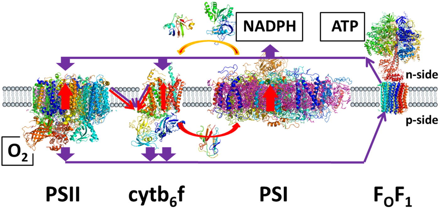
Figure 1 illustrates the present concept of the key elements of the primary processes of photosynthesis in green plants (photosystem II (PSII), PSI, cytb6f and the adenosine triphosphate (ATP) synthase). It is meant as a scaffold for the reader when travelling through the originally foggy, and then increasingly detailed and complicated terrain that follows.
Key proteins of the primary processes of photosynthesis in green plants (PSII, PSI, cytb6f and the ATP synthase), electron transfer (red), proton transfer (purple) and products (O2, NADPH and ATP) (Junge and Nelson, Reference Junge and Nelson2015).

Pioneering studies
Photosynthetic oxygen production from water
In 1772 Joseph Priestley reported a first systematic study on gas turnover between plants and animals (Priestley, Reference Priestley1772). A mouse confined in a sealed vessel suffocated if not backed up by a plant. It has meant that the mouse produces ‘bad air’ and the plant restores ‘vital air’. In modern terms, plants take up carbon dioxide (CO2) and produce oxygen (O2), while animals inhale O2 and exhale CO2. Hundred years after Priestley Theodor Engelmann (Engelmann, Reference Engelmann1881) introduced a less harmful bioassay for oxygen, namely the flocking behaviour of oxygen-loving, motile bacteria around illuminated chloroplasts. It yielded the first action spectrum of oxygenic photosynthesis (with typical chlorophyll peaks). Half a century later the great biochemist Otto Warburg (Nobel Prize 1931) studied by manometry the turnover of O2 and CO2 in respiring cells and tissues (Warburg, Reference Warburg1922). Robert Emerson and William Arnold used his technique to detect O2-production by illuminated algae (Emerson and Arnold, Reference Emerson and Arnold1932). They pioneered excitation of photosynthesis by short flashes of light (duration some 10 µs). Varying both the repetition rate of flashes and their energy they obtained two seminal results (Emerson and Arnold, Reference Emerson and Arnold1932). (i) The rate of oxygen evolution is limited by a temperature-sensitive (dark) step of some 10 ms duration, and (ii) the maximum production per flash is 1 mol O2 per 2500 mol chlorophyll. The relevance of these findings could only later be fully appreciated (see below).
The minimal balanced equation of oxygenic photosynthesis is 2H2O + CO2 = O2 + (CH2O). The substrate for oxygen production, water or carbon dioxide, was under debate. Robin Hill studied photosynthetic oxygen production in freshly isolated chloroplasts by another bio-assay, the spectral shift of added oxy-/deoxy-haemoglobin. Upon ageing of chloroplasts both, oxygen production and carbon dioxide consumption faded out. The addition of ferric ions as oxidant restored oxygen production but not carbon dioxide uptake. Hill took the decoupling of the former from the latter as evidence that plants ‘do not evolve oxygen from carbon dioxide’ (Hill, Reference Hill1939). Using heavy oxygen as a tracer Martin Kamen and collaborators found ‘the 18O/16O-ratio of the evolved oxygen identical with that of the water’. The obvious conclusion has been that O2 originates solely from water (Ruben et al., Reference Ruben, Kamen and Hyde1941) (for a modern remake see Clausen et al. (Reference Clausen, Beckmann, Junge and Messinger2005a)). Otto Warburg had meanwhile perfected the manometric monitoring the exchange of CO2 and O2 in photosynthesis and respiration (Burk et al., Reference Burk, Schade, Hunter and Warburg1951). Having observed a stimulating role of bicarbonate on photosynthetic oxygen production he claimed that oxygen results from bicarbonate and not from water (Warburg et al., Reference Warburg, Krippahl and Jetschma1965). The origin of the stimulating effect of CO2 on PSII that drove Warburg to this erroneous view, has only been settled much later (reviewed by Govindjee and his colleagues (Shevela et al., Reference Shevela, Eaton-Rye, Shen and Govindjee2012)) and the regulatory function of CO2 was detailed (Koroidov et al., Reference Koroidov, Shevela, Shutova, Samuelsson and Messinger2014; Brinkert et al., Reference Brinkert, De Causmaecker, Krieger-Liszkay, Fantuzzi and Rutherford2016).
Electron transport, oxygen production, proton transport and ATP synthesis
Lou Duysens (Duysens et al., Reference Duysens, Huiskamp, Vos and Vanderhart1956), Bessel Kok (Kok, Reference Kok1956) and Horst Witt (Witt and Moraw, Reference Witt and Moraw1959) started to characterize a wealth of spectroscopic signatures of photosynthetic electron transfer. Excitation with short flashes allowed resolving partial reactions in time (Kok, Reference Kok1956; Witt et al., Reference Witt, Mueller and Rumberg1961a). Two PSs operate in series (Duysens et al., Reference Duysens, Kamp and Amesz1961; Witt et al., Reference Witt, Mueller and Rumberg1961b). PSII produces oxygen and delivers electrons to PSI, which in turn provides electrons for the reduction of CO2 (see Fig. 1). The rate limiting step of the whole electron transport chain is located between the two PSs. Its duration (about 10 ms) is compatible with the one found for oxygen production by Emerson and Arnold (Emerson and Arnold, Reference Emerson and Arnold1932). The sensitizers of both PSs are chlorophyll-a constructs, which have been coined after the wavelength of their respective red absorption peak. P700 (Kok and Gott, Reference Kok and Gott1960) drives PSI and P680 (Döring et al., Reference Döring, Stiehl and Witt1967) drives PSII, respectively. These biophysical data were then not readily appreciated by some biochemists. When Warburg was confronted with Witt's detailed reaction scheme in 1962, he mused: ‘Could you tell us how the chemical mechanism of photosynthesis can be described on the basis of your spectroscopic observations?’ Witt countered his eminent critic, the pioneer of oxygen detection, by observing that ‘it would be difficult to deduce the mechanism of a combustion engine based only on sniffing the exhaust’ (see Junge and Rutherford (Reference Junge and Rutherford2007)). From their then limited perspective both were of course right (see below).
At that time it had been accepted that the chlorophylls and therewith the primary photosynthetic activity is embedded in the tightly folded inner membranes of chloroplasts, forming thylakoids (Menke, Reference Menke1962). Peter Mitchell (Nobel Prize 1978) hypothesized a new role of this inner membrane system of chloroplasts and likewise the cristae membrane of mitochondria (Mitchell, Reference Mitchell1961, Reference Mitchell1966). He envisaged that the membrane is proton tight such that zigzagging electron/hydrogen transport generates a pH-difference (Mitchell, Reference Mitchell1961), and transmembrane voltage (Mitchell, Reference Mitchell1966), in sum coined as protonmotive force. The backflow of protons is confined to the ATP synthase where it drives ATP-synthesis. Essentials of Mitchell's hypothesis were soon experimentally established for the thylakoid membrane. André Jagendorf subjected broken chloroplasts to an acid/base-jump (pH-jump) and obtained ATP (Jagendorf and Uribe, Reference Jagendorf and Uribe1966). The author with Horst Witt discovered the electrochromic origin of certain absorption transients of intrinsic chloroplast pigments (Junge and Witt, Reference Junge and Witt1968). Upon excitation with flashing light they observed very rapid voltage generation by both PSs and slower proton pumping (Schliephake et al., Reference Schliephake, Junge and Witt1968). Subsequent studies of the author with Bernd Rumberg have revealed that the flash generated voltage decays in synchrony with ATP synthesis (Junge, Reference Junge1970; Junge et al., Reference Junge, Rumberg and Schroeder1970). Titration of the voltage decay with the extremely powerful ionophore gramicidin revealed that the electrified membrane contains at least 105 chlorophyll molecules (Junge and Witt, Reference Junge and Witt1968), i.e. more than 100 electron transport chains. This number was later raised up to more than 107 chlorophyll molecules (Schönknecht et al., Reference Schönknecht, Althoff and Junge1990). In other words, the intricately folded thylakoid membrane within a chloroplast forms one contiguous, i.e. simply connected sheet (Junge, Reference Junge1977; Schönknecht et al., Reference Schönknecht, Althoff and Junge1990). It implies that almost all copies of the electron transport chain contribute driving force to all copies of ATP synthase. The lateral separation of PSII and PSI (and ATP synthase) in different areas of the folded thylakoid membrane (Boardman and Anderson, Reference Boardman and Anderson1964) apparently does not impair their ability for concerted electron and proton transport. Losses of the chemical portion of protonmotive force between laterally separated proton pumps and ATP synthase were only later discovered. They are minor both in mitochondria (Rieger et al., Reference Rieger, Junge and Busch2014) and in chloroplasts (Sjoholm et al., Reference Sjoholm, Bergstrand, Nilsson, Sachl, Von Ballmoos, Widengren and Brzezinski2017). Top efficiency of ATP synthesis has apparently been sacrificed for the sake of tight packing of the coupling membrane, both in chloroplasts and mitochondria.
Photosystem II with the oxygen evolving complex (BC)
Four-electron chemistry of water oxidation linked to one-electron photo-physics
In 1968 Anne and Pierre Joliot have lifted studies of photosynthetic oxygen evolution to a kinetic level by introducing a rapidly responding oxygen electrode (Joliot and Joliot, Reference Joliot and Joliot1968). When they excited dark adapted Chlorella cells with a series of short light flashes oxygen production peaked at flash number 3 and continued with damped oscillations of period four (Joliot et al., Reference Joliot, Barbieri and Chabaud1969). The release of oxygen occurs in 1 ms, i.e. a 10 times shorter time-interval than the overall bottleneck of the full electron transport chain (Emerson and Arnold, Reference Emerson and Arnold1932). In 1970 Bessel Kok concluded that PSII contains an accumulator of four positive ‘charges’ that links the one-electron-progression of the photo-physical reaction centre to the four electron/proton-abstraction from two molecules of water to yield dioxygen (Kok et al., Reference Kok, Forbush and Mcgloin1970). From there on, the catalytic centre has been conceived as stepwise progressing in a cycle over five oxidation states, S0 to S4. With S1 being the most stable state in dark adapted material, oxygen is only produced after three steps of the cycle during the transition S4 → S0. Two most important features of this four-stepped mechanism were soon recognized (see Renger (Reference Renger1977)): (a) the levelling of the energy demand of the four successive one-electron abstractions from water to match the fixed energy input provided by red quanta of light, and (b) the control (by sequestration and/or short live-time) of potentially harmful intermediates on the way from water to dioxygen (‘cryptoradicals’; Renger, Reference Renger1987). The cleavage of water to produce dioxygen was expected to liberate protons into the lumen of thylakoids. The stoichiometric H+/e−-pattern over the four-stepped progression, S0 → S1 → S2 → S3 → S4 + O2 was 1:0:1:2 (Förster et al., Reference Förster, Hong and Junge1981; Förster and Junge, Reference Förster and Junge1985). For some time it was controversial between various labs (reviewed in Lavergne and Junge, Reference Lavergne and Junge1993). Different patterns were observed in partially fragmented PSII-preparations. The seeming ambiguity has later been settled in favour of a proton release pattern of 1:0:1:2. It has required the discrimination by kinetic markers between transient electrostatic proton release/uptake at the membrane periphery (membrane Bohr effect) and chemical proton production from the catalytic core of PSII (Haumann and Junge, Reference Haumann and Junge1994) (see review in Junge et al. (Reference Junge, Haumann, Ahlbrink, Mulkidjanian and Clausen2002)). It has been early noted that the release of two protons during the terminal, oxygen evolving reaction is kinetically biphasic with one proton appearing at about 100 µs and the other one at 1 ms half rise (Förster et al., Reference Förster, Hong and Junge1981; Förster and Junge, Reference Förster and Junge1985). The release of a first proton appears as a primer for the terminal reaction cascade involving four electron transfers (see Fig. 3 farther down).
George Cheniae established the necessity of manganese for oxygen evolution (Cheniae and Martin, Reference Cheniae and Martin1967, Reference Cheniae and Martin1970). Ken Sauer's lab in Berkeley has focused on characterizing its function. Starting with electron paramagnetic resonance (EPR) (Blankenship and Sauer, Reference Blankenship and Sauer1974), they turned with Mel Klein into X-ray spectroscopy (Kirby et al., Reference Kirby, Goodin, Wydrzynski, Robertson and Klein1981a, Reference Kirby, Robertson, Smith, Thompson, Cooper and Klein1981b) and, till today, into advanced X-ray diffraction (see below). When exciting dark adapted chloroplasts with repetitive flashes of light they detected by EPR that some manganese was released into solution with period of four. It suggested that manganese associated with PSII undergoes valence-changes with a period of four (Wydrzynski and Sauer, Reference Wydrzynski and Sauer1980). From then on it was accepted that manganese is involved in Kok's ‘charge’ accumulator (reviewed in Sauer (Reference Sauer1980)). When generating Kok's state S2 by excitation of dark adapted material with a single short flash of light Charles Dismukes and Yona Siderer observed a multiline EPR-signal which they attributed to a mixed-valence pair of manganese, Mn(III)Mn(IV), in a dimeric or tetrameric Mn-cluster (Dismukes and Siderer, Reference Dismukes and Siderer1981). For quite a while this attribution has been the stronghold for the valence of the manganese cluster during the four transitions from S0 to S4.
Enrichment and purification of PSII: P680, the Mn-cluster and tyrosine (Yz)
Further progress required enrichment if not purification of PSII. Per Ake Albertson's lab pioneered the isolation of PSII-enriched membrane vesicles from spinach (Akerlund et al., Reference Akerlund, Andersson and Albertsson1976; Andersson et al., Reference Andersson, Akerlund and Albertsson1977). A number of consecutive studies demonstrated that the enrichment was facilitated by the intrinsic segregation between PSII in the appressed portions and PSI in the extended portions of stacked thylakoid membranes (Andersson and Anderson, Reference Andersson and Anderson1980, Reference Anderson and Andersson1988).
Purification of a manganese containing, oxygen evolving PSII was desirable. A first claim (Spector and Winget, Reference Spector and Winget1979) was not generally reproducible and probably fraudulent. A great step forward were membrane fragments from spinach chloroplasts that revealed high rates of oxygen evolution and were virtually uncontaminated by PSI (Berthold et al., Reference Berthold, Babcock and Yocum1981). They were coined BBY-particles after their parents, Berthold, Babcock and Yocum. The absolute content of PSII was quantified via the previously found, and chemically still unidentified EPR-signal II (Babcock and Sauer, Reference Babcock and Sauer1973), then coined Z, supposedly an electron donor to P680+ (Babcock and Sauer, Reference Babcock and Sauer1975). In 1984 a fully competent PSII-core particle from spinach was isolated by Demetrios Ghanotakis with Gerald Babcock and Charles Yocum (Ghanotakis et al., Reference Ghanotakis, Babcock and Yocum1984b). It contained four Mn-ions and one Z per 250 chlorophyll molecules. In addition to the hetero-dimeric core proteins of PSII, D1 and D2, and further proteins with antennae function (CP43, CP47) the complex contained peripheral proteins of 17, 23 and 33 kDa molecular mass. The removal of the 17 and 23 kDa proteins facilitated the access of external reductants to Z. It also impaired oxygen production which was however reconstituted by added Ca (Ghanotakis et al., Reference Ghanotakis, Babcock and Yocum1984a, Reference Ghanotakis, Babcock and Yocum1985a, Reference Ghanotakis, Babcock and Yocum1985b).
At this time (i.e. before crystallization, BC) structural information on the mutual arrangement of the four Mn-ions, and a role of Ca in relation to the Mn-cluster was still lacking (for reviews see Yocum, Reference Yocum1991; Debus, Reference Debus1992).
Kinetics of electron transfer between P680+, Yz and the Mn-cluster
P680 is photo-oxidized in less than 300 ps (Schatz et al., Reference Schatz, Brock and Holzwarth1987, Reference Schatz, Brock and Holzwarth1988). In PSII-preparations with small size of the antennae system the time of photon capture is shorter than in those with large antennae complement. In other words, PSII hosts an energetically ‘shallow trap’ where many transfers of excitation between the trap and its antennae complement precede the eventual charge separation (Schatz et al., Reference Schatz, Brock and Holzwarth1988; Barter et al., Reference Barter, Bianchietti, Jeans, Schilstra, Hankamer, Diner, Barber, Durrant and Klug2001). P680+ is then reduced in nanoseconds. For lack of time resolution in the earlier work the reduction of P680+ was first detected in functionally impaired PSII where it was much delayed compared to normal (Döring et al., Reference Döring, Stiehl and Witt1967). In likewise impaired material Gerald Babcock's group found that the slow reduction of P680+ kinetically matched the oxidation of the EPR-visible radical intermediate, Z (Boska et al., Reference Boska, Sauer, Buttner and Babcock1983). In fully competent, oxygen evolving PSII, on the other hand, P680+ is reduced in nanoseconds as later found in Horst Witt's lab. The rate of P680+-reduction depends on the state of Kok's ‘charge accumulator’ before photon-absorption. The half-rise time ranges around 20–40 ns when starting from S0 and S1 and rises to about 300 ns when starting from S2 and S3 (Brettel et al., Reference Brettel, Schlodder and Witt1984; Schlodder et al., Reference Schlodder, Brettel, Schatz and Witt1984; Meyer et al., Reference Meyer, Schlodder, Dekker and Witt1989). According to its UV-difference spectrum the electron donor to P680+ was identified as a tyrosine, and the rate of its oxidation in nanoseconds matched the one for the reduction of P680+ (Gerken et al., Reference Gerken, Brettel, Schlodder and Witt1988). By site-directed mutagenesis the electron donor was eventually identified as tyrosine-161 on the D1-polypeptide of PSII (Debus et al., Reference Debus, Barry, Babcock and Mcintosh1988a, Reference Debus, Barry, Sithole, Babcock and Mcintosh1988b). It has been coined Yz since then. While Yz is oxidized by P680+ in nanoseconds, it is reduced by the Mn-cluster in micro- to milli-seconds. The half-rise time of Yz+-reduction and Mn-oxidation depends on the state Si before photon absorption, namely 30, 110, 350 and 1300 µs when starting from S0, S1, S2 and S3, respectively (Dekker et al., Reference Dekker, Plijter, Ouwehand and Van Gorkom1984).
Around 1990 the three players of oxygen evolution, namely P680, Yz and a Mn4(Ca)Ox-entity, had been identified, and the kinetic constants of electron transfer between them quantified. The challenge was to elucidate the mutual arrangement of P680/Yz/Mn4Ca, the role of Ca, and, of course, how the dioxygen bond is formed.
Tentative structural models of the catalyst
The X-ray crystal structure of the reaction centre of purple bacteria (BRC), the first of a membrane protein ever, was published in 1985 (Deisenhofer et al., Reference Deisenhofer, Epp, Miki, Huber and Michel1985) (Nobel Prize to J. Deisenhofer, R. Huber and H. Michel in 1988). It was soon evident that its subunits L and M share sequence similarity with D1 and D2 of PSII (see Nitschke and Rutherford, Reference Nitschke and Rutherford1991 for review). For quite a while the bacterial reaction centre served as scaffold for discussing the structure of PSII of oxygenic photosynthesis. Without any structural model of PSII proper, several labs tried to distil structural information from spectroscopic data.
Gary Brudvig's group compared the EPR-signals, attributable to state S2 of the oxygen evolving centre, with EPR-signals of certain Fe4S4 proteins of known structure. Because of the similarity of their ferro- and antiferromagnetic exchange couplings they tentatively conceived a tetrameric Mn4O4-cluster in a cubane-like arrangement as one possibility for PSII (Depaula et al., Reference Depaula, Beck and Brudvig1986). Based thereupon they hypothesized a comprehensive reaction scheme (Brudvig and Crabtree, Reference Brudvig and Crabtree1986). A ‘naked’ Mn4O4-cluster (the protein was not considered!) was proposed to cycle over the five oxidation states S0 to S4, evolve oxygen during the transition S4 → S0, release protons with the known stoichiometric pattern of 1:0:1:2, accommodate two water molecules to yield an Mn4O6-cluster in an adamantane-like conformation, and eventually to form the dioxygen bond (see Fig. 1 in Brudvig and Crabtree (Reference Brudvig and Crabtree1986)). Among three possible sets for the oxidation states of the four Mn ions the authors considered a very high-valence (HV) state, namely Mn(IV)3Mn(V) in S4, as probable (Brudvig and Crabtree, Reference Brudvig and Crabtree1986).
Mel Klein started investigations of the state of manganese in photosynthesis by X-ray absorption spectroscopy (XAS) (Kirby et al., Reference Kirby, Goodin, Wydrzynski, Robertson and Klein1981a, Reference Kirby, Robertson, Smith, Thompson, Cooper and Klein1981b), Ken Sauer's group joined this field (Goodin et al., Reference Goodin, Yachandra, Britt and Sauer1984), and they went on together in Berkeley. Using synchrotron radiation that was tuned to the K-edge of manganese and calcium ions they studied the X-ray absorption near edge structure (XANES), and the extended X-ray absorption fine structure (EXAFS). The former gives information on valence states, the latter yields the distance between the primarily excited ion and its neighbours. A set of Mn–Mn distances was determined (with very high (0.1 Å) precision) for the S1- and S2-states (Yachandra et al., Reference Yachandra, Guiles, Mcdermott, Cole, Britt, Dexheimer, Sauer and Klein1987a, Reference Yachandra, Guiles, Mcdermott, Cole, Britt, Dexheimer, Sauer and Klein1987b). The most prominent Mn–Mn distances were two times 2.7 and one time 3.3 Å. Their K-edge showed shifts that were indicative of Mn-oxidation during S0 → S1 and S1 → S2, and much less so during the transition S2 → S3 (Roelofs et al., Reference Roelofs, Liang, Latimer, Cinco, Rompel, Andrews, Sauer, Yachandra and Klein1996). Ligand centred as opposed to Mn-centred oxidation was considered as a possibility (Messinger et al., Reference Messinger, Robblee, Bergmann, Fernandez, Glatzel, Visser, Cinco, Mcfarlane, Bellacchio, Pizarro, Cramer, Sauer, Klein and Yachandra2001). The absolute valence of Mn4 could then not be assigned unequivocally, except for S2 with probable configuration of (III, IV, IV, IV) (Roelofs et al., Reference Roelofs, Liang, Latimer, Cinco, Rompel, Andrews, Sauer, Yachandra and Klein1996; Iuzzolino et al., Reference Iuzzolino, Dittmer, D'RNER, Meyer-Klaucke and Dau1998; Schiller et al., Reference Schiller, Dittmer, Iuzzolino, Dorner, Meyer-Klaucke, Sole, Nolting and Dau1998).
The results of the Berkeley group were presented in a series of reviews (Yachandra et al., Reference Yachandra, Derose, Latimer, Mukerji, Sauer and Klein1993, Reference Yachandra, Sauer and Klein1996). The Mn–Mn distance of 2.7 Å was attributed to di-μ-oxo-linked Mn–Mn and of 3.3 Å to mono-μ-oxo-linked Mn–Mn, respectively, by comparison with data on Mn-model complexes of known structure (e.g. Christou, Reference Christou1989; Wieghardt, Reference Wieghardt1989). The Berkeley group proposed a Mn4-model of two di-μ-oxo-linked Mn-dimers that are linked by one mono-μ-oxo bridge (the dimer of dimers model) (Yachandra et al., Reference Yachandra, Derose, Latimer, Mukerji, Sauer and Klein1993, Reference Yachandra, Sauer and Klein1996). However, a number of other structures for the four oxygen-linked Mn-ions were also compatible with the same set of Mn–Mn distances. In an attempt to resolve the structural ambiguity Holger Dau studied linear dichroism of X-ray absorption in oriented membrane fragments (Schiller et al., Reference Schiller, Dittmer, Iuzzolino, Dorner, Meyer-Klaucke, Sole, Nolting and Dau1998) (for further dichroism studies see Yano et al. (Reference Yano, Kern, Sauer, Latimer, Pushkar, Biesiadka, Loll, Saenger, Messinger, Zouni and Yachandra2006)). The additional information did not lead to an unequivocal structural model. The high precision of the Mn–Mn distances as determined by EXAFS (<0.1 Å) has, however, served to gauge the correctness of structural models by X-ray diffraction and computational chemistry.
The proximity of Ca to the Mn-ions was eventually established (Latimer et al., Reference Latimer, Derose, Mukerji, Yachandra, Sauer and Klein1995), and the distance between strontium (the only functional substitute for calcium) and Mn was determined by EXAFS (Cinco et al., Reference Cinco, Robblee, Rompel, Fernandez, Yachandra, Sauer and Klein1998). Sr neighbours two Mn-ions at the same distance, 3.5 Å. From there on the catalytic centre of oxygen production has been conceived as a hetero-nuclear Mn4Ca-cluster. It has been coined ‘oxygen evolving complex’, in short the OEC.
Various approaches to elucidate the structure and valence of the Mn4-cluster based on the multiline CW-EPR signal of state S2 had produced largely different models (e.g. Ahrling and Pace, Reference Ahrling and Pace1995; Zheng and Dismukes, Reference Zheng and Dismukes1996) because of a highly under-determined data set. Jeffrey Peloquin and David Britt increased the number of observable parameters by pulsed EPR/ENDOR (Peloquin and Britt, Reference Peloquin and Britt2001). A trinuclear Mn3 with one appended Mn (coined ‘dangler’) was better compatible with their data than the Mn4-cubanoid that was previously suggested. They noted that ‘each manganese has a unique coordination environment’ and may serve ‘a specific mechanistic purpose’. Based on their EXAFS studies on the S0-state the Berkeley group considered a cluster with three di-μ-oxo-bridged Mn plus one outlier Mn as a possibility (Robblee et al., Reference Robblee, Messinger, Cinco, Mcfarlane, Fernandez, Pizarro, Sauer and Yachandra2002).
Tentative mechanistic models for catalysis
Several authors have speculated about the chemical mechanism of water oxidation. Most of them focused on the metal ions with one exception. Gerald Babcock emphasized an active role of Tyr161 of D1 (‘Yz’), namely as hydrogen acceptor for bound ‘water’ (Hoganson and Babcock, Reference Hoganson and Babcock1997; Tommos and Babcock, Reference Tommos and Babcock1998). Following Lev Krishtalik (Krishtalik, Reference Krishtalik1989, Reference Krishtalik1990) they argued that a neutral tyrosyl radical abstracts hydrogen from bound water (or OH) because concerted transfer of H required less reorganization energy than the sequential transfer of an electron and proton. Babcock presented a detailed reaction scheme that was based on Brudvig's concept of a Mn4-cubanoid. The question was whether or not tyrosine, when oxidized by P680+, was a neutral radical. Michael Haumann in the author's group investigated electrostatic relaxation versus chemical production of protons (Haumann and Junge, Reference Haumann and Junge1994) plus local electrochromism (Haumann et al., Reference Haumann, Mulkidjanian and Junge1997b) in the reaction cascade between P680, Yz and the Mn4Ca-cluster. In intact PSII the oxidation of Yz revealed strong local electrochromism and no proton release into the bulk, as if the oxidation of Yz left a positive charge on tyrosine or closely nearby. It has been proposed that the tyrosine-proton is shifted to a neighbouring base, B, to which Yz is hydrogen-bonded (Ahlbrink et al., Reference Ahlbrink, Haumann, Cherepanov, Bogershausen, Mulkidjanian and Junge1998; Junge et al., Reference Junge, Haumann, Ahlbrink, Mulkidjanian and Clausen2002). B was then identified as D1-His190 (Hays et al., Reference Hays, Vassiliev, Golbeck and Debus1998). The sequestration of the tyrosine-proton in its vicinity holds only in intact PSII, it does not hold if smaller subunits of PSII or Ca are removed (see (Ahlbrink et al., Reference Ahlbrink, Haumann, Cherepanov, Bogershausen, Mulkidjanian and Junge1998; Junge et al., Reference Junge, Haumann, Ahlbrink, Mulkidjanian and Clausen2002). The priming reaction for water oxidation is then conceived as follows: P680+YzHis → P680Yz&HisH+.
Taken together it has implied that oxidized Yz can act as an electron but not as a hydrogen acceptor to the catalytic metal cluster.
Vincent Pecoraro and his coworkers had studied the reactivity of synthetic manganese complexes. Based thereupon they proposed a mechanistic model for photosynthetic water oxidation (Pecoraro et al., Reference Pecoraro, Baldwin, Caudle, Hsieh and Law1998). ‘An essential feature of the model is the nucleophilic attack by calcium-ligated hydroxide on an electrophilic oxo group ligated to high-valent manganese to achieve the critical 0–0 bond formation step.’
The involvement of Mn(V) = oxo had been previously discussed by Johannes Messinger based on studies on slow water exchange (Messinger et al., Reference Messinger, Badger and Wydrzynski1995). The proposed pivotal role of Mn(V) = oxo for the catalytic mechanism has been taken up by others, and it is highly debated till today (see below).
By density functional theory (DFT) Per Siegbahn and Robert Crabtree scrutinized possible mechanisms for the formation of an O–O bond (Siegbahn and Crabtree, Reference Siegbahn and Crabtree1999) in line with Babcock's and Pecoraro's proposals. By calculations of energy surfaces they found that hydrogen abstraction from Mn–OH by a neutral tyrosine radical probably could not directly produce a reactive, manganese bound oxyl. As a way out they proposed an indirect pathway involving Ca that is chelated to the manganese.
Crystal structures of PSII with the Mn4CaO5-cluster
Starting from his PhD-work in Göttingen (right after World War II) the mechanism of water oxidation to yield dioxygen has remained Horst Witt's career-long preoccupation, his ‘spröde Geliebte’ (prudish beloved, see Junge and Rutherford (Reference Junge and Rutherford2007)). But it was only after retirement that he focused on crystallization of PSII, using a PSII complex from the thermophilic cyanobacterium Synechococcus sp. (Rögner et al., Reference Rögner, Dekker, Boekema and Witt1987). Witt joined forces with the crystallographer Wolfram Saenger, and, on their way to the structure of PSII, the Berlin group arrived first at crystals and a structural model of PSI ‘of water splitting photosynthesis’ (Witt et al., Reference Witt, Witt, Difiore, Rogner, Hinrichs, Saenger, Granzin, Betzel and Dauter1988). (Three years later it was improved to a resolution of 2.5 Å (Jordan et al., Reference Jordan, Fromme, Witt, Klukas, Saenger and Krauss2001).) Thereafter Athina Zouni characterized PSII-crystals capable of water oxidation (Zouni et al., Reference Zouni, Jordan, Schlodder, Fromme and Witt2000). The long desired first structural model of PSII was presented (Zouni et al., Reference Zouni, Witt, Kern, Fromme, Krauss, Saenger and Orth2001). At a resolution of 3.8 Å it revealed a dimeric structure of PSII, the relative positions of its large subunits, the transmembrane helices, the position and orientation of cofactors (chlorophylls, Yz, quinones, cytochromes), and the position and shape of Mn4Ca as a whole, however without detail on the mutual arrangement of the metal ions.
Jian-Ren Shen and Nobuto Kamiya followed very closely. They characterized oxygen evolving crystals of PSII (Shen and Kamiya, Reference Shen and Kamiya2000) and determined their structure at 3.7 Å resolution (Kamiya and Shen, Reference Kamiya and Shen2003). The results were much alike those of the Berlin group with new features on ligands to the metal-cluster, and certain carotenoids. The inner structure of the metal-cluster was again presented as pear-shaped and without detail.
James Barber in London has not less been determined than the former two groups to solve the crystal structure of PSII (see Andersson (Reference Andersson2005)). In 1998, in close collaboration with Werner Kühlbrandt and by electron crystallography of 2D crystals they derived a structural model of a PSII-subcomplex that was capable of photochemical electron transport but not of water oxidation. The location of four subunits and a total of 23 transmembrane helices was resolved at 8 Å (Rhee et al., Reference Rhee, Morriss, Barber and Kuhlbrandt1998). The structural similarity between the reaction centre of purple bacteria, PSI and PSII has been evident (see their Fig. 5). James Barber then turned to X-ray crystallography in collaboration with So Iwata, and they obtained a fully refined structural model of PSII at 3.5 Å resolution (Ferreira et al., Reference Ferreira, Iverson, Maghlaoui, Barber and Iwata2004). Over 5000 side chains of the hetero-dimeric protein complex were assigned.
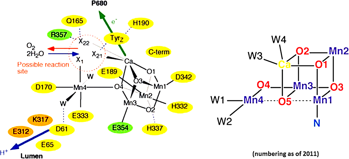
Most important has been their model for the metal-cluster (see Fig. 2). In conceiving this far sighted structural model the authors relied on their own diffraction data, including the anomalous diffraction aiming at Mn (X-ray wavelength 1.89 Å) and Ca (2.25 Å), the Mn–Mn and Mn–Ca distances from EXAFS-studies of the Berkeley group (Yachandra et al., Reference Yachandra, Guiles, Mcdermott, Cole, Britt, Dexheimer, Sauer and Klein1987b), knowledge on μ-oxo-linked Mn-model-clusters as previously discussed (Brudvig and Crabtree, Reference Brudvig and Crabtree1989), and the proposed Mn3 + Mn(dangler)-structure (Peloquin and Britt, Reference Peloquin and Britt2001).
Left: The first structural model of the catalytic centre (Ferreira et al., Reference Ferreira, Iverson, Maghlaoui, Barber and Iwata2004). Right: Schematic representation of the Mn4CaO5(H2O)4-complex with the numbering as used after the first model at high-resolution (Umena et al., Reference Umena, Kawakami, Shen and Kamiya2011).
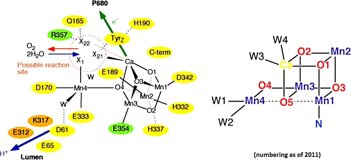
The Barber-model of the metal cluster had three Mn plus one Ca forming a cubanoid and the fourth Mn dangling out. The ions were bridged by four oxygen atoms and ligated by four water molecules (though not clearly resolved). A bicarbonate was postulated to bridge the Ca-ion and the dangling Mn. Four protein residues ligated the Mn4CaO4-cluster, with Yz = D1-Tyr161 and its hydrogen-bonded partner, D1-His190, in close vicinity to the former. In essence, this model has set the path until today. One year later the Berlin group presented a model at slightly higher resolution of 3 Å, where all carboxylate residues were now bi-dentate bridging. Even the one that was terminally bound to one manganese in Barber's model was now bridging two metals (Loll et al., Reference Loll, Kern, Saenger, Zouni and Biesiadka2005). Again the resolution was not sufficient to resolve bridging oxygen atoms and water.
Johannes Messinger reviewed the status of agreements and diverging views up to 2004. He discussed two possible mechanisms for O–O bond formation, namely involving the dangler Mn and one Ca-bound water on the one hand, and involving a bridging oxygen and a dangler-bound water (Messinger, Reference Messinger2004).
Crystals exposed to a CW-X-ray beam from a synchrotron undergo radiation damage. By X-ray spectroscopy Holger Dau had detected dose-dependent Mn-reduction in PSII (Dau et al., Reference Dau, Dittmer, Iuzzolino, Schiller, Dorner, Heinze, Sole and Nolting1997). The larger Mn–Mn bond length in Barber's crystal structure as compared with the one inferred from EXAFS (Dau et al., Reference Dau, Liebisch and Haumann2004) raised serious doubt on the valence-state of the Mn4Ca-cluster in Barber's model. The Berlin group joined forces with the Berkeley group and they clearly demonstrated that X-ray exposure (doses by order of magnitude lower than causing loss of diffractivity) reduced Mn4 from the expected valence of state S1 (III,III,IV,IV) to (II,II,II,II) (Yano et al., Reference Yano, Kern, Irrgang, Latimer, Bergmann, Glatzel, Pushkar, Biesiadka, Loll, Sauer, Messinger, Zouni and Yachandra2005). It explained why the Mn–Mn distances of the structural model were larger than very precisely determined by EXAFS. At the same time the valence-set of the four Mn-ions was determined by 55Mn-pulse-NMR in Wolfgang Lubitz’ lab. It was (III,III,III,IV) in S0 and (III,IV,IV,IV) in S2 (Kulik et al., Reference Kulik, Epel, Lubitz and Messinger2005).
Soon thereafter Per Siegbahn took Barber's arrangement of the metals and their ligands (with modifications by the Berlin group) as the basis for modelling Mn4CaOx-clusters in silico (Siegbahn, Reference Siegbahn2006). By DFT and energy minimization a particular configuration of Mn4CaO5 was selected to represent the HV state S4 from where the O–O bond is formed. The Mn–Mn bond lengths in the energy minimized structure were shorter than in Barber's. In this respect Siegbahn's modelling was predictive for radiation damage and not only post mortem descriptive as common in computational biochemistry/biophysics.
It was not astounding that, for a while, Barber's structure of the metal cluster was met with reservations, because it has been all but trivial, that the metal and ligand positions in PSII are the same if the valence of Mn4 is (III,III,IV,IV), as supposed for S1, or (II,II,II,II), as after radiation damage.
Subsequent work on the atomic structure has been aimed at (a) minimization of radiation damage, (b) improved spatial resolution, (c) selective characterization of the five functional states S0 to S4 and (d) time resolution of structural transitions between them. The latter necessitates X-ray diffraction at room temperature.
The first high-resolution structure of PSII at 1.9 Å was published by Jian-Ren Shen and Nobuo Kamiya (Umena et al., Reference Umena, Kawakami, Shen and Kamiya2011). Barber's cubanoid for Mn3Ca with the dangling Mn (Ferreira et al., Reference Ferreira, Iverson, Maghlaoui, Barber and Iwata2004), and the ligand structure of the Berlin group (Loll et al., Reference Loll, Kern, Saenger, Zouni and Biesiadka2005) were corroborated in essence. For the first time, the bridging oxygen atoms and four water molecules were clearly resolved, and former Mn4CaO4 became Mn4CaO5(H2O)4, as schematically illustrated in Fig. 2. Barber's numbering of the metal ions and oxygen atoms has been changed accordingly. The position of the dangler-Mn relative to the cubane was corrected by 3 Å, and Asp 170 was now a bidentate ligand (bridging Mn4 and Ca) and not monodentate, as before. This structural model was obtained by continuous synchrotron radiation (CW-XRD), and at cryogenic temperature. The Mn–Mn distances were still slightly longer than in both, EXAFS experiments and DFT calculations, and hence still indicative of some radiation damage.
A damage-free structure was desirable. X-ray diffraction with ultra-short pulses (typ. duration, <50 fs) of a free electron laser (XFEL) seemed to offer a way out. The pioneering study was published in 2012 by the joined groups of Berlin (i.a. Athina Zouni), Berkeley (i.a. Jan Kern, Junko Yano, Vittal Yachandra) and Umeå (Johannes Messinger). Very many stochastically oriented micro-crystals of PSII were each exposed to a single X-ray shot to ‘diffract before destruction’ (Kern et al., Reference Kern, Alonso-Mori, Hellmich, Tran, Hattne, Laksmono, Glockner, Echols, Sierra, Sellberg, Lassalle-Kaiser, Gildea, Glatzel, Grosse-Kunstleve, Latimer, Mcqueen, Difiore, Fry, Messerschmidt, Miahnahri, Schafer, Seibert, Sokaras, Weng, Zwart, White, Adams, Bogan, Boutet, Williams, Messinger, Sauter, Zouni, Bergmann, Yano and Yachandra2012). It followed a series of such XFEL studies (e.g. Kern et al., Reference Kern, Alonso-Mori, Tran, Hattne, Gildea, Echols, Glockner, Hellmich, Laksmono, Sierra, Lassalle-Kaiser, Koroidov, Lampe, Han, Gul, Difiore, Milathianaki, Fry, Miahnahri, Schafer, Messerschmidt, Seibert, Koglin, Sokaras, Weng, Sellberg, Latimer, Grosse-Kunstleve, Zwart, White, Glatzel, Adams, Bogan, Williams, Boutet, Messinger, Zouni, Sauter, Yachandra, Bergmann and Yano2013, Reference Kern, Tran, Alonso-Mori, Koroidov, Echols, Hattne, Ibrahim, Gul, Laksmono, Sierra, Gildea, Han, Hellmich, Lassalle-Kaiser, Chatterjee, Brewster, Stan, Gloeckner, Lampe, Difiore, Milathianaki, Fry, Seibert, Koglin, Gallo, Uhlig, Sokaras, Weng, Zwart, Skinner, Bogan, Messerschmidt, Glatzel, Williams, Boutet, Adams, Zouni, Messinger, Sauter, Bergmann, Yano and Yachandra2014; Hellmich et al., Reference Hellmich, Bommer, Burkhardt, Ibrahim, Kern, Meents, Muh, Dobbek and Zouni2014; Kupitz et al., Reference Kupitz, Basu, Grotjohann, Fromme, Zatsepin, Rendek, Hunter, Shoeman, White, Wang, James, Yang, Cobb, Reeder, Sierra, Liu, Barty, Aquila, Deponte, Kirian, Bari, Bergkamp, Beyerlein, Bogan, Caleman, Chao, Conrad, Davis, Fleckenstein, Galli, Hau-Riege, Kassemeyer, Laksmono, Liang, Lomb, Marchesini, Martin, Messerschmidt, Milathianaki, Nass, Ros, Roy-Chowdhury, Schmidt, Seibert, Steinbrener, Stellato, Yan, Yoon, Moore, Moore, Pushkar, Williams, Boutet, Doak, Weierstall, Frank, Chapman, Spence and Fromme2014) carried out at room temperature and aiming at improved resolution.
Shen and his colleagues also conducted XFEL experiments. In contrast to the former group they used large crystals with volume-per-volume exposure at cryogenic temperature. In 2015 they presented a ‘damage-free’ structural model at a resolution of 1.95 Å (Suga et al., Reference Suga, Akita, Hirata, Ueno, Murakami, Nakajima, Shimizu, Yamashita, Yamamoto, Ago and Shen2015). The Mn–Mn distances were shorter than previously obtained by conventional CW-XRD (Ferreira et al., Reference Ferreira, Iverson, Maghlaoui, Barber and Iwata2004; Umena et al., Reference Umena, Kawakami, Shen and Kamiya2011), and they were better compatible with those from EXFAS and DFT (for the remaining slight difference, see Fig. 1 in Siegbahn (Reference Siegbahn2013b)). The 1.95 Å-structure has become the standard for the following discussions.
A long hedged dream of inorganic chemists came true when Chunxi Zhang and his colleagues synthesized a biomimetic analogue of nature's Mn4Ca-cluster (Zhang et al., Reference Zhang, Chen, Dong, Shen, Dau and Zhao2015).
The obvious challenge was now to determine the damage-free structure of the catalytic centre at room temperature, in its different oxidation states (S0, …, S4), and, if possible, time resolved.
In 2016 the Berkeley–Berlin team published a XFEL-study on structural differences between states S1 and S3 of the metal cluster, as function of the temperature, and with and without ammonia bound (Young et al., Reference Young, Ibrahim, Chatterjee, Gul, Fuller, Koroidov, Brewster, Tran, Alonso-Mori, Kroll, Michels-Clark, Laksmono, Sierra, Stan, Hussein, Zhang, Douthit, Kubin, De Lichtenberg, Pham, Nilsson, Cheah, Shevela, Saracini, Bean, Seuffert, Sokaras, Weng, Pastor, Weninger, Fransson, Lassalle, Brauer, Aller, Docker, Andi, Orville, Glownia, Nelson, Sikorski, Zhu, Hunter, Lane, Aquila, Koglin, Robinson, Liang, Boutet, Lyubimov, Uervirojnangkoorn, Moriarty, Liebschner, Afonine, Waterman, Evans, Wernet, Dobbek, Weis, Brunger, Zwart, Adams, Zouni, Messinger, Bergmann, Sauter, Kern, Yachandra and Yano2016). With ammonia the authors mapped those water binding sites in the metal-cluster that are not involved in the formation of the O–O bond. While temperature affected some positions of transmembrane α-helices and redox-cofactors, the Mn4CaO5-cluster, proper, was practically invariant to temperature. At a resolution of 2.25 Å the authors did not detect differences between the dark state (S1) and the ‘two flashes advanced’ state (mainly S3) (Young et al., Reference Young, Ibrahim, Chatterjee, Gul, Fuller, Koroidov, Brewster, Tran, Alonso-Mori, Kroll, Michels-Clark, Laksmono, Sierra, Stan, Hussein, Zhang, Douthit, Kubin, De Lichtenberg, Pham, Nilsson, Cheah, Shevela, Saracini, Bean, Seuffert, Sokaras, Weng, Pastor, Weninger, Fransson, Lassalle, Brauer, Aller, Docker, Andi, Orville, Glownia, Nelson, Sikorski, Zhu, Hunter, Lane, Aquila, Koglin, Robinson, Liang, Boutet, Lyubimov, Uervirojnangkoorn, Moriarty, Liebschner, Afonine, Waterman, Evans, Wernet, Dobbek, Weis, Brunger, Zwart, Adams, Zouni, Messinger, Bergmann, Sauter, Kern, Yachandra and Yano2016).
Photosystem II with the oxygen evolving complex (AC)
Mechanistic models for O–O bond formation
Adopting a chemical mechanism that had been proposed by V. Pecoraro and his colleagues (Pecoraro et al., Reference Pecoraro, Baldwin, Caudle, Hsieh and Law1998) James Barber speculated that the dioxygen-bond is formed by nucleophilic attack of a water ligand to the Ca-ion on a highly electrophilic oxo supposedly bound to the dangler-Mn(V) (Ferreira et al., Reference Ferreira, Iverson, Maghlaoui, Barber and Iwata2004; Iwata and Barber, Reference Iwata and Barber2004). Barber has favoured this concept until today (Barber, Reference Barber2016, Reference Barber2017). His view is in line with earlier computational studies by P. Siegbahn (Siegbahn and Crabtree, Reference Siegbahn and Crabtree1999; Siegbahn, Reference Siegbahn2000), and it has been shared recently by others (McEvoy et al., Reference Mcevoy, Gascon, Batista and Brudvig2005; Sproviero et al., Reference Sproviero, Gascon, Mcevoy, Brudvig and Batista2006, Reference Sproviero, Gascon, Mcevoy, Brudvig and Batista2008; Askerka et al., Reference Askerka, Brudvig and Batista2017).
After the first structural model of the metal-cluster had been published Per Siegbahn investigated by DFT the energy profile of various mechanistic models for the formation of the O–O bond. Calculations of the energy demand for an O–O-bonded intermediate in the Mn4CaO5-structure led him to reject the nucleophilic attack mechanism. He has since then proposed a radical attack mechanism involving a new bridging oxygen (O5) within an open cubanoid (coined oxyl-oxo-mechanism) (Siegbahn, Reference Siegbahn2006) that was not included in Barber's structural model. O5 resulted from ‘predictive computational chemistry’, and it was only later observed in diffraction experiments at higher resolution. Siegbahn has extended this work later on (Siegbahn, Reference Siegbahn2006, Reference Siegbahn2008a, Reference Siegbahn2017; Siegbahn and Blomberg, Reference Siegbahn and Blomberg2014).
Yu Guo and his coworkers embarked on similar DFT calculations and likewise favoured an oxo-oxyl mechanism involving the same bridging oxygen (O5) in an open cubanoid (Guo et al., Reference Guo, Li, He, Zhao, Gong and Yang2017). On first inspection Guo's results are in line with Siegbahn's, however a closer look reveals that their calculated energy profile for the last transition (S3 → S0) violates the experimentally established fact that oxygen liberation and Yz+-reduction are both kinetically enslaved by the same rate limiting step. In his calculated energy profile these two reactions are separated by an energy notch, 12 kcal/mol deep (see their Fig. 4; Guo et al., Reference Guo, Li, He, Zhao, Gong and Yang2017). Siegbahn's calculated energy profile on the other hand is compatible with the kinetic data.
The proponents of the leading concepts for the formation of the O–O bond, nucleophilic attack versus oxo-oxyl, have presented their diverging views in series of publications starting from 2004 (Ferreira et al., Reference Ferreira, Iverson, Maghlaoui, Barber and Iwata2004) and 2006 (Siegbahn, Reference Siegbahn2006) until today. While James Barber most recently argued in favour of nucleophilic attack by analogy with the known mechanism of carbon dioxide dehydrogenase (Barber, Reference Barber2017), Per Siegbahn (Siegbahn, Reference Siegbahn2017) repeated his previous (Siegbahn, Reference Siegbahn2006) exclusion of nucleophilic attack on energetic grounds.
In addition to the above two concepts for the formation of the O–O bonds several modified or even alternative mechanisms have been proposed (see e.g. Messinger, Reference Messinger2000; Gao et al., Reference Gao, Akermark, Liu, Sun and Akermark2009; Yamanaka et al., Reference Yamanaka, Isobe, Kanda, Saito, Umena, Kawakami, Shen, Kamiya, Okumura, Nakamura and Yamaguchi2011; Saito et al., Reference Saito, Yamanaka, Kanda, Isobe, Takano, Shigeta, Umena, Kawakami, Shen, Kamiya, Okumura, Shoji, Yoshioka and Yamaguchi2012; Cox and Messinger, Reference Cox and Messinger2013).
Experimental tests of proposed mechanisms
The light driven stepping over the intermediate states of the ‘charge accumulator’ (Mn4CaO5) has been kinetically well characterized (for a comprehensive review, see Dau et al. (Reference Dau, Zaharieva and Haumann2012)). The eventually fourfold oxidized YzMn4Ca-entity reacts with two molecules of water (-derivatives) which involves the transfer of four electrons. The terminal four-electron-cascade is kinetically enslaved by a bottle neck of approximately 1 ms duration. Characterizing the short-lived intermediates is difficult if not impossible. The formation of a peroxide intermediate in the penultimate state S3 has been in focus.
The valence of the four manganese ions in states Si (i = 0–4)
A first stronghold for the assignment of oxidation states to the four Mn-atoms has been the EPR-multiline EPR-signal of the S2-state. It has been attributed to the interaction between Mn(III) and Mn(IV) (Dismukes and Siderer, Reference Dismukes and Siderer1981). Two valence configurations of the four Mn-ions are compatible with this notion, namely Mn(III)3Mn(IV) and Mn(III)Mn(IV)3 (de Paula and Brudvig, Reference De Paula and Brudvig1985). These assignments have been extended into two schemes for the Si-series which are usually referred to as low-valence (LV) and HV models. An enormous amount of work by different techniques had been devoted to discriminate between these models, for a while without reaching consent (reviewed in Krewald et al. (Reference Krewald, Retegan, Cox, Messinger, Lubitz, Debeer, Neese and Pantazis2015)). The comparison of NEXAFS-data with DFT-calculations supported the HV-model, although a few complications seemed to prevent an unequivocal attribution (Brena et al., Reference Brena, Siegbahn and Agren2012). Related to the above controversy has been the one over ligand-centred versus Mn-centred oxidation in the transition S2 → S3. The former was advocated in Yachandra et al. (Reference Yachandra, Sauer and Klein1996) and Messinger et al. (Reference Messinger, Robblee, Bergmann, Fernandez, Glatzel, Visser, Cinco, Mcfarlane, Bellacchio, Pizarro, Cramer, Sauer, Klein and Yachandra2001) and the latter in Iuzzolino et al., Reference Iuzzolino, Dittmer, D'RNER, Meyer-Klaucke and Dau1998; Dau et al., Reference Dau, Liebisch and Haumann2003; Siegbahn, Reference Siegbahn2009; Brena et al., Reference Brena, Siegbahn and Agren2012. 55Mn-ENDOR studies on states S0 and S2 (Kulik et al., Reference Kulik, Epel, Lubitz and Messinger2005) and XAS-studies on S2 and S3 (Dau et al., Reference Dau, Liebisch and Haumann2003) have established the HV concept for the four manganese ions, namely, S0 → S1 → S2 → S3 = (III,III,III,IV) → (III,III,IV,IV) → (III,IV,IV,IV) → (IV,IV,IV,IV).
The oxidation of manganese during S2 → S3 and the existence of a Mn4(IV)-state has been corroborated by X-ray emission spectroscopy (Zaharieva et al., Reference Zaharieva, Chernev, Berggren, Anderlund, Styring, Dau and Haumann2016a; Schuth et al., Reference Schuth, Zaharieva, Chernev, Berggren, Anderlund, Styring, Dau and Haumann2018).
Two lines of computational studies have agreed on that the S2-state is interconvertible between two sub-states. In one state, coined S2B, the only trivalent ion is Mn4(III), the dangler outside of the cubane. In the other state, coined S2A, it is Mn1(III) within the cubane. Both these studies agreed in that only S2B can proceed to S3 owing to the presence of a titratable ligand (Bovi et al., Reference Bovi, Narzi and Guidoni2013; Retegan et al., Reference Retegan, Krewald, Mamedov, Neese, Lubitz, Cox and Pantazis2016). While S2A is more stable than S2B the oxidation of Yz inverts this situation such that S2B passes by proton-coupled electron transfer into state S3 (Mn(IV)4) (Narzi et al., Reference Narzi, Bovi and Guidoni2014).
Krewald et al. cast the available data, including their own on hyperfine coupling constants, into one consistent theoretical framework (Krewald et al., Reference Krewald, Retegan, Cox, Messinger, Lubitz, Debeer, Neese and Pantazis2015). They found that the most stable states, the open cubane structure of S2 and the closed cubane of S3, correspond to structures proposed earlier (based on DFT) by Siegbahn (Siegbahn, Reference Siegbahn2013a). In contrast to speculation in the literature their simulations yielded no evidence for ligand centred oxidation between S2 and S3. And they showed that only the HV-model complies with the data but not the LV-model (Krewald et al., Reference Krewald, Retegan, Cox, Messinger, Lubitz, Debeer, Neese and Pantazis2015). In light of these results, state S3 is to be conceived as YzMn4(IV) or Yz+°Mn3(IV)Mn(III) (Cox et al., Reference Cox, Retegan, Neese, Pantazis, Boussac and Lubitz2014; Retegan et al., Reference Retegan, Cox, Lubitz, Neese and Pantazis2014), and state S4 asYz+°Mn4(IV). The latter initiates the formation of the O–O bond.
The entry of water into the reaction cycle
The points of entry of solvent water into the reaction sequence of oxygen evolution were at first characterized by mass spectroscopy (Messinger et al., Reference Messinger, Badger and Wydrzynski1995; Hillier et al., Reference Hillier, Messinger and Wydrzynski1998). The location of the binding sites was then unknown. The μ-oxo-bridges were initially considered as too strong for solvent exchange and catalytic relevance. Hydrogen exchange studies and 17O-NMR spectroscopy has recently demonstrated that at least one μ-oxo bridge in the metal cluster, either O4 or O5, is solvent exchangeable (Rapatskiy et al., Reference Rapatskiy, Cox, Savitsky, Ames, Sander, Nowaczyk, Rogner, Boussac, Neese, Messinger and Lubitz2012). The latter μ-oxo might participate in O–O bond formation as previously postulated by Siegbahn (Siegbahn, Reference Siegbahn2006).
Dimitrios Pantazis and colleagues (Retegan et al., Reference Retegan, Krewald, Mamedov, Neese, Lubitz, Cox and Pantazis2016) in Wolfgang Lubitz’ lab in Mülheim discussed water exchange in the light of experiments on the Mn-coordination number in the S3 state (Haumann et al., Reference Haumann, Muller, Liebisch, Iuzzolino, Dittmer, Grabolle, Neisius, Meyer-Klaucke and Dau2005) and of recent work on water exchange, when progressing through the Si states (Suzuki et al., Reference Suzuki, Sugiura and Noguchi2008). One water molecule binds after oxygen release. It has been implied to form the di-μ-oxo bridge in the S0-state (Krewald et al., Reference Krewald, Retegan, Cox, Messinger, Lubitz, Debeer, Neese and Pantazis2015) and a fluxional bridge in the S2-state (Bovi et al., Reference Bovi, Narzi and Guidoni2013). The second water binds in the S2 → S3 transition. It is probably not the reacting water in the next turnover (Suzuki et al., Reference Suzuki, Sugiura and Noguchi2008; Cox and Messinger, Reference Cox and Messinger2013; Nilsson et al., Reference Nilsson, Krupnik, Kargul and Messinger2014a). Pantazis proposed that W2 on Mn4, it pivots close to O5, might act as reaction partner in the formation of the O–O bond (Retegan et al., Reference Retegan, Krewald, Mamedov, Neese, Lubitz, Cox and Pantazis2016). This view is however at odds with ammonia mapping of reactive water by the Berlin–Berkeley team which has let them to exclude W2 in this role (Young et al., Reference Young, Ibrahim, Chatterjee, Gul, Fuller, Koroidov, Brewster, Tran, Alonso-Mori, Kroll, Michels-Clark, Laksmono, Sierra, Stan, Hussein, Zhang, Douthit, Kubin, De Lichtenberg, Pham, Nilsson, Cheah, Shevela, Saracini, Bean, Seuffert, Sokaras, Weng, Pastor, Weninger, Fransson, Lassalle, Brauer, Aller, Docker, Andi, Orville, Glownia, Nelson, Sikorski, Zhu, Hunter, Lane, Aquila, Koglin, Robinson, Liang, Boutet, Lyubimov, Uervirojnangkoorn, Moriarty, Liebschner, Afonine, Waterman, Evans, Wernet, Dobbek, Weis, Brunger, Zwart, Adams, Zouni, Messinger, Bergmann, Sauter, Kern, Yachandra and Yano2016).
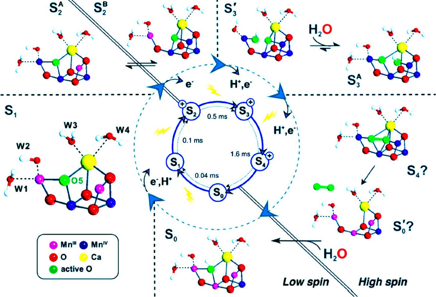
Christopher Kim and Richard Debus investigated the vibrational modes of bound water by FTIR-spectroscopy (Kim and Debus, Reference Kim and Debus2017). The D–O–D bending-mode of one water molecule is altered upon functional substitution of Sr for Ca. This particular hydrogen-bonded water molecule is eliminated during the transition S2 → S3. The authors proposed that this water molecule, probably W3, is deprotonated and moves to a position close to O5 as partner for O–O bond formation in the next transition (S3 → S4–S0). During S2 → S3 the vacant water position on Ca is refilled by the incoming substrate water (Kim and Debus, Reference Kim and Debus2017). Their view is compatible with the reorganization of hydrogen bonds as inferred from a large H/D-kinetic isotope effect on the reduction of Yz& as monitored by UV- (Haumann et al., Reference Haumann, Bogershausen, Cherepanov, Ahlbrink and Junge1997a), X-ray- (Zaharieva et al., Reference Zaharieva, Dau and Haumann2016b) and FTIR-spectroscopy (Sakamoto et al., Reference Sakamoto, Shimizu, Nagao and Noguchi2017). Nicholas Cox and colleagues (Krewald et al., Reference Krewald, Retegan, Neese, Lubitz, Pantazis and Cox2016) cast the available spectroscopic evidence into a spin-geared reaction scheme where the O–O bond is formed within the cubane. Their scheme is illustrated in Fig. 3.
Tentative scheme for the cyclic stepping of the catalytic metal cluster over Kok's states S0 → S1 → S2 → S3 → S4 and the formation of the dioxygen bond (Krewald et al., Reference Krewald, Retegan, Neese, Lubitz, Pantazis and Cox2016).

The above spectroscopic results from Wolfgang Lubitz lab and other laboratories are in line with P. Siegbahn's oxyl-oxo-mechanism (Siegbahn, Reference Siegbahn2006, Reference Siegbahn2017) and at odds with J. Barber's nucleophilic attack mechanism (Ferreira et al., Reference Ferreira, Iverson, Maghlaoui, Barber and Iwata2004; Barber, Reference Barber2017). They qualify alternative computational models (Kusunoki, Reference Kusunoki2007, Reference Kusunoki2011), in particular those based on LV of the Mn-cluster (Sproviero et al., Reference Sproviero, Gascon, Mcevoy, Brudvig and Batista2008; Gatt et al., Reference Gatt, Stranger and Pace2011; Petrie et al., Reference Petrie, Gatt, Stranger and Pace2012; Li et al., Reference Li, Sproviero, Ryde, Batista and Chen2013). The challenge has been back to structural biology.
Structural evidence for/against proposed mechanisms of O–O bond formation
In 2016 the Berkeley–Berlin team published a XFEL-study on structural differences between states S1 and S3 of the metal cluster at room temperature (Young et al., Reference Young, Ibrahim, Chatterjee, Gul, Fuller, Koroidov, Brewster, Tran, Alonso-Mori, Kroll, Michels-Clark, Laksmono, Sierra, Stan, Hussein, Zhang, Douthit, Kubin, De Lichtenberg, Pham, Nilsson, Cheah, Shevela, Saracini, Bean, Seuffert, Sokaras, Weng, Pastor, Weninger, Fransson, Lassalle, Brauer, Aller, Docker, Andi, Orville, Glownia, Nelson, Sikorski, Zhu, Hunter, Lane, Aquila, Koglin, Robinson, Liang, Boutet, Lyubimov, Uervirojnangkoorn, Moriarty, Liebschner, Afonine, Waterman, Evans, Wernet, Dobbek, Weis, Brunger, Zwart, Adams, Zouni, Messinger, Bergmann, Sauter, Kern, Yachandra and Yano2016). At a resolution of 2.25 Å no difference in electron density was detected between the dark state (S1) and the ‘two flashes advanced’ state (mainly S3), as mentioned.
In 2017 Shen and coworkers in Japan reported structural differences of the metal-cluster between states S1 and S3 up to a resolution of 2.1 Å (Suga et al., Reference Suga, Akita, Sugahara, Kubo, Nakajima, Nakane, Yamashita, Umena, Nakabayashi, Yamane, Nakano, Suzuki, Masuda, Inoue, Kimura, Nomura, Yonekura, Yu, Sakamoto, Motomura, Chen, Kato, Noguchi, Tono, Joti, Kameshima, Hatsui, Nango, Tanaka, Naitow, Matsuura, Yamashita, Yamamoto, Nureki, Yabashi, Ishikawa, Iwata and Shen2017). Their study was carried out at room temperature and under excitation with 10 fs pulses of a free electron laser. They found an ‘apparent positive peak’ in the difference Fourier map close to oxygen atom O5 which they interpreted as new oxygen, coined O6. O5 and O6 were taken as candidates for the formation of the O–O bond (Suga et al., Reference Suga, Akita, Sugahara, Kubo, Nakajima, Nakane, Yamashita, Umena, Nakabayashi, Yamane, Nakano, Suzuki, Masuda, Inoue, Kimura, Nomura, Yonekura, Yu, Sakamoto, Motomura, Chen, Kato, Noguchi, Tono, Joti, Kameshima, Hatsui, Nango, Tanaka, Naitow, Matsuura, Yamashita, Yamamoto, Nureki, Yabashi, Ishikawa, Iwata and Shen2017) in line with Siegbahn's computational results (Siegbahn, Reference Siegbahn2006, Reference Siegbahn2008b, Reference Siegbahn2009). The authors discussed that Glu189, the only mono-dentate ligand to the cluster, had to move away from the cubane to accommodate O6.
When taken at face value this result could put an end to the major controversy over the mechanism of the O–O bond formation (nucleophilic attack versus oxyl-oxo).
At present, however, several features call for scrutiny: (a) the ‘apparent positive peak’ in Shen's difference map (interpreted as O6) is hardly elevated over noise and, at the given resolution, superimposed by electron density of Mn. (b) It is not yet clear to what extent it depends on particular software for the evaluation of raw data. (c) The purity of the dark state (100% S1?) and of the ‘2-flash-advanced state’ (45% S3 in Shen's implies 55% S2!) has been difficult to assess in crystals of PSII. (d) Spectroscopic evidence disfavours the presence of a peroxide intermediate in state S3. (e) The supposedly ‘moving’ residue Glu189 has been point mutated into Arg, Lys and Gln without influence on the rates of various electron transfer reactions at the donor side of PSII (Clausen et al., Reference Clausen, Winkler, Hays, Hundelt, Debus and Junge2001).
The Berlin–Berkeley group has advanced the search technique for intermediates of the O–O bond by combining serial femtosecond X-ray crystallography with simultaneous X-ray spectroscopy under multi-flash visible laser excitation (Kern et al., Reference Kern, Alonso-Mori, Tran, Hattne, Gildea, Echols, Glockner, Hellmich, Laksmono, Sierra, Lassalle-Kaiser, Koroidov, Lampe, Han, Gul, Difiore, Milathianaki, Fry, Miahnahri, Schafer, Messerschmidt, Seibert, Koglin, Sokaras, Weng, Sellberg, Latimer, Grosse-Kunstleve, Zwart, White, Glatzel, Adams, Bogan, Williams, Boutet, Messinger, Zouni, Sauter, Yachandra, Bergmann and Yano2013). In principle, this technique allows time resolution of structural transitions by variation of the time delays between the X-ray and the visible flashes. The major challenge is to obtain the necessary spatial resolution (<2 Å) to reveal the expected displacement of water (oxygen) molecules in 100 µs during the transition from S2 towards S3 and onwards via ‘S4’ to S0 in a millisecond. Recently, this team has reported the structure of all states of Kok's cycle at 2.04–2.08 Å resolution including two transient states during S2 → S3 at 150 and 400 µs after excitation by a flash of visible light (Kern et al., Reference Kern, Chatterjee, Young, Fuller, Lassalle, Ibrahim, Gul, Fransson, Brewster, Alonso-Mori, Hussein, Zhang, Douthit, De Lichtenberg, Cheah, Shevela, Wersig, Seuffert, Sokaras, Pastor, Weninger, Kroll, Sierra, Aller, Butryn, Orville, Liang, Batyuk, Koglin, Carbajo, Boutet, Moriarty, Holton, Dobbek, Adams, Bergmann, Sauter, Zouni, Messinger, Yano and Yachandra2018). Their data exclude the formation of a peroxide intermediate already in state S3. In S3 they found the binding of a new ‘Ox’ (a water derivative) in a position between Mn1 and Ca. In the next step, S3 → S4–S 0 it could either form an O–O bond with O5 or replace O5 after formation of such a bond between O5 and another Ox. These results are in perfect agreement with the above cited results obtained by X-ray and magnetic resonance spectroscopy. There is no evidence that a peroxide intermediate is already formed in state S3. But it is expected that one intra-cubanoid oxo-group, namely O5, is involved in the formation of the O–O bond in the next step, S3 → S4–S0.
Perspective for a rigorous resolution of the molecular mechanism
The rigorous resolution (in space and time) of the four one-electron steps and the eventual four-electron reaction cascade of this pivotal reaction for life on earth is complicated by two features: (a) the great stability of the (Mn4CaO5cage)-structure and (b) the seeming kinetic enslaving of six partial reactions by one particular step.
(a) The Mn4CaO5-cluster is very stable, and its expected structural changes during the terminal reaction cascade are probably subtle. Even gross valence changes of Mn like those caused by radiation damage in CW-X-ray diffraction (Dau et al., Reference Dau, Dittmer, Iuzzolino, Schiller, Dorner, Heinze, Sole and Nolting1997; Yano et al., Reference Yano, Kern, Irrgang, Latimer, Bergmann, Glatzel, Pushkar, Biesiadka, Loll, Sauer, Messinger, Zouni and Yachandra2005) increase the bond length between metal ions only by small amount (see Siegbahn (Reference Siegbahn2009)). This is why mechanistic insight can hardly be expected from tracking the motion of the metal ions proper.
As already mentioned, the effects of radiation damage have been minimized by either extremely low dose in conventional synchrotron X-ray diffraction (Tanaka et al., Reference Tanaka, Fukushima and Kamiya2017) and by femtosecond irradiation in XFEL-diffraction experiments (Kern et al., Reference Kern, Tran, Alonso-Mori, Koroidov, Echols, Hattne, Ibrahim, Gul, Laksmono, Sierra, Gildea, Han, Hellmich, Lassalle-Kaiser, Chatterjee, Brewster, Stan, Gloeckner, Lampe, Difiore, Milathianaki, Fry, Seibert, Koglin, Gallo, Uhlig, Sokaras, Weng, Zwart, Skinner, Bogan, Messerschmidt, Glatzel, Williams, Boutet, Adams, Zouni, Messinger, Sauter, Bergmann, Yano and Yachandra2014; Tran et al., Reference Tran, Kern, Hattne, Koroidov, Hellmich, Alonso-Mori, Sauter, Bergmann, Messinger, Zouni, Yano and Yachandra2014; Suga et al., Reference Suga, Akita, Hirata, Ueno, Murakami, Nakajima, Shimizu, Yamashita, Yamamoto, Ago and Shen2015). Another type of radiation damage, namely a ‘coulomb-explosion’ of the metal cluster in some 10 fs has only recently been considered. MD-simulations have revealed that it is minimized if the X-ray pulse length is shorter than 10 fs (Amin et al., Reference Amin, Askerka, Batista, Brudvig and Gunner2017, Reference Amin, Badawi and Obayya2016).
It came as a surprise that not only the metal-cluster is extremely stable but its protein cage as well. The Berlin group deleted the metal-cluster from PSII, and they found the protein cage very little altered, as if waiting for the insertion of di-μ-oxo-bridged pairs of manganese (Zhang et al., Reference Zhang, Bommer, Chatterjee, Hussein, Yano, Dau, Kern, Dobbek and Zouni2017).
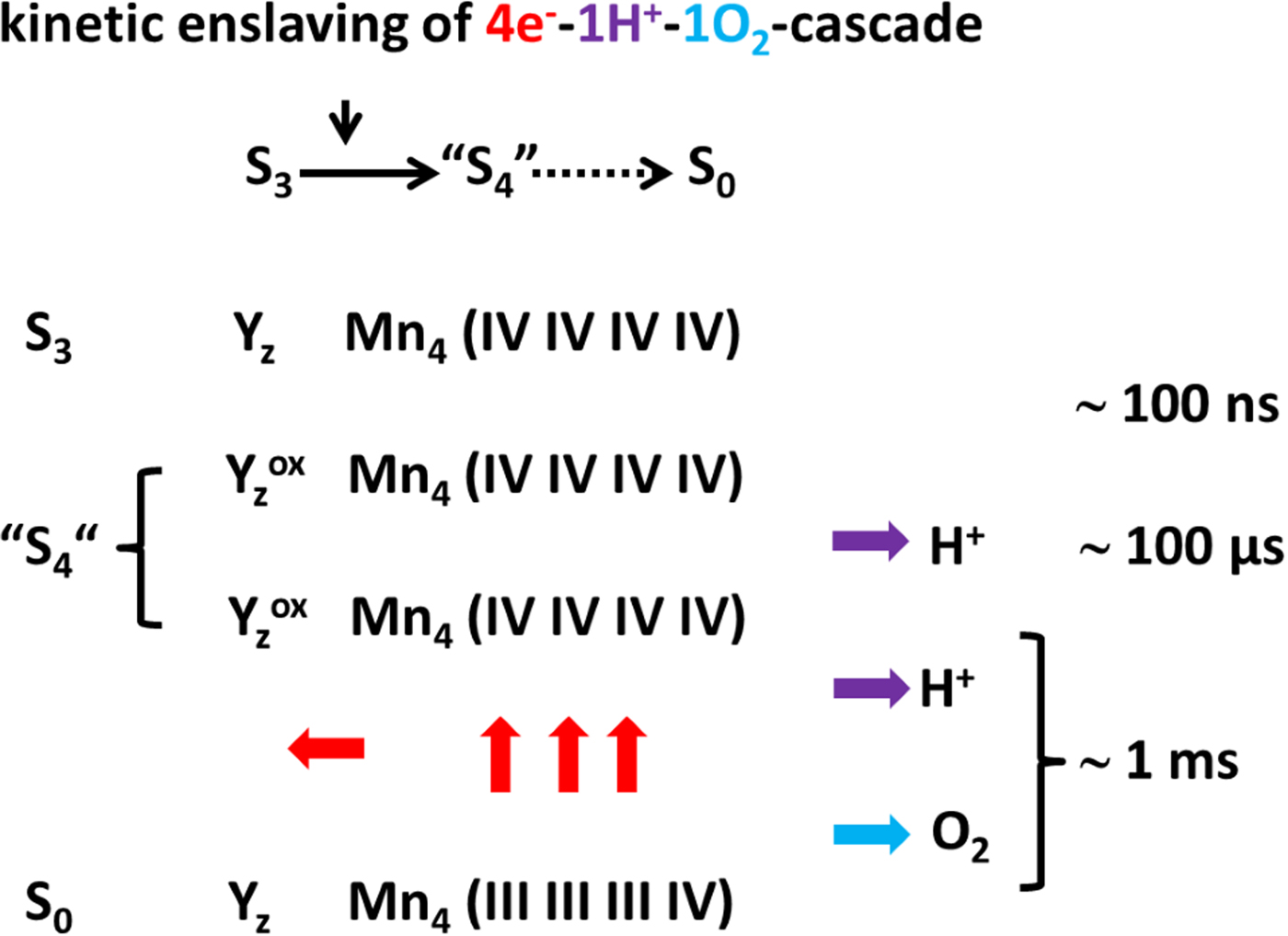
(b) The key to fully understand the mechanism of water oxidation are the trajectories of water, hydroxyl, oxygen, proton and electron. The motion of the former three might be tracked in the near future by time-resolved XFEL-crystallography at <2 Å resolution. Spatial tracking of protons (and electrons) is however not yet in sight. Here one is restricted to kinetic evidence based on spectroscopic data. Here, the kinetic enslaving of six partial reactions by one particular step represents a major challenge as illustrated in Fig. 4.
The terminal reaction cascade of oxygen formation (see the text).

If the valence of Mn4 in state S2 is taken as a reference, namely (IV, IV, IV, III), the penultimate state before oxygen release possibly is: S3 = [Mn(IV)4CaO5(H2O)4Ligands]+. The next photo-excitation creates the sequence of events that is illustrated in Fig. 4. Oxygen is released plus two protons and one water molecule is taken up. The release of two protons is kinetically bipartite, a first step in the time range of 100 µs is followed by a slower step at 1 ms (Förster et al., Reference Förster, Hong and Junge1981). The fast release of one proton seems to be the priming event for the subsequent cascade of six reactions which are kinetically enslaved by one and the same bottle neck of 1 ms half-duration, namely (1) the transfer of one electron to Yz°HisH+, (2) the reduction of three manganese ions, (3) the release of a second proton, (4) the release of di-oxygen and (5) the uptake of one molecule of water. The exact sequence of these events is subject to ongoing research. Michael Haumann and Holger Dau ventured into time-resolved calorimetry using photo-thermal beam deflection (Krivanek et al., Reference Krivanek, Dau and Haumann2008; Klauss et al., Reference Klauss, Krivanek, Dau and Haumann2009). They obtained a wealth of signal transients that were interpreted to show the release of one proton first, oxygen release and water intake second and the release of another proton third (see Fig. 8 in Klauss et al. (Reference Klauss, Haumann and Dau2015)). It seems worthwhile to take up such studies with certain mutant-PSII were oxygen release is dramatically slowed down (Hundelt et al., Reference Hundelt, Hays, Debus and Junge1998; Clausen et al., Reference Clausen, Debus and Junge2004; Nilsson et al., Reference Nilsson, Rappaport, Boussac and Messinger2014b).
Energetics of oxygen production by PSII
Aiming at the stabilization of transient intermediates of the terminal reaction cascade PSII was exposed to high oxygen pressure (Clausen and Junge, Reference Clausen and Junge2004). Certain UV-absorption transients that clearly showed a phase of ~1 ms half-time during S3 → S4–S0 were monitored. These transients had been previously attributed to the reduction of the manganese cluster (Dekker et al., Reference Dekker, Plijter, Ouwehand and Van Gorkom1984). If the oxygen pressure was increased 100-fold over atmospheric this phase was virtually eliminated. Half-inhibition was reached at ≈10-fold increase. It suggested a rather low driving force for photosynthetic oxygen production, implying ‘little leeway for photosynthetic organisms to push the atmospheric oxygen concentration much above the present level’ (Clausen and Junge, Reference Clausen and Junge2004) (for possible ecological and geochemical impact see Raven and Larkum (Reference Raven and Larkum2007)). At first this observation seemed to be corroborated by another technique, delayed chlorophyll-luminescence (Clausen et al., Reference Clausen, Junge, Dau and Haumann2005b). Later however, no stalling effect of increased oxygen pressure was detected by (a) time-resolved X-ray absorption aiming at the K-edge of Mn (Haumann et al., Reference Haumann, Grundmeier, Zaharieva and Dau2008), (b) chlorophyll-fluorescence (Kolling et al., Reference Kolling, Brown, Ananyev and Dismukes2009) and (c) direct detection by membrane-inlet mass spectroscopy of 18O2 produced from H218O against a background of pressurized 16O2 (Shevela et al., Reference Shevela, Beckmann, Clausen, Junge and Messinger2011). In the latter experiments the oxygen pressure was raised up to 20 bar without any indication for stalling of dioxygen liberation. It implies that the driving force is at least 200 meV. Recently this number was driven up by another approach. In one cyanobacterial mutant-PSII the S0-state is stabilized for days. Johannes Messinger and his colleagues incubated this mutant for up to 3 days in H216O and 18O2 without detecting the formation of any amount of 16,18O2 (Nilsson et al., Reference Nilsson, Cournac, Rappaport, Messinger and Lavergne2016). They concluded that the driving force for dioxygen release during S3 → S4–S0 must be greater than 400 meV. They understood this large driving force in terms of Lev Krishtalik's pioneering discussion of the energetics of photosynthetic oxygen evolution in terms of thermodynamic principles (Krishtalik, Reference Krishtalik1989, Reference Krishtalik1990). Jérome Lavergne in Nilsson et al. (Reference Nilsson, Cournac, Rappaport, Messinger and Lavergne2016) attributed the experimentally established high driving force to the large entropic effect of dilution of bound oxygen into the solution (see free energy profile in Fig. 6 of Nilsson et al. (Reference Nilsson, Cournac, Rappaport, Messinger and Lavergne2016)). Their conclusion was that the protein has no grip on this ‘extra enzyme’ contribution to the driving force.
In oxygenic photosynthesis the entropy of dilution favours the release of freshly formed and still bound dioxygen. What about cell respiration where cytochrome oxidase has to overcome this huge entropic barrier?
The ‘equilibrium constant’ for oxygen in cytochrome oxidase was studied by Marten Wikström's group (Krab et al., Reference Krab, Kempe and Wikstrom2011). The authors determined the apparent K m(O2) for steady turnover of the enzyme, at neutral pH and in the absence of protonmotive force, K m(O2) = 25 nM (see their Fig. 4A). The free energy of oxygen binding from O2-saturated water is in the order of −450 mV, very strong indeed, when considering the more than the +300 mV of Krishtalik's entropic contribution to oxygen dilution (see discussion in Nilsson et al. (Reference Nilsson, Rappaport, Boussac and Messinger2014b)).
Oxygenic photosynthesis and a sustainable future of mankind
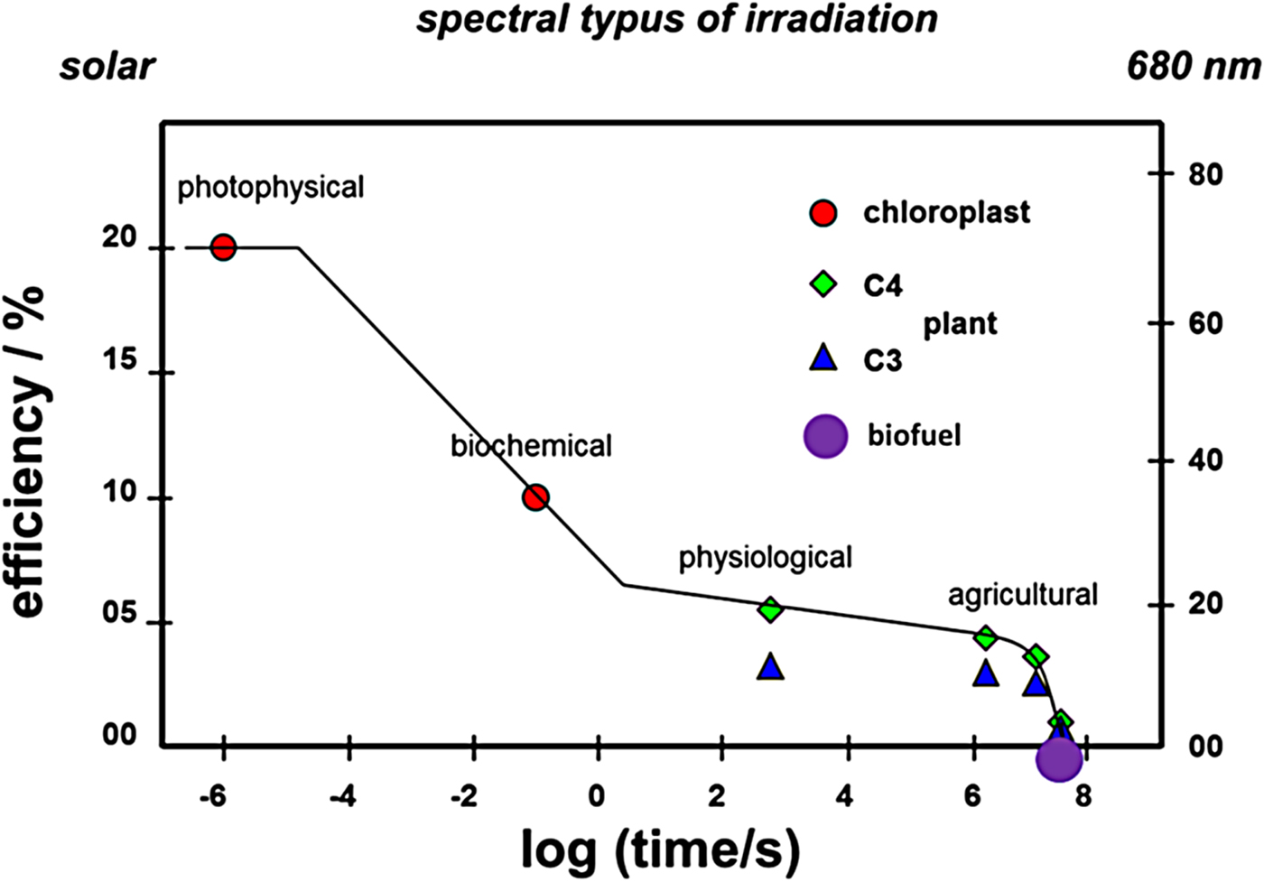
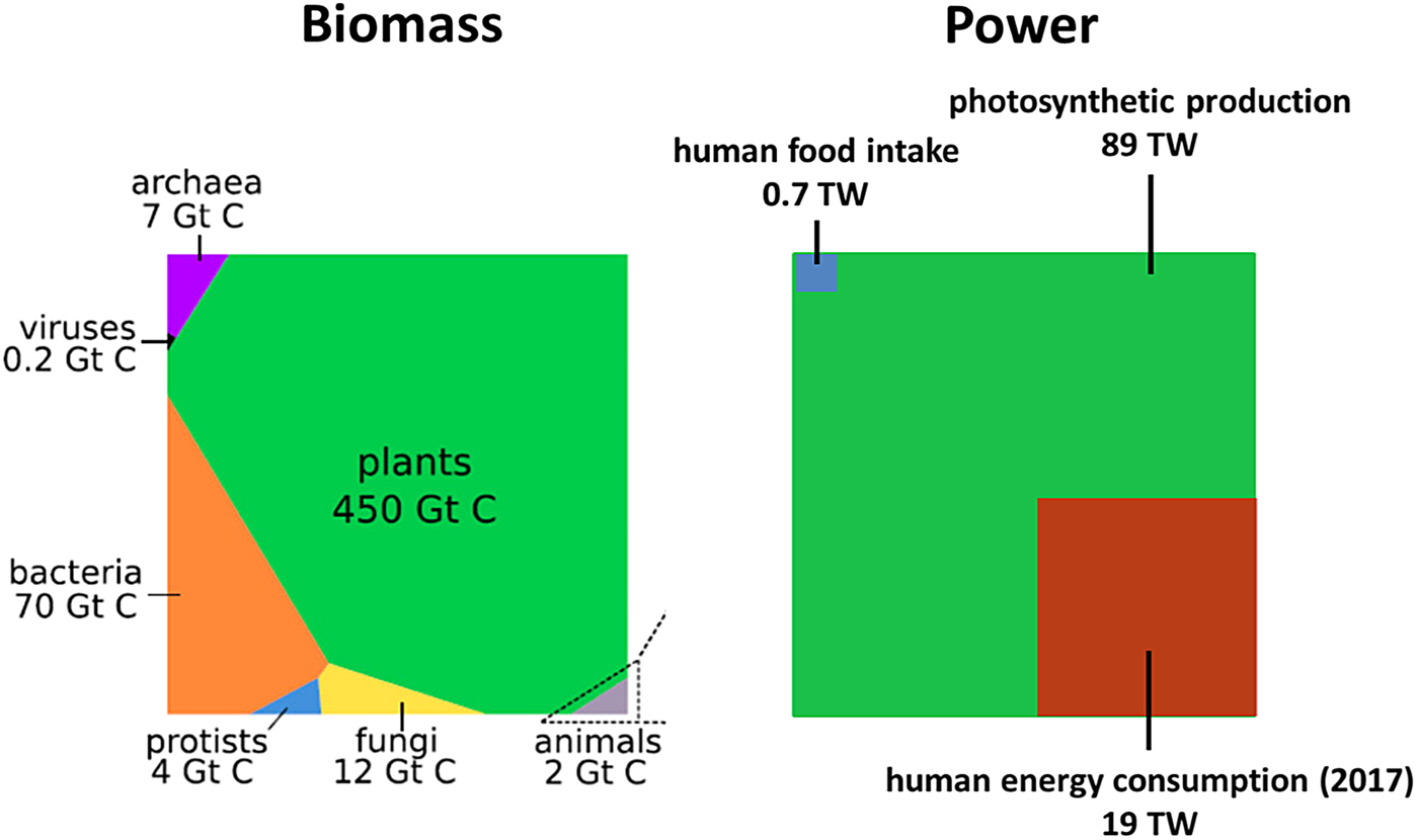
Oxygenic photosynthesis has produced the oxygen we breathe and the biomass serving as food, feed, fibre and fuel, as mentioned. Its driving force is sunlight. The mean power of sunlight at the surface of earth is huge, 124 × 1015 W = 124 PW. Only a very small fraction thereof is captured by photosynthetic organisms, 89 × 1012 W = 89 TW, about half and half on land and in the sea (Field et al., Reference Field, Behrenfeld, Randerson and Falkowski1998; Falkowski et al., Reference Falkowski, Scholes, Boyle, Canadell, Canfield, Elser, Gruber, Hibbard, Hogberg, Linder, Mackenzie, Moore, Pedersen, Rosenthal, Seitzinger, Smetacek and Steffen2000). The low proportion of solar energy capture is partially owed to unfavourable climate, substrate, seasons and the day–night cycle. Another loss of energy capture is intrinsic to the physico-chemical mechanism of photosynthesis, as described above. The solar energy conversion efficiency of photosynthesis is limited even under the most favourable conditions of agriculture, and even more so, if one aims at processed products like biofuels (see Fig. 5).
The efficiency of solar energy conversion by oxygenic photosynthesis as a function of time (see the text).

In the range of microseconds after absorption of a quantum of light the efficiency reaches up to 20% if related to the solar spectrum at the sea level (right scale in Fig. 5). It is about 80% under excitation with red light (see right scale in Fig. 5). These figures refer to a thin canopy (for a thick canopy, see Dau and Zaharieva (Reference Dau and Zaharieva2009)). Therewith the efficiency matches the one of photovoltaic cells (Blankenship et al., Reference Blankenship, Tiede, Barber, Brudvig, Fleming, Ghirardi, Gunner, Junge, Kramer, Melis, Moore, Moser, Nocera, Nozik, Ort, Parson, Prince and Sayre2011). The initial drop (to 20%) is mainly caused by (a) limited use of the full solar spectrum (~50%) and (b) internal energy conversion (‘blue → red’) within chlorophyll. In the test tube and within the time range of 100 ms to yield NADPH the efficiency drops by another factor of two. William Rutherford has coined this drop as ‘sacrificing energy efficiency for directionality’ (Rutherford et al., Reference Rutherford, Osyczka and Rappaport2012). In other words, wasteful back-reactions are overcome by high speed of the forward reaction which requires an energy drop. On the physiological time scale (h) the efficiency drops further from the best vegetative month of the year to the average over a full year. The maximum yearly averaged efficiency at the given level of atmospheric CO2 (380 ppm in 2010) has been estimated by Don Ort's group as 4.6% for C3- and 6% for C4-plants (Zhu et al., Reference Zhu, Long and Ort2008). The efficiency of crops in the field is mostly lower. Take Brazil's large scale energy farming as an example. In 2010 the area specific yield of sugarcane was 80 tdwt ha−1 yr−1. It implies a solar energy conversion efficiency of about 2%. The consecutive conversion of sugarcane into bio-ethanol yielded 6300 l ha−1 yr−1 EthOH, equivalent of an overall energy efficiency of about 0.2%. Slops and slurry were utilized in the process, and the fossil energy input was not counted in this estimate. Various well-to-wheels analyses for biofuel production in the USA and in Europe (e.g. by the JRC in 2012: ISBN 978-9279-21395-3, ISSN 1831-9424, EUR 24952 EN, doi:10.2788/79018 and Torchio and Santarelli, Reference Torchio and Santarelli2010; Wang et al., Reference Wang, Han, Dunn, Cai and Elgowainy2012; Orsi et al., Reference Orsi, Muratori, Rocco, Colombo and Rizzoni2016) came to the same conclusion. The overall energy yield of biofuel production is at best 10% of the energy content of the biomass if not being negative (i.e. requiring higher fossil fuel input than biofuel output). Energy farming for biofuels is a questionable option, at least for densely populated countries that cannot waste arable and/or ecologically valuable land.
In a recent meta-study Ron Milo and his colleagues reviewed the distribution over taxa of the biomass on earth (Bar-On et al., Reference Bar-On, Phillips and Milo2018). Figure 6 (left) shows their result. In terms of fixed carbon, plants (83%) dominate over bacteria (13%) and animals (0.4%). Homo sapiens account for less than 0.01% of total. (By the way, the total amount of fixed carbon in sapiens is by one order of magnitude greater than that of all wild mammals taken together.) Confrontation of the biomass distribution over taxa (Fig. 6, left) with the energy provision by photosynthesis (Fig. 6, right, green) and the global primary energy consumption of mankind (Fig. 6, right, brown), shows that mankind, summed over all nations, uses 22% of what is provided by photosynthesis. Mankind's metabolic energy intake (Fig. 6, right, blue) is much less than the global energy consumption by technical civilization (Fig. 6, right, brown). It is noteworthy that the energy consumption per capita in industrialized countries like Japan and Germany is twice the global average, and in the USA it is more than four times greater. In other words, if the present global population cared to live in the style of USA citizens, they had to use the whole biomass production at almost 100% energy conversion efficiency, an absurd scenario.
Left: Distribution over taxa of the biomass on earth in terms of Gt of fixed carbon (Bar-On et al., Reference Bar-On, Phillips and Milo2018). Right: Solar energy fixation by global photosynthesis in TW (green) and energy consumption of mankind, both metabolically (blue) and technically (brown).

Accordingly the engineering of plants and algae for higher efficiency (see e.g. Ort et al. (Reference Ort, Merchant, Alric, Barkan, Blankenship, Bock, Croce, Hanson, Hibberd, Long, Moore, Moroney, Niyogi, Parry, Peralta-Yahya, Prince, Redding, Spalding, Van Wijk, Vermaas, Von Caemmerer, Weber, Yeates, Yuan and Zhu2015); South et al. (Reference South, Cavanagh, Liu and Ort2018)) should be directed towards improved supply of food, feed, fibre and platform chemicals rather than of fuel. Having reached the Anthropocene, mankind cannot rely on oxygenic photosynthesis to satisfy its energy needs.
Acknowledgements
The writing of this paper was triggered during a meeting in Singapore in honour of Bertil Andersson, the past president of NTU. So it is for you, Bertil.
I am very grateful for constructive criticism by my friends and colleagues James Barber, Holger Dau, Michael Haumann, Johannes Messinger, Per Siegbahn, Mårten Wikström and Athina Zouni.