Introduction
Anatomy is a visual science which requires mastery of structure, function and relationships.
Computer-based, three-dimensional, interactive models of human anatomy have evolved over the last decade with advances in computer technology and web-based education curricula.Reference Florance1 Proposed advantages of computer-based anatomy education include the use of three-dimensional (3D) illustrations that can provide essentially unlimited depictions of anatomical structures. This may be especially beneficial for the study of anatomical areas for which cadaveric dissection access is limited, but which have particularly complex functional and spatial relationships. Three-dimensional computer models may also enable alteration of important parameters such as anatomical variability, disease state and viewing angle.Reference Florance1 Additionally, 3D computer models lend themselves to the creation of teaching programmes that enable students to actively interact with the model.
As a result, medical education has recently begun a dramatic shift toward introducing digital representations into its learning programmes. Multiple studies have been conducted; interestingly, results have been mixed.
In a prior study conducted by our team, a 3D model of the human larynx was created based on imaging of human cadavers.Reference Hu, Wilson, Ladak, Haase and Fung2 A prototype, World Wide Web based, instructional lecture was created using this 3D laryngeal model, and tested on a heterogeneous group of students (including medical, physiotherapy and dentistry students), early in their training.Reference Hu, Wilson, Ladak, Haase, Doyle and Fung3 The results of the study did not reveal a significant educational benefit of the 3D model when factual and functional anatomy knowledge was tested with an anatomy quiz immediately following the tutorial. Possible explanations included the fact that the students were novices with limited anatomical experiences and thus may have been overwhelmed by the high-fidelity 3D model. Additionally, their varied background, and consequent differences in learning experiences and styles, may have overshadowed the positive educational effects of the 3D model.
In the current study, we attempted to overcome some of the limitations of our previous study and to retest the efficacy of our 3D model. Firstly, our current study participants comprised junior doctors in surgical and anaesthetic training positions. We expected that all these doctors would have basic anatomical knowledge, together with similar educational backgrounds and learning experiences. They were more senior students, and a more homogeneous group. Therefore, we expected that a high-fidelity 3D anatomical model should not represent an overwhelming volume of visual or factual information for these students. Additionally, surgeons are known to rely strongly on internal representation and transformation of spatial information. We assumed that most of our participants had either chosen their desired future specialty, or had been selected for their training position, based on a particular interest in anatomy and adequate spatial abilities. To test this assumption, we administered a mental rotation test to assess participants' spatial ability and to investigate any associations with functional anatomy learning.
Methods
This study was approved by the research ethics board at the University of Western Ontario (ethics approval number 13454E).
Participants
Participants were recruited from amongst first and second year surgical and anaesthesia medical residents at the University of Western Ontario.
Model development
The 3D model had been previously developed in a pilot study.Reference Hu, Wilson, Ladak, Haase and Fung2
In summary, adult male and female cadaveric necks were imaged using computed tomography and magnetic resonance imaging. Amira version 4.1 computer segmentation software (Visage Imaging, Inc., Carlsbad, CA, USA) was used to outline key structures. The resulting model was imported into Microsoft Powerpoint software (Microsoft, Redmond, Washington, USA), enhanced with colour, audio, video clips and clinical vignettes, and launched on WebCT, a password-protected, online website at the University of Western Ontario.
Study design
The study was a prospective, randomised, controlled trial.
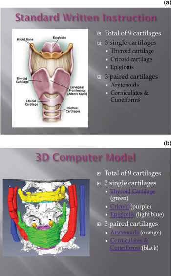
Participants were randomised to one of two instructional arms, using an online random number generator. The same textual content was used in both arms. One group received a computer-based tutorial with two-dimensional (2D) diagrams from various anatomy textbooks (the ‘standard’ group) (Figure 1a). The other group received the same computer-based tutorial, but with 3D interactive models instead of 2D diagrams (the 3D group) (Figure 1b).
Example of tutorial material containing (a) a standard two-dimensional illustration, and (b) a computer-generated, three-dimensional model.

Each participant was given 45 minutes to study laryngeal anatomy, using the computer-based tutorial. Immediately afterwards, a link to an anatomy examination was provided, in order to evaluate what the student had learned. Following this, a survey was distributed to evaluate each student's overall impression and satisfaction with their tutorial. Additionally, a mental rotation test was administered to students, within a ‘principles of surgery’ session, in order to assess their spatial ability.Reference Peters, Laeng, Latham, Jackson, Zaiyouna and Richardson4
Outcome measurement
Laryngeal anatomy test
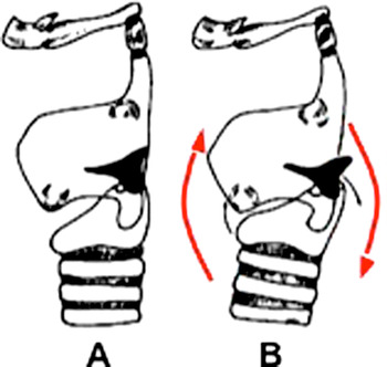
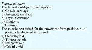
This test was used as in our previous study.Reference Hu, Wilson, Ladak, Haase, Doyle and Fung3 The first 13 questions were factual, while the last seven questions involved 3D concepts, with the aim of evaluating participants' understanding of 3D spatial relationships (Table I).
Figure accompanying the anatomy questions used to assess participants' understanding of 3D spatial relationships.

Sample anatomy test questions

3D = three-dimension spatial relationship
Mental rotation test
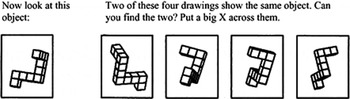
The modified Vanderberg and Kuse mental rotation test was used to assess participants' spatial ability. This is a validated questionnaire which had been used in previous studies to assess participants' mental rotation ability (Figure 3).Reference Peters, Laeng, Latham, Jackson, Zaiyouna and Richardson4 It contains 20 items in five sets of four items. Each item consists of a criterion figure, two correct alternatives, and two incorrect ones or ‘distractors’. Corrected alternatives are identical to the criterion figure in structure but are shown in various views, rotated around one axis in steps of 20°. The distractors usually show the criterion figure rotated in a mirror-image sense, or show a different figure. A point is scored only if both choices are correct; this eliminates the need to adjust for guessing. The total possible score ranges from zero to 24. A mean time of 10 minutes was given for completion.
An item from the modified Vandenberg and Kuse mental rotation test.

Opinion survey
We created a questionnaire to assess participants' opinions of the 2D and 3D imagery used (see Appendix 1). The questionnaire evaluated participants' perceptions of the clarity, ‘user-friendliness’, comprehensibility and effectiveness of the different image types, and also evaluated their preference for one or the other image type, using a Likert-style scale from 1 to 5, with 1 = ‘strongly disagree’ and 5 = ‘strongly agree’.
Statistical analysis
In order to obtain a power (1 − β) of 0.80, an α of 0.05, and an effective difference in anatomy scores of 2, we calculated that we required a sample size of 20 participants per group. Statistical comparisons were performed by using paired and grouped Student t-tests and repeated measures analysis of variance. Results are presented as means ± standard deviations (SDs). A probability level of p < 0.05 was considered statistically significant. Statistical analysis was conducted using the SAS commercial software package (SPSS Version 16, SPSS Inc., 2007). Pearson's correlation was used to assess the association between spatial ability and functional anatomy learning. Participants' perceptions of the two imagery strategies were assessed by calculating the mean and SDs for the survey questionnaire item scores.
Results
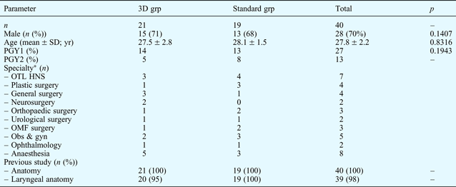
Forty students participated in the study, 19 in the standard group and 21 in the 3D group. The demographics and baseline characteristics of the two groups were compared using the unpaired t-test (Table II). There were no significant differences in age, gender or previous laryngeal anatomy knowledge, comparing the two groups.
Participants' demographic data
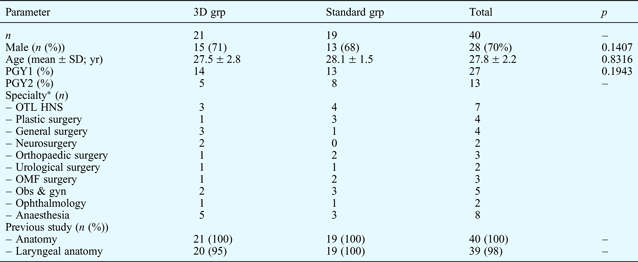
*p = 1.0000, 3D group vs standard group, all specialties. 3D grp = three-dimensional model group; standard grp = 2D imagery group; SD = standard deviation; yr = years; PGY1 = postgraduate year 1; PGY2 = postgraduate year 2; OTL HNS = otolaryngology head & neck surgery; OMF = oromaxillary facial; obs & gyn = obstetrics & gynaecology
Primary outcome: efficacy of two- vs three-dimensional imagery
The results of the laryngeal anatomy test are summarised in Table III. There was no significant difference between the two groups (p = 0.722; t-test).
Test score results

Data represent means ± standard deviations unless specified otherwise. *n=21; †n = 19. ‡Scored out of 20; **scored out of 7. §n = 12; ¥n = 17; an = 29. 3D grp = three-dimensional model group; standard grp = 2D imagery group; 3D Qs = 3D spatial relationship questions; rotn = rotation
Secondary outcome: association between spatial ability and functional anatomy learning
Twenty-nine participants completed both the mental rotation test and the laryngeal anatomy test. As stated above, the last seven of the 20 laryngeal anatomy test questions were designed to assess spatial abilities.
The mental rotation test scores ranged from 17 to 24. With α = 0.05, 27 degrees of freedom and a two-tailed test, Pearson's correlation coefficient was 0.47919; this indicated an insignificant association (p = 0.085).
Tertiary outcome: participants' opinions
There was a significant difference in participants' perceptions of the clarity and user-friendliness of the computer-generated, 3D laryngeal images. Additionally, participants indicated a preference for 3D imagery when learning anatomical concepts, if given the opportunity.
Discussion
Primary outcome: efficacy of two- vs three-dimensional imagery
Many medical professionals believe that computer-generated 3D models could significantly enhance medical education, as they may be particularly effective in providing external visualisation of anatomy that is hard to conceptualise otherwise.Reference Florance1 Additionally, good visualisation skills may help learners to develop mental representations that can later be used to draw inferences in different settings (e.g. during functional anatomy testing, or during surgical training when detailed anatomical understanding is required).
However, interestingly, most studies have not reported a superior effect of using 3D computer-generated imagery, compared with standard 2D imagery, when teaching anatomy.
Khalil et al. compared computer-based, interactive imagery and paper-based imagery in teaching pelvic anatomy.Reference Khalil, Johnson and Lamar5 They administered pre- and post-teaching tests to 64 first year undergraduate veterinary students, and found no significant difference in immediate recall of anatomical information. However, students perceived computer-based, interactive imagery as being a better strategy for the assimilation of anatomical information.
Garg and colleagues carried out a series of three studies of anatomy teaching methods which investigated the utility of computer-generated 3D anatomical models of carpal bone reconstruction, with the facility for multiple views and for learner-control of reconstructions.Reference Garg, Norman and Sperotable6–Reference Garg, Norman, Eva, Spero and Sharan8 They concluded that learners with poor spatial ability did poorly regardless of the type of learning module used, but especially poorly when presented with multiple 3D views. They suggested that perhaps their novice students had been overwhelmed by the high fidelity 3D model used, and needed to learn the basics of anatomy first. Students' spatial understanding was enhanced when they were allowed to digitally rotate the models themselves and to select the views of interest; the authors suggested that such learner control contributes to the successful integration of complex spatial information. Additionally, Garg and colleagues found that, when given the opportunity to rotate the models, most students still preferred the cardinal views to the multiple additional views available; the authors suggested that learners initially form a key view of a structure, and then gather information and inferences by rotating that key view in their mind.
Various other studies have shown that greater fidelity of the model, and a greater degree of approximation to real-life conditions, do not necessarily lead to additional educational benefits, especially for junior learners.Reference Chandra, Savoldelli, Joo, Weiss and Naik9–Reference Friedman, You-Ten, Bould and Naik11 At our centre, Hu et al. initially tested the efficacy of a 3D laryngeal model, compared with a written tutorial using standard 2D laryngeal images, in a heterogeneous group of medical and dental students in the early stages of their training. No significant difference was found in the outcome measured (i.e. the laryngeal test score).Reference Hu, Wilson, Ladak, Haase and Fung2
We attempted to overcome the limitations of previous studies by including a homogeneous population of more advanced learners, and by using only computer-based instructional methods. We did not observe any beneficial effect of the 3D model on immediate recall, compared with 2D imagery, as measured by the laryngeal anatomy test score.
The equivocal results in our study may be attributable to several factors; these correspond to the limitations of this study.
• Medical education now includes many digital images
• This study compared a three-dimensional (3D) model with two-dimensional (2D) images, for teaching laryngeal anatomy
• Learning outcomes did not differ significantly
• Little is currently known about improving the medical learning potential of digital imagery
• Benefit from 3D models is affected by model interactivity and students' learning styles and experiences
• Learners prefer 3D models to 2D images
Firstly, our primary outcome measure was merely a surrogate measure of 3D anatomical understanding. We did not extensively assess the validity of our laryngeal anatomy test; however, the construct validity was verified by three anatomy instructors.
Secondly, it was not possible to measure the participants' effort level during the study. Participants were all volunteers with no incentive to perform well, so some may have made less than maximal effort, commensurate with their motivation.
Lastly, although the computer tutorial with the 3D model had additional interactivity in comparison to the standard, 2D computer tutorial, in terms of the depth of the model and the added video and audio clinical vignettes, participants were not able to control the views of the model. The Amira program used to create the 3D model requires a special software platform with which to view the models, and the average computer does not have this capability. In exchange for the convenience of participants viewing the module from their home computer, this aspect of the model was sacrificed.
Only one previous study, by Nicholson et al. has reported a positive outcome for the use of computer-generated, 3D anatomical models in anatomical learning.Reference Nicholson, Chalk, Funnell and Daniel12 These authors reported that a computer-based, 3D anatomical model enhanced medical students' learning of ear anatomy. This study's 3D model was fully interactive and rotated in all three planes. The authors suggested that the anatomy of the middle and inner ear was far more complicated than the anatomy addressed by other studies of 3D models (e.g. carpal bone and shoulder anatomy). They concluded that the observed positive effect on participants' learning may have been due to their 3D model's greater level of interactivity, and to the greater complexity of the modelled anatomy.
Secondary outcome: association between spatial ability and functional anatomy learning
During functional anatomy learning, students need to understand how space is organised (in order to describe anatomical structures in different planes), to visualise structures in isolation and in relation to other organs, and to understand function. Thus, efficient functional anatomy learning requires accurate ‘visualisation of spatial reasoning’, and the ability to visualise images with and without rotation.Reference Corton, McIntire, Wai, Ling and Wendel13 This suggests that students with more developed visio-spatial abilities should obtain better results in anatomy examinations. Previous evidence suggests that spatial ability is related to successful learning of anatomy and of surgical (and dental) procedures; however, few formal studies have been undertaken.Reference Risucci, Geiss, Gellman, Pinard and Rosser14, Reference Wanzel, Hamstra, Anastakis, Matsumoto and Cusimano15
In Rochford's study, anatomy test questions were classified by experts as either spatial or non-spatial; spatial ability tests were then developed which involved rotation, visualisation, synthesis and sectioning of geometrical shapes.Reference Rochford16 These spatial ability tests reliably predicted anatomical learning achievement: medical students with a lower level of spatial ability obtained consistently lower marks than those with a higher level of spatial ability, for spatial-type anatomy test questions. By contrast, students with all levels of spatial ability performed equally well on tests of non-spatial anatomical knowledge.
Guillot et al. confirmed the complex relationship between spatial ability and functional anatomy learning by testing 184 students, using a mental rotation test and a 220-question anatomy test, after 14 hours spent learning basic skeletal and muscular anatomy (via lectures and other forms of training).Reference Guillot, Champely, Batier, Thiriet and Collet17 A strong relationship was found between mental rotation test scores and anatomy test results (significance not stated).
Hoyek and colleagues' study involved training participants in mental rotation, using 12 sessions of 20 minutes each, three times a week, in order to assess whether specific mental rotation training could improve students' scores for anatomy questions requiring mental rotation skills.Reference Hoyek, Collet, Rastello, Fargier, Thiriet and Guillot18 The trained group showed a tendency to score more highly than the untrained group.
Our study found no significant association between spatial ability and functional anatomy learning. Our study results differed from those of Guillot et al. their participants' mental rotation test scores ranged widely (i.e. 8–18; total possible score = 24), enabling the separation of participants with good and poor spatial abilities. In contrast, the mental rotation test scores of our 32 participants in surgical training programmes varied far less (range, 17–24), thus making it difficult to differentiate those with good and poor spatial ability. A much higher number of participants would be needed to establish a valid association between spatial ability and anatomical test performance. However, this result should be interpreted cautiously; we conducted a randomised, controlled study, and the intervention itself may have resulted in distorted correlations.
Tertiary outcome: participants' opinions
Assessment of participants' opinion survey responses showed that, overall, those using the 3D model gave higher ratings than those using the 2D images, for all five survey statements. There were statistically significant differences for statements regarding the clarity and comprehensibility of the 3D model, and regarding participants' preference for 3D images. This overall preference for the 3D model, compared with more traditional teaching images, is consistent with results from previous studies.Reference Hariri, Rawn, Srivastava, Youngblood and Ladd19–Reference Grober, Hamstra, Wanzel, Reznick, Matsumoto and Sidhu20
Conclusion
Computer-generated, 3D imagery has a promising role in future medical training.
However, not all interfaces with such models are equally effective. Despite much optimism, we currently know relatively little about how learners interact with 3D imagery, and about how best to design such imagery to aid students' acquisition of functional anatomy knowledge and spatial skills. Different learners have different learning styles. For example, more senior learners may benefit from a higher fidelity model, while more junior learners may not. Various factors may make 3D learning resources more effective; for example, higher levels of interactivity and learner-control may be beneficial.
Regardless of such factors, it is clear that students enjoy learning with 3D computer models, and that such models will continue to be warmly received by students.
Appendix 1. Opinion survey
Please rate the following statements by assigning a rating based on the following scale: 1 = strongly disagree; 2 = somewhat disagree; 3 = neutral; 4 = somewhat agree; 5 = strongly agree