Introduction
Computed tomography (CT) and conventional magnetic resonance imaging (MRI) using spin-echo T1- and T2-weighted images are extensively used at present for the evaluation of laryngeal lesions. However, it is not uncommon to encounter lesions that have indeterminate findings on cross-sectional imaging and necessitate further investigation. Diffusion-weighted imaging with the calculation of apparent diffusion coefficient values has been investigated in several studies, in an attempt to distinguish between benign and malignant head and neck lesions.Reference Wang, Takashima, Takayama, Kawakami, Saito and Matsushita1–Reference Schafer, Srinivan and Mukherji4
Diffusion-weighted imaging was recently introduced to the field of head and neck cancer for differential diagnosis,Reference Thoeny5–Reference Perrone, Guerrisi, Izzo, D'Angeli, Sassi and Mele8 monitoring of the treatment responseReference Thoeny5, Reference Vandecaveye, Dirix, De Keyzer, Op de Beeck, Vander Poorten and Hauben9 and differentiation of recurrence from post-radiotherapy changes.Reference Rashed, Elwan, Abdurrahman, El-Fiky, Shehata and Basyouni10 Diffusion-weighted imaging uses the movement of water molecules to produce images that indirectly reflect information regarding cell density and microstructures in living tissues.Reference Galbán, Lemasson, Williams, Li, Heist and Johnson11 This can be estimated and quantified in terms of apparent diffusion coefficients.Reference Galbán, Lemasson, Williams, Li, Heist and Johnson11, Reference Thoeny, De Keyzer and King12
However, there are limitations in the performance of laryngeal diffusion-weighted imaging. For instance, the larynx has a complex anatomical structure, causing susceptibility effects. Hence, the value of diffusion-weighted imaging in differentiating benign from malignant laryngeal lesions has only been investigated in a few studies.Reference Tshering Vogel, Zbaeren, Geretschlaeger, Vermathen, De Keyzer and Thoeny13–Reference Vandecaveye, de Keyzer, Vander Poorten, Deraedt, Alaerts and Landuyt16 Today, these limitations have gradually been overcome as a result of the steady improvement in MRI.Reference Thoeny, De Keyzer and King12, Reference Tshering Vogel, Zbaeren, Geretschlaeger, Vermathen, De Keyzer and Thoeny13 In head and neck cancer cases, previous studies have confirmed a significant difference in the apparent diffusion coefficient values between benign and malignant lesions.Reference Thoeny, De Keyzer and King12, Reference Abdel Razek, Gaballa, Elhawarey, Megahed, Hafez and Nada17
An alternative to the mean apparent diffusion coefficient approach is the apparent diffusion coefficient histogram. In this approach, the region of interest of a lesion is identified in multiple scans, and a plot of the number of voxels at each apparent diffusion coefficient value is depicted as a histogram.Reference Schafer, Srinivan and Mukherji4
This study aimed to determine whether a threshold apparent diffusion coefficient value may help to differentiate laryngeal carcinomas from benign lesions.
Materials and methods
This prospective study was carried out between May 2011 and November 2013, following approval of the Institutional Review Board of Ain Shams University Hospitals. An informed consent form was signed by all participants.
Patients were included if they were scheduled to have surgery for a de novo laryngeal lesion. All patients underwent routine laboratory investigations, chest X-ray, contrast-enhanced CT and diffusion-weighted MRI. Microlaryngosurgery was conducted in all patients for lesion mapping purposes and in order to take a biopsy for histopathological examination. Total or partial laryngectomy was performed for malignant masses according to the extent of the pathology, while excision biopsy was carried out for benign lesions.
Radiological findings were compared to histological findings. (The reporting radiologist was blind to the post-resection histopathology reports and pre-operative staging until the end of the study.) Following this comparison, the diagnostic value of diffusion-weighted MRI for the identification of malignant lesions was determined. In addition, the agreement between diffusion-weighted MRI and histopathology (with regard to the identification of malignant lesions) was assessed. Moreover, the sensitivity, specificity, efficiency, and negative and positive predictive values of the apparent diffusion coefficient in detecting benign and malignant lesions were analysed.
Magnetic resonance imaging
The study was performed using an Achieva 1.5T XR MRI scanner and software release 3.2 (Royal Philips Electronics, Eindhoven, the Netherlands). Using surface coil coverage from the skull base to the supraclavicular fossa, the following sequences were obtained: axial and coronal T1-weighted and T2-weighted imaging, and axial proton density with fat suppression imaging. A dose of 0.1 ml/kg of Dotarem® (0.5 mmol/ml) was injected. This was followed by fat-saturated, T1-weighted, axial, coronal and sagittal (post-contrast) imaging. The MRI parameters were as follows: field of view = 20–22 cm, slice thickness = 4 mm, inter-slice gap = 0.5–1 mm and matrix = 192 × 256.
Diffusion-weighted imaging
Single-shot, echo-planar, diffusion-weighted imaging (with three applied b-values of 0, 500 and 1000 s/mm2) was conducted, with the following parameters: repetition time/echo time = 5100/137 ms, slice thickness = 3 mm and matrix = 96 × 128.
Diffusion-weighted MRI images were obtained with different b-values simultaneously, to avoid misregistration in computing the different apparent diffusion coefficient values. Higher b-values produce more diffusion weighting and therefore higher contrast between laryngeal lesions and normal tissue. However, higher b-values also produce more susceptibility distortions, and could increase the noise in the diffusion-weighted images because the distortions are different depending on the gradient directions. (The b value is the diffusion sensitivity factor defined by the formula: b = γReference Sumi, Sakihama, Sumi, Morikawa, Uetani and Kabasawa2GReference Sumi, Sakihama, Sumi, Morikawa, Uetani and Kabasawa2δ Reference Sumi, Sakihama, Sumi, Morikawa, Uetani and Kabasawa2 (Δ − δ/3), in which γ is the gyromagnetic ratio, G is the strength of the gradients applied along the given axes to assess for diffusion, Δ is the width of those gradients and δ is their time).
Apparent diffusion coefficient measurement
The mean apparent diffusion coefficient value, measured in square millimetres per second (reflecting the average diffusion of a proton in a given area per unit of time), was determined by manual outlining of the lesion or its components (e.g. solid enhancing part, necrotic part) and calculating the average value of that volume.
The location of the region of interest was determined according to the anatomical images. The region of interest was drawn by an electronic cursor around the laryngeal lesion in the apparent diffusion coefficient map. The region of interest ranged from 20 to 60 mm2, depending on the lesion size.
The obscuration of lesion heterogeneity with a single average value is a primary limitation of the mean apparent diffusion coefficient value. Therefore, an apparent diffusion coefficient histogram was also produced. The histogram analysis considers the potential heterogeneity of the lesion and alters the mean apparent diffusion coefficient value accordingly. The region of interest of a lesion is identified in multiple scans and a plot of the number of voxels for each apparent diffusion coefficient value is depicted as a histogram. The x-axis on the histogram represents the apparent diffusion coefficient values and the y-axis represents the number of voxels within the lesion for a particular apparent diffusion coefficient value.
Image evaluation
A single, specialised consultant radiologist, who was blinded to the cases and was unaware of the histopathological diagnosis, reviewed the MRI images, including the anatomical and diffusion-weighted images. The subsite, size, borders, invasion to the surrounding structures, necrosis and solid enhancing components of lesions were evaluated. Calculation of the apparent diffusion coefficient values was performed using the software available on workstations. The observer making the evaluations noted the presence or absence of image distortion (associated with susceptibility artefacts) on the diffusion-weighted trace images and especially on the apparent diffusion coefficient maps. (Four patients were excluded because of image distortion associated with susceptibility artefacts.) The average apparent diffusion coefficient values were obtained from the mean apparent diffusion coefficient and histogram.
Statistical methods
Statistical analysis was conducted on a personal computer using MedCalc for Windows, version 12.5 (MedCalc Software, Ostend, Belgium) and DAG-Stat software.Reference Mackinnon18 The D'Agostino–Pearson test was performed to test the normality of numerical data distribution. The chi-square test was used for the comparison of qualitative data.
Receiver-operating characteristic curve analysis was used to determine the best cut-off apparent diffusion coefficient value for the identification of malignant lesions. This was achieved using two-by-two contingency tables, with histopathological diagnosis considered as the reference standard. The following quality indices were calculated: sensitivity, specificity, efficiency, positive predictive value and negative predictive value. Agreement between histopathological diagnosis and apparent diffusion coefficient diagnosis was examined by calculation of Cohen's kappa coefficient and the prevalence and bias adjusted kappa (‘PABAK’).Reference Altman19 All p values are two-tailed; p < 0.05 was considered as statistically significant.
Results
Fifty-three patients were included in this study (41 males and 12 females). Four were excluded on account of susceptibility artefacts (due to linear blurring, geometric distortion or imaging distortion), which compromised image quality.
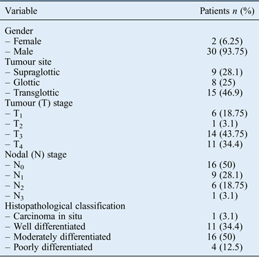
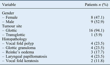
The ages of the remaining 49 patients ranged from 5 to 82 years (median, 53 years). Thirty-nine (79.6 per cent) of these patients were male and 10 (20.4 per cent) were female. The histopathological findings of the laryngeal lesions indicated malignant tumours in 32 patients (65.3 per cent) and benign lesions in 17 patients (34.7 per cent). The histopathological classifications of the lesions and characteristics of patients are shown in Tables I and II.
Characteristics of patients with malignant lesions

Characteristics of patients with benign lesions
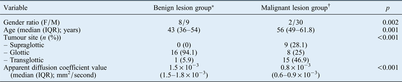
The apparent diffusion coefficient values ranged from 0.5 × 10⁻³ to 2 × 10⁻³ mm2/second (mean, 1.07 ± 0.45 mm2/second). The median apparent diffusion coefficient value for malignant lesions was 0.8 × 10⁻³ mm2/second, compared with a value of 1.5 × 10⁻³ mm2/second for benign lesions; this difference was highly significant (p < 0.001). A comparison of patients with benign and malignant laryngeal lesions is shown in Table III.
Comparison of patients with benign and malignant lesions
*n = 17; †n = 32. F = female; M = male; IQR = interquartile range
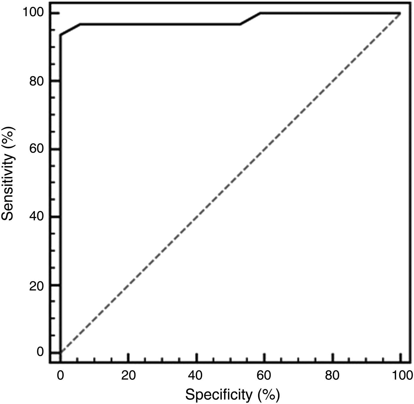
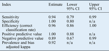
An apparent diffusion coefficient value of 1.1 × 10⁻³ mm2/second gave the best result when used as the cut-off point to differentiate malignant from benign masses. Sensitivity, specificity, efficiency, and positive and negative predictive values were 94, 100, 96, 100 and 89 per cent respectively (Table IV). The level of agreement between diffusion-weighted MRI and histopathology for lesion classification was 92 per cent, indicating very good agreement between both variables. The sensitivity, specificity, and positive and negative predictive values of the receiver-operating characteristic curve for the diagnosis of malignant lesions using the apparent diffusion coefficient value were 93.8, 100, 100 and 89.5 per cent respectively (Figure 1).
Receiver-operating characteristic curve for the diagnosis of malignant lesions using the apparent diffusion coefficient value. Area under the receiver-operating characteristic curve was 0.98 (95 per cent confidence interval = 0.89 to 1.0). The best cut-off apparent diffusion coefficient value was ≤1.1 × 10⁻³ mm2/second (sensitivity of 93.8 per cent, specificity of 100 per cent).
Diagnostic value of apparent diffusion coefficient of ≤1.1 × 10⁻³ mm2/second
CI = confidence interval; n/a = not applicable
As mentioned, the analyses revealed a highly significant difference in apparent diffusion coefficient values between benign and malignant lesions. Furthermore, there was little variability in apparent diffusion coefficient values within each group. When the threshold apparent diffusion coefficient of 1.1 × 10⁻³ mm2/second was applied in our study, there were no outliers in the benign category and only two outliers (of 32 patients) in the malignant category. The highest apparent diffusion coefficient value among malignant lesions (1.6 × 10⁻³ mm2/second) was seen in a patient with laryngeal squamous cell carcinoma (SCC); the apparent diffusion coefficient map mistakenly indicated that this patient had a benign lesion.
The results also revealed no significant difference between the apparent diffusion coefficient value and apparent diffusion coefficient histogram in differentiating laryngeal malignancies from benign lesions.
Discussion
To date, there have been few reports on diffusion-weighted imaging for laryngeal lesions.Reference Hatakenaka, Nakamura, Yabuuchi, Shioyama, Matsuo and Ohnishi14, Reference de Bree, van, Putten, Brouwer, Castelijns, Hoekstra and Leemans15, Reference Abdel Razek, Gaballa, Elhawarey, Megahed, Hafez and Nada17, Reference Taha, Hassan, Amir, Taha and Riad20 No previous report has investigated the pre-operative discrimination of laryngeal carcinomas from benign lesions. Diffusion-weighted MRI is a non-invasive technique capable of depicting the extent of random movement of water protons in biological tissues.Reference Stejskal and Tanner21 The amount of signal loss over the range of b-values correlates with the mobility of protons, and is quantified by means of the apparent diffusion coefficient.Reference Le Bihan, Breton, Lallemand, Aubin, Vignaud and Laval-Jeantet22 Several studies have already shown the benefit of diffusion-weighted imaging in distinguishing malignant from benign tumours in the head and neck.Reference Thoeny5, Reference Razek6, Reference Thoeny, De Keyzer and King12, Reference Hatakenaka, Nakamura, Yabuuchi, Shioyama, Matsuo and Ohnishi14 These abilities are aided by quantitative apparent diffusion coefficient values. In theory, hypercellular tumour tissue leads to impeded diffusion and, subsequently, a lower apparent diffusion coefficient value.Reference Vandecaveye, De Keyzer, Dirix, Lambrecht, Nuyts and Hermans7 Thus, malignant tumours usually show lower apparent diffusion coefficient values than benign tumours.Reference Razek6
The apparent diffusion coefficient cut-off value in our study was 1.1 × 10⁻³ mm2/second. This was associated with a sensitivity of 94 per cent, specificity of 100 per cent, efficiency of 96 per cent, positive predictive value of 100 per cent and negative predictive value of 89 per cent. This threshold did not markedly overlap for laryngeal carcinomas and benign lesions. The sensitivity, specificity, and positive and negative predictive values of the receiver-operating characteristic curve for the diagnosis of malignant lesions using the apparent diffusion coefficient value were 93.8, 100, 100 and 89.5 per cent respectively. These findings suggest that an apparent diffusion coefficient threshold of 1.1 × 10⁻³ mm2/second is optimal for distinguishing laryngeal carcinomas from benign lesions.
Srinivasan et al. established an optimal apparent diffusion coefficient threshold of 1.36 × 10⁻³ mm2/second for the diagnosis of lesions in the head and neck.Reference Srinivasan, Dvorak, Perni, Rohrer and Mukherji23 Abdel Razek et al. found that an apparent diffusion coefficient of 1.25 × 10⁻³ mm2/second was useful as a threshold for differentiating malignant from benign head and neck lesions.Reference Abdel Razek, Gaballa, Elhawarey, Megahed, Hafez and Nada17 Sasaki et al. reported that a lower apparent diffusion coefficient cut-off of 0.84 × 10⁻³ mm2/second was best for differentiating sinonasal benign or inflammatory lesions from malignant tumours; the corresponding sensitivity, specificity and accuracy values were 61, 94 and 79 per cent respectively.Reference Sasaki, Eida, Sumi and Nakamura24
The highest apparent diffusion coefficient value among those with malignant laryngeal lesions in the current study was seen in a patient with a T2 SCC of the larynx (1.6 × 10−Reference Habermann, Gossrau, Graessner, Arndt, Cramer and Reitmeier3 mm2/second), as calculated from the apparent diffusion coefficient map, which was misdiagnosed as a benign lesion. This finding could be due to the smaller size of the tumour and the predominance of areas of high apparent diffusion coefficient values compared to the limited zones of low apparent diffusion coefficient values, in concordance with Eida et al.Reference Eida, Sumi, Sakihama, Takahashi and Nakamura25
• Diffusion-weighted magnetic resonance images indirectly reflect cell density and microstructures in living tissues, estimated and quantified in terms of apparent diffusion coefficients
• There are limitations to laryngeal diffusion-weighted imaging
• Previous studies have revealed differences in apparent diffusion coefficient values between benign and malignant head and neck lesions
• In this study, an apparent diffusion coefficient threshold of 1.1 × 10⁻³ mm2/second was optimal for distinguishing laryngeal carcinomas from benign lesions
• Apparent diffusion coefficient values were lower for patients with laryngeal carcinomas than for those with benign lesions
To our knowledge, there are four previous reports on diffusion-weighted imaging in laryngeal carcinoma. One study showed the importance of diffusion-weighted MRI in decision making for the management of laryngeal carcinoma, emphasising its role in predicting thyroid cartilage invasion and its ability to differentiate between inner and outer thyroid lamina invasion.Reference Taha, Hassan, Amir, Taha and Riad20 In another study, receiver-operating characteristic analysis showed that the area under the curve was 0.956 and that the optimum threshold for the apparent diffusion coefficient to differentiate malignant from pre-cancerous laryngeal lesions was 1.45 × 10⁻³ mm2/second.Reference Shang, Ruan, Zhou, Bao, Cheng and Wang26 In the third study, the authors found that diffusion-weighted MRI was capable of differentiating tumoural tissue from radiotherapy-induced tissue alterations.Reference Eida, Sumi, Sakihama, Takahashi and Nakamura25 In the last study, by Tshering Vogel et al., the optimal apparent diffusion coefficient cut-off value for differentiating tumour recurrence after chemoradiotherapy from non-tumoural changes was 1.3 × 10⁻³ mm2/second. However, the authors found a larger overlap between benign and malignant outcomes. They suggested that this was because the larynx is subject to more susceptibility and movement artefacts than other locations in the head and neck; furthermore, the ratio of T1 patients in their study was relatively large, so the sizes of the tumours were smaller.Reference Tshering Vogel, Zbaeren, Geretschlaeger, Vermathen, De Keyzer and Thoeny13
In our study, there was no significant difference between the apparent diffusion coefficient value and the apparent diffusion coefficient histogram in differentiating laryngeal malignancy from benign lesions. This suggests that the apparent diffusion coefficient value provides an adequate method of differentiating malignant from benign lesions.
This study was conducted using a single-centre registry of patients with laryngeal carcinoma, and the number of patients was small. Thus, additional studies that comprise greater numbers of patients with pre-operative laryngeal carcinoma and benign lesions are needed to confirm the results of this investigation.
Conclusion
An apparent diffusion coefficient threshold of 1.1 × 10⁻³ mm2/second is optimal for distinguishing laryngeal carcinomas from benign lesions. Apparent diffusion coefficient values were lower for patients with laryngeal carcinomas than for those with benign lesions.