1. Introduction
The classical genus of Ediacaran macroorganisms, Dickinsonia, had a low, flat body, was from almost round to elongated, bilateral or almost bilateral, and divided into numerous transverse elements. There is a small sub-triangular lobe at one end of the body of Dickinsonia. A gradual decrease in the size of the transverse elements is observed towards the opposite end of the body.
Fossil remains of Dickinsonia were found in the upper Ediacaran deposits of South Australia (Flinders Ranges) and Eastern Europe (Podolia, southeastern White Sea area, Middle Urals) (Sprigg, Reference Sprigg1947; Keller & Fedonkin, Reference Keller and Fedonkin1976; Fedonkin, Reference Fedonkin, Velikanov, Aseeva and Fedonkin1983; Krivosheev & Polenov, Reference Krivosheev and Polenov2001). The habitat of these organisms is usually reconstructed as hydrodynamically active shallow-marine settings between fair weather and storm wave base, sporadically covered with siliciclastic suspension (Seilacher, Reference Seilacher1989; Grazhdankin, Reference Grazhdankin2004; Droser & Gehling, Reference Droser and Gehling2015; Droser et al. Reference Droser, Tarhan and Gehling2017, Reference Droser, Tarhan, Evans, Surprenant and Gehling2020). There are observations that Ural Dickinsonia could inhabit shallower parts of the basin with deviating salinity (Bobkov et al. Reference Bobkov, Kolesnikov, Maslov and Grazhdankin2019; Mitchell et al. Reference Mitchell, Bobkov, Bykova, Dhungana, Kolesnikov, Hogarth, Liu, Mustill, Sozonov, Rogov, Xiao and Grazhdankin2020). Retallack (Reference Retallack2007, Reference Retallack, Noffke and Chafetz2012) reported on the confinement of the Australian Dickinsonia to the horizons of palaeosols; however, other researchers refute this proposition (Callow et al. Reference Callow, Brasier and McIlroy2013; Xiao, Reference Xiao2013; Tarhan et al. Reference Tarhan, Droser, Gehling and Dzaugis2015; Evans et al. Reference Evans, Droser and Gehling2015, Reference Evans, Gehling and Droser2019 a).
The fossil remains of Dickinsonia and other numerous macroorganisms are preserved in situ under the covers of silty-sandy sediments in burial event horizons of the Flinders–Belomorian style (Grazhdankin, Reference Grazhdankin2004; Narbonne, Reference Narbonne2005; Droser et al. Reference Droser, Gehling and Jensen2006, Reference Droser, Tarhan, Evans, Surprenant and Gehling2020; Droser & Gehling, Reference Droser and Gehling2015; Ivantsov & Zakrevskaya, Reference Ivantsov and Zakrevskaya2018). It was shown that microbial mats represent the substrate inhabited by the Flinders–Belomorian community (Seilacher, Reference Seilacher1989, Reference Seilacher1999; Gehling, Reference Gehling1999; Steiner & Reitner, Reference Steiner and Reitner2001; Bottjer & Clapham, Reference Bottjer, Clapham, Xiao and Kaufman2006; Callow & Brasier, Reference Callow and Brasier2009; Gehling & Droser, Reference Gehling and Droser2009; Laflamme et al. Reference Laflamme, Schiffbauer, Narbonne and Briggs2011; Tarhan et al. Reference Tarhan, Droser, Gehling and Dzaugis2017). The high activity of the environment during the formation of the Flinders–Belomorian localities and the low density of the soft-bodied organisms in comparison with mineral sediment should theoretically lead to the separation of bodies from the substrate, their transfer and their burial inside the more fine-grained part of the sedimentary cycle. This, apparently, happened to a part of the community (Ivantsov & Zakrevskaya, Reference Ivantsov and Zakrevskaya2018; Droser et al. Reference Droser, Tarhan, Evans, Surprenant and Gehling2020). However, the preservation of the majority of macrofossils here is exactly the opposite case, in the most coarse-grained part of the cycle, right on the bottom of the layer. This indicates the adhesion of the Flinders–Belomorian macroorganisms, including Dickinsonia, to the substrate at the moment of burial (Schopf & Baumiller, Reference Schopf and Baumiller1998; Retallack, Reference Retallack2007; Ivantsov, Reference Ivantsov2011 a).
Dickinsonia has been described as mobile pelagic or benthic Eumetazoa: jellyfishes (Sprigg, Reference Sprigg1947, Reference Sprigg1949; Harrington & Moore, Reference Harrington, Moore and Moore1956), flatworms (Termier & Termier, Reference Termier and Termier1968; Fedonkin, Reference Fedonkin1981) or annelids (Glaessner, Reference Glaessner1959; Glaessner & Wade, Reference Glaessner and Wade1966; Wade, Reference Wade1972; Runnegar, Reference Runnegar1982; Conway Morris, Reference Conway Morris1989; Jenkins, Reference Jenkins, Lipps and Signor1992) from the very beginning of its research. At first, little evidence was offered for these interpretations. In the framework of the ‘annelid’ hypothesis, only clear anteroposterior asymmetry and visible body segmentation can be considered such evidence. Other described features and structures that could indicate the complexity of the Dickinsonia body plan, such as dorsoventral polarization of the body, intestinal tract, prostomium, intestinal caeca or folds of muscle contractions (Glaessner & Wade, Reference Glaessner and Wade1966; Wade, Reference Wade1972; Gehling, Reference Gehling1991; Jenkins, Reference Jenkins, Lipps and Signor1992; Dzik, Reference Dzik2000) were not well documented and were hardly taken into account in subsequent studies. A displacement of the right and left halves of the ‘segments’ along the longitudinal axis of the body of Dickinsonia is often observed on the imprints. Whether such a shift was of primary or secondary nature as a result of taphonomic processes is still being debated (Ivantsov, Reference Ivantsov2001 b, Reference Ivantsov2008, Reference Ivantsov2011 a, Reference Ivantsov2013; Ivantsov & Malakhovskaya, Reference Ivantsov and Malakhovskaya2002; Ivantsov et al. Reference Ivantsov, Fedonkin, Nagovitsyn and Zakrevskaya2019 a, c, Reference Ivantsov, Zakrevskaya, Nagovitsyn, Krasnova, Bobrovskiy and Luzhnaya (Serezhnikova)2020). The authors of this article consider displacement as a stable anatomical feature of Dickinsonia and similar organisms and refer to these ‘half-segments’ as isomers (Ivantsov, Reference Ivantsov2001 b). Another peculiar feature of Dickinsonia is the posterior terminal location of the growth zone. This observation, made by Wade (Reference Wade1972) and recently disputed (Hoekzema et al. Reference Hoekzema, Brasier, Dunn and Liu2017; Dunn et al. Reference Dunn, Liu and Donoghue2018), is supported by our study of the characteristics of regeneration of the posterior end of the body in Dickinsonia from Lyamtsa locality (Ivantsov et al. Reference Ivantsov, Zakrevskaya and Nagovitsyn2019 c, Reference Ivantsov, Zakrevskaya, Nagovitsyn, Krasnova, Bobrovskiy and Luzhnaya (Serezhnikova)2020). Fedonkin (Reference Fedonkin, Sokolov and Iwanowski1990) listed both mentioned features among the reasons for placing Dickinsonia in the Proarticulata phylum that he described. It occupies an intermediate position between ‘Coelenterata’ and Bilateria (Fedonkin, Reference Fedonkin, Sokolov and Iwanowski1990).
Due to the unusual and simple morphology of the Dickinsonia fossil remains, accessible for public discussion, as well as the peculiarity of the taphonomy, a fantastic variety of hypotheses on the affinities of these macroorganisms was suggested: from giant syncytial protists (Zhuravlev, Reference Zhuravlev1993; Seilacher et al. Reference Seilacher, Grazhdankin and Legouta2003), to terrestrial lichens and fungi (Retallack, Reference Retallack1994, Reference Retallack2007), to the independent Kingdom Vendobionta (Seilacher, Reference Seilacher1989, Reference Seilacher1992).
An important stage in the study of the genus Dickinsonia and the close genus Yorgia was the discovery of peculiar fossils in the Vendian deposits of the White Sea, interpreted as resting traces (Ivantsov, Reference Ivantsov, Ponomarenko, Rozanov and Fedonkin2001 a), and then as feeding traces (Ivantsov & Fedonkin, Reference Ivantsov and Fedonkin2001) and later described as a special ichnogenus Epibaion (Ivantsov & Malakhovskaya, Reference Ivantsov and Malakhovskaya2002; Ivantsov, Reference Ivantsov2011 a). The same trace fossils were abundantly found in the Ediacaran deposits of Australia (Gehling et al. Reference Gehling, Droser, Jensen, Runnegar and Briggs2005; Evans et al. Reference Evans, Gehling and Droser2019 a). A characteristic feature of these trace fossils is the association in groups, often arranged in long chains. Moreover, there is usually a body imprint of an individual tracer-maker at one end of such chains. The orientation of the traces in a chain shows that the end of the body that bore the sub-triangular (crescent-shaped in the case of Yorgia) lobe was the anterior one in the direction of movement (Gehling et al. Reference Gehling, Droser, Jensen, Runnegar and Briggs2005; Ivantsov, Reference Ivantsov2011 a, Reference Ivantsov2013; Evans et al. Reference Evans, Gehling and Droser2019 a). It is believed that in the act of creating a trace, the organisms were spreading over the mat surface, affecting it in some way and destroying its upper layer (Ivantsov & Malakhovskaya, Reference Ivantsov and Malakhovskaya2002; Gehling et al. Reference Gehling, Droser, Jensen, Runnegar and Briggs2005; Seilacher, Reference Seilacher, Vickers-Rich and Komarower2007; Sperling & Vinther, Reference Sperling and Vinther2010; Ivantsov, Reference Ivantsov2011 a; Evans et al. Reference Evans, Gehling and Droser2019 a). The discovery of traces of the impact of Yorgia and Dickinsonia on the microbial mats has strengthened the position of the supporters of the animal nature of these Ediacaran macroorganisms. The discovery of the complex structure of the integument and internal structures, as well as the biochemical analysis of organic matter preserved on the imprints of Dickinsonia, also support this hypothesis (Dzik & Ivantsov, Reference Dzik and Ivantsov2002; Bobrovskiy et al. Reference Bobrovskiy, Hope, Ivantsov, Nettersheim, Hallman and Brocks2018; Ivantsov et al. Reference Ivantsov, Zakrevskaya and Nagovitsyn2019 c). The spectrum of considered hypothesis diversity has narrowed but still remains extremely wide, spreading from the early metazoans similar to placozoans (Rozhnov, Reference Rozhnov2009; Sperling & Vinther, Reference Sperling and Vinther2010), to eumetazoans and bilaterians (Gehling et al. Reference Gehling, Droser, Jensen, Runnegar and Briggs2005; Ivantsov, Reference Ivantsov2011 a; Gold et al. Reference Gold, Runnegar, Gehling and Jacobs2015; Evans et al. Reference Evans, Droser and Gehling2017, Reference Evans, Gehling and Droser2019 a, b; Ivantsov et al. Reference Ivantsov, Nagovitsyn and Zakrevskaya2019 b, c).
The fact that the feeding traces, belonging to one individual, form groups and elongated chains gives evidence of the ability of Dickinsonia to move along the sea bottom, which to some extent contradicts the fact of their adhesion to the bottom during a burial event. Additional observations show the absence of the structures connecting individual feeding traces in a group, as well as a lack of information on the actual traces of Dickinsonia locomotion. This is partly why the ideas about the belonging of these organisms to “Coelenterata” (Ctenophora) still exist (Brasier & Antcliffe, Reference Brasier and Antcliffe2008), and attempts to compare Dickinsonia with lichens continue (Retallack, Reference Retallack2013, Reference Retallack2014; Retallack & Broz, Reference Retallack and Broz2020). This article is intended to fill an existing information gap. Here, we demonstrate recently discovered fossil structures that can be interpreted as traces of motor activity of Dickinsonia associated with a movement along the substrate, temporary adhesion to it, self-caused shift and bending of the margins of the body, as well as an escape from the place of burial.
2. Material and locality
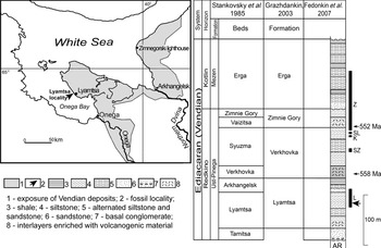
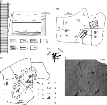
The studied collection of Dickinsonia originates from the Lyamtsa Beds of the upper Vendian Ust-Pinega Formation of the southeastern White Sea area (north of European Russia). It was collected on the southern coast of the Onega Peninsula within the Lyamtsa locality from a single bedding plane of the burial event horizon, marked as L2(XII) (Fig. 1). This accumulation of macrofossils is confined to the bottom of the layer of grey fine-grained polymictic (quartzo-feldspathic) clayey sandstone, gradually turning into siltstone upwards. The total thickness of the layer varies from 3 to 17 cm. Numerous mudstone pellets are observed in areas, with the maximum thickness of the layer at a height of 0.3–1 cm from the base of the layer. The pellets are flat, polygonal in plan with rounded corners, up to 7 cm along the long axis, and up to 3–4 mm thick (Fig. 2a). Depressions that appeared after the diagenetic compaction of the pellets are clearly visible on the lower surface of the layer (Fig. 2b). Imprints of redeposited fragments of the microbial mat are widespread at a height of 2–3 cm from the bottom of the layer. It is important to note the stratigraphic sequence of the location of these objects within the layer: the remains of the macrobiota are located at the bottom, mudstone pellets are located above and redeposited fragments of the microbial mat are even higher. In the middle part of the layer, there is a level with several adjacent surfaces of small ripple marks with a distance of about 0.5 cm between the ridges. The sole of the layer is smoothed and has a microbially induced texture, consisting of low flat-topped tubercles separated by winding grooves. In some places, it is covered with a net of the finest imprints of presumable filamentous algae. The layer is overlain by a half-metre stratum of siltstones, and all are enclosed a thick clayey package, characteristic of the upper part of the Lyamtsa Beds. The casts of small spindle-shaped and drop-shaped flat-bottomed gutters can quite often be observed on the bottom of the layer (about 30 cm along the long axis and up to 1–1.5 cm deep) (Fig. 2b). Based on the preserved remains, the margins of the mat around the gutter were torn into pieces, which were partially transferred into the overlying layer. On one side of the gutters, these pieces of mat lifted into the overlying sediment were normally arranged; on the other side, they were turned up. The direction of erosion was determined by the position of the convex end of the drop-shaped gutters, and the direction of sediment transfer and filling of the gutters was determined by the dip of sandy thin layers and the position of the lifted scraps of the microbial mat. Moreover, it appeared to be that the erosion and filling of gutters with the sediment occurred in two mutually perpendicular directions (Fig. 2c).
The geographical and stratigraphic position of the Lyamtsa locality and the L2(XII) burial event horizon (arrow). Dark vertical stripes on the right correspond to the intervals of the White Sea localities of macrofossils: L – Lyamtsa; SZ – Suzma; K – Karakhta; SL – Solza; Z – Zimnie Gory (Ivantsov et al. Reference Ivantsov, Zakrevskaya, Nagovitsyn, Krasnova, Bobrovskiy and Luzhnaya (Serezhnikova)2020). Source: Stankovsky et al. (Reference Stankovsky, Verichev, Dobeiko, Sokolov and Iwanowski1985), Grazhdankin (Reference Grazhdankin2003) and Fedonkin et al. (Reference Fedonkin, Ivantsov, Leonov, Serezhnikova and Semikhatov2007).

The fossiliferous layer of the L2(XII) burial event horizon in the area with maximum thickness: (a) position of the layer in the section of the lower part of the Lyamtsa Formation and lithological features; (b) schematic drawing of one of the gutters; (c) a rose diagram showing the orientation of the anterior ends of 30 Dickinsonia specimens relative to the directions of erosion and filling of gutters; (d) schematic drawing of a surface area showing the location of the fossils; (e) a fragment of a gutter cast with drag marks and casts of small colonies of Beltanelliformis brunsae (Ivantsov et al. Reference Ivantsov, Zakrevskaya, Nagovitsyn, Krasnova, Bobrovskiy and Luzhnaya (Serezhnikova)2020). Legend: 1 – sandstone; 2 – siltstone; 3 – mudstone; 4 – parallel joint; 5 – cross-bedding and mudstone pellets; 6 – several levels of ripple marks; 7 – redeposited fragments of microbial mats; 8 – the edge of the mat visible on the bedding surface; 9 – the outer edge of the chipped-off section of the gutter cast; 10 – the edge of the area of the microbial mat lifted into the sediment; 11 – the central area of the gutter; 12 – the inner edge of the chipped-off section of the gutter cast; 13 – normal and lifted position of the sections of the microbial mat; 14 – casts of the drag furrows; 15 – depressions under mudstone pellets; 16 – casts of Beltanelliformis brunsae colonies. A – Aspidella terranovica; D – Dickinsonia cf. menneri; P – Parvancorina minchami; U – unclassified remains of macroalgae.

The assemblage of macrofossils of the L2(XII) burial event horizon is extremely poor. It contains one species of Dickinsonia, resembling D. menneri (Keller & Fedonkin, Reference Keller and Fedonkin1976; Ivantsov et al. Reference Ivantsov, Nagovitsyn and Zakrevskaya2019 b, Reference Ivantsov, Zakrevskaya, Nagovitsyn, Krasnova, Bobrovskiy and Luzhnaya (Serezhnikova)2020), Parvancorina minchami, Beltanelliformis brunsae, fragments of band-like algae (Fig. 2d), small structures of palaeopascichnids, rare Epibaion axiferus traces, as well as the other two or three poorly diagnosed forms. The remains of Beltanelliformis were located below the final surface of the microbial mat and are better seen in the gutter casts (Ivantsov et al. Reference Ivantsov, Zakrevskaya, Nagovitsyn, Krasnova, Bobrovskiy and Luzhnaya (Serezhnikova)2020) (Fig. 2e). Dickinsonia in the L2(XII) layer is represented by several forms of preservation of remains and fossil structures, including an actual body imprint, a film of mummified organic matter on a body imprint, a feeding trace, Epibaion axiferus, long furrows (imprints interpreted as a locomotion trace), a halo of grooves (structures of stretching and lift-off from the substrate surface of a body part) and a ring of mineral particles (an assumed trace of adhesion). A specific feature of the burial event horizon, as well as the whole Lyamtsa locality, is that the sandstone within the boundaries of the Dickinsonia body imprint is soaked or covered with a film of dark brown mummified organic material, which is a remainder of the modified substance of the body itself. We discovered the phenomenon of partial preservation of the substance of macroorganisms in the burial event horizons of the Flinders–Belomorian style of the southeastern White Sea area during excavations of the Zimnie Gory localities in the 1990s (Fedonkin, Reference Fedonkin2002; Ivantsov, Reference Ivantsov2011 b). A study of biomarkers from the substance of Dickinsonia and some other organisms was recently performed, which established their biochemical affinity with metazoans (Bobrovskiy et al. Reference Bobrovskiy, Hope, Ivantsov, Nettersheim, Hallman and Brocks2018).
We examined about 200 imprints of Dickinsonia from the L2(XII) burial event horizon, one-quarter of which (64 specimens) is represented by medium and large specimens. The division into two size groups, in this case into a group of small individuals with a body length of 4.1–13.2 mm and a group of medium and large individuals with a length of 18.3–122 mm, could indicate that there were two or more generations of Dickinsonia of different ages (Zakrevskaya, Reference Zakrevskaya2014; Ivantsov et al. Reference Ivantsov, Zakrevskaya, Nagovitsyn, Krasnova, Bobrovskiy and Luzhnaya (Serezhnikova)2020). The trace fossils and structures described in this publication are more or less developed in all medium and large specimens, and these specimens were specifically studied. Although some of the structures, such as the siltstone ring, are present on small specimens, their size does not allow detailed examination.
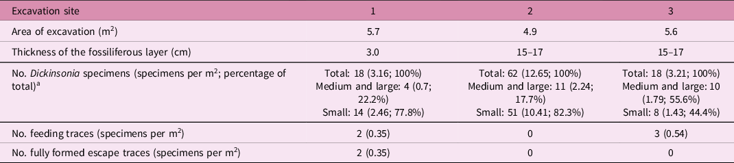
The concentration of Dickinsonia fossil remains was calculated at three sampling sites, which uncovered a total of 16.2 m2 of productive surface (only about one-quarter of the entire studied area). One site was made in the sector with the minimum thickness of the fossiliferous layer, and the other two were located in the sector with the maximum thickness (Table 1).
A number of Dickinsonia remains of different size groups in the excavation sites of the L2(XII) burial event horizon

a Small: body length of 4.1–13.2 mm; medium and large: body length of 18.3–122 mm.
In 30 specimens of Dickinsonia from excavations no. 2 and 3, it was possible to approximately measure the orientation of the anterior end of the body relative to the longitudinal axis of the erosional channel. The distribution of the orientation turned out to be rather chaotic (Fig. 2c). Moreover, the cases of a diametrically opposite arrangement of closely adjacent specimens were observed (Fig. 2d). Despite the small sampling and, accordingly, low validity, it can be assumed that there is no definite orientation in the Dickinsonia imprints from the L2(XII) burial event horizon.
The distribution of the structures by body sectors was determined on 32 large Dickinsonia specimens.
The studied material is deposited in the Borissiak Paleontological Institute of the Russian Academy of Sciences (PIN RAS), Moscow, Russia and is included in collection no. 4716.
3. Methods
The unique material that became the basis for this publication was obtained through the use of gentle washing, which we had never previously used for the preparation of Precambrian macrofossils. The extremely poor compaction of the clays in contact with the fossiliferous surface of the L2(XII) burial event horizon made it possible in the process of finishing preparation to wash off the imprints with plain fresh water sprayed with low-powered household pulverizers. AL Nagovitsyn (Arkhangelsk Regional Lore Museum, Arkhangelsk) developed the method and adapted it to the fieldwork in the Vendian localities of the southeastern White Sea area. The use of brushes of various stiffness and hydrogen peroxide, common for the White Sea excavations, would obviously lead to the loss of siltstone coat, representing a part of the structures described in the article.
4. Evidence of Dickinsonia mobility
4.a. Long furrows of deformed specimens
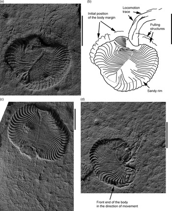
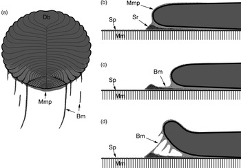
These fossil structures are represented by groups of furrows of various widths extending from the posterior end of the Dickinsonia imprint along the surface of the fossil-bearing layer (Fig. 3). Some of the furrows directly reach the imprint, while the others end at some distance from it. The furrows can be more or less straight or strongly curved, but they remain parallel to each other (Fig. 3a, b). The total length of the group of furrows is close to the length of the adjacent imprint. These structures are associated exclusively with peculiar Dickinsonia individuals that have experienced deep intravital deformations (Ivantsov et al. Reference Ivantsov, Nagovitsyn and Zakrevskaya2019 b). We had never before observed such groups of furrows located separately from the body imprints. It is evident that the grooves are imprints of some thin and rather dense material lying on the surface of the microbial mat. These structures are similar to the bodies of macroorganisms in terms of their ability to form imprints, as well as the lack of remains of the material that served as a matrix for these imprints. This suggests that the matrix that created the furrows, as well as the body, were formed by a material of the same kind, that is, an organic matter. In the described case, such a substance could be mucus secreted by the organisms (Fig. 4a). Previously, we made such an assumption in relation to the movement traces of Kimberella (Ivantsov, Reference Ivantsov2009, Reference Ivantsov2010, Reference Ivantsov, Fyodorov and Yakovlev2012), and then extended it to the traces of Tribrachidium and Dickinsonia (Ivantsov et al. Reference Ivantsov, Nagovitsyn and Zakrevskaya2019 b). The sufficient density of the mucus could be provided by inclusions of small mineral particles precipitated at the initial stage of the taphonomic event. The mineral component of this mixture has probably been preserved in the depths of the furrows to the present day. However, it is indistinguishable from the clay from the underlying layer, which is difficult to remove with our washing method. The curvature of the furrows observed in a number of specimens clears the suspicion that they could have an abiogenic origin (for instance, that either the bodies themselves or the sediment behind them moved due to directional lateral pressure). We therefore interpret the furrows as imprints of the groups of mucus ridges left by Dickinsonia moving along the surface of the microbial mat, that is, as specific traces of locomotion (Ivantsov et al. Reference Ivantsov, Nagovitsyn and Zakrevskaya2019 b). Since the ridges are associated with highly deformed individuals, it is assumed that they were formed due to the loss of matter through damaged parts of the body (Ivantsov et al. Reference Ivantsov, Nagovitsyn and Zakrevskaya2019 b). However, while poorly pronounced, these structures can also accompany undamaged areas (Fig. 3c); it should therefore be recognized that the reason for the selective formation of the ridges is still unclear.
Locomotion traces of Dickinsonia: (a, b) specimen no. 4716/5178 (a, plan view; b, drawing); (c) specimen no. 4716/5270; and (d) specimen no. 4716/5181. All photographs were taken with the coating with ammonium chloride; all scale bars represent 1 cm (modified from Ivantsov et al. Reference Ivantsov, Nagovitsyn and Zakrevskaya2019 b).

Schematic reconstruction of the mechanism of formation of various structures demonstrating the mobility of Dickinsonia: (a) locomotion traces; (b) silt-sandy rim; (c) pulling structures; and (d) tear-off structures (Mm – microbial mat; Mmp – mixture of mucus and mineral particles; Sp – silt powder; Db – Dickinsonia’s body; Sr – siltstone ring; Bm – bundles of a mixture of mucus and mineral particles).

The small length of the traces and the close contact with the bodies indicate that they were formed immediately before the death of the trace-producing organisms. Apparently, their preservation is somehow connected with the taphonomic event that led to the formation of the burial event horizon. We assume that the traces of locomotion were preserved because they were created during the taphonomic event itself when the sediment burying the bottom community had already begun to precipitate, but its layer was still very thin, and some of Dickinsonia could move (Ivantsov et al. Reference Ivantsov, Nagovitsyn and Zakrevskaya2019 b).
4.b. Halo of grooves
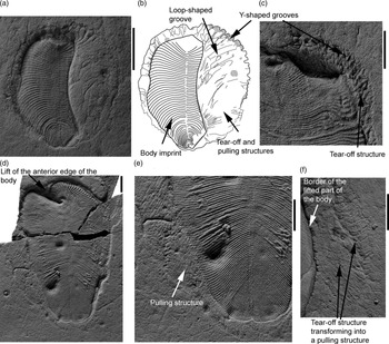
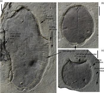
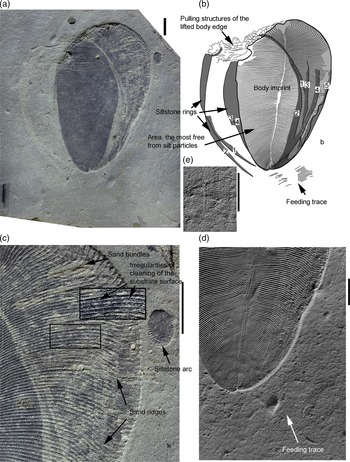
Many medium- to large-sized Dickinsonia imprints from the L2(XII) burial event horizon, of standard morphology and intravitally damaged, are surrounded by a halo of grooves of different width and shape (Figs 5–8). On the outer margin, such a halo is limited by a winding ledge, behind which the main surface of the layer extends with an elevation. Imprints of the end parts of the isomers can be observed in the bends of the ledge (Figs 3a, b, 5a, b). The halo is usually asymmetric, more developed near one margin of the body imprint, and less developed or absent at the opposite margin (Fig. 5d, e). The grooves of the halo are straight and curved, Y-shaped, and of more complex shapes, often oblique to each other and intersecting (Fig. 5a–c). Sometimes only the margin and depression of the halo itself is preserved, and the grooves are absent (Fig. 6b). Sometimes the halo can only be detected by a subtle change in the shade of the rock (Fig. 6c, top of the image).
Tear-off and pulling structures: (a, b) specimen no. 4716/5180 (a, plan view; b, drawing); (c) specimen no. 4716/5282; (d, e) specimen no. 4716/5209 (d, plan view; e, fragment with higher magnification); and (f) a fragment of the halo of tear-off and pulling structures of specimen no. 4716/5175. All photographs were taken with the coating with ammonium chloride; all scale bars represent 1 cm.

Silt-sandy rim: (a) specimen no. 4716/5165; (b) specimen no. 4716/5191; and (c) specimen no. 4716/5190; all scale bars represent 1 cm.

Some specimens of Dickinsonia from Lyamtsa demonstrate the lack of one or more parts of their bodies. These ‘lost’ parts vary in size from a small notch that occupies only a few percent of the imprint area (Figs 7a, b, 8a, b) to almost half of it (Fig. 5a, b). Sometimes it is only a deep fold along which part of the Dickinsonia body is drawn into the overlying layer (Fig. 5d). Such structures are interpreted by Evans et al. (Reference Evans, Droser and Gehling2015) as the result of the lifting of a portion of the Dickinsonia body. The fact that the part of the body was lifted into the fossil-bearing layer of sandstone, rather than torn off in some way, is evident in the White Sea samples. They have this portion preserved within the layer in a form of pyrite pseudomorph (Bobrovskiy et al. Reference Bobrovskiy, Krasnova, Ivantsov, Luzhnaya (Serezhnikova) and Brocks2019). In the Australian locality, any structures are absent from the bottom of the layer, where the part of the imprint disappeared. However, a sandy hillock could appear there formed as a result of the deposition of the sand flow beneath the body of Dickinsonia (Evans et al. Reference Evans, Droser and Gehling2015). In the majority of Lyamtsa imprints, the place of the missing part is taken by the grooves of the halo, which become even deeper there (Figs 6a, 7a, b). Sometimes, if the halo around the whole imprint was not clearly developed, the grooves are visible only in this place (Fig. 8a, b).
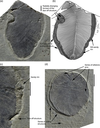
Silt-sandy rim, tear-off and pulling structures: (a, b) specimen no. 4716/5272 (a, plan view; b, drawing); (c) specimen no. 4716/5289, a section of a sandy rim; and (d) specimen no. 4716/5290; all scale bars represent 1 cm.

Silt-sandy rim, tear-off and pulling structures, feeding traces: (a–d) specimen no. 4716/5252: (a) plan view; (b) drawing; (c) a fragment of a sandy rim (an area with an irregular distribution of clastic material is shown in the frame with magnification); and (d) the posterior end of the body and the poorly developed feeding trace. (e) An isolated feeding trace, Epibaion axiferus, specimen no. 4716/5003. Photographs (d, e) were made with the coating with ammonium chloride; all scale bars represent 1 cm.

The fact that the halo and its variety – formed where the portion of the body was lifted – are embedded in the layer indicates that they, like the main fossil remain, represent imprints but not the imprints of the body of the animal itself. We assume that these structures are only indirectly related to the Dickinsonia body, being the imprints of a mucous membrane formed under the body during a taphonomic event. The shifted margins of the body or lifted parts revealed this film. The halo of grooves barely differs from the surrounding surface in a greenish and slightly lighter shade of the rock (Figs 6c, 7a), which in this case is a sign of a slight increase in the admixture of clay or silty particles. According to our interpretation, this additional admixture is the remains of the main material which created the traces, a mixture of mucus and silty particles, after the decomposition of its organic component. The morphology of the halo reflects the sculpture of the surface of the mucous membrane, composed of the ridges, representing the imprints of the grooves of the ventral side, and various bulges formed when the margin of the body was pulled along the film and the body was detached from this film (Fig. 4c, d).
Only the individual parts of Dickinsonia could be exposed to pulling rather than its entire body, and all these parts were stretched to varying degrees. When the margin of the body, which already had a raised area, was displaced, a characteristic deep and straight groove was formed on the imprint, sharply bordering the pulling structure (Fig. 6a, c).
The tear-off structure differs from the pulling structure in the greater roughness of the grooves. The winding, dividing and branching grooves are associated with it. The grooves on the pulling structure are more numerous, parallel to each other and usually straight. In the case of an arcuate bend, the furrows on the tear-off structure can be radially arranged (Fig. 7a, b). However, the tearing off of the margin was often accompanied by its pulling, and therefore the formed structures can merge into each other (Figs 5a, b, 8a, b). Moreover, in the case of a change in the movement direction of the body margin, the curved grooves were formed (Fig. 5f).
4.c. Rim of mineral grains
The basis of this structure is a fine powder of silty or sandy grains, bordering the Dickinsonia imprint with a light greenish-grey stripe (Figs 3, 6–8). On the surface of the grey fine-grained sandstone of the fossil-bearing layer, it is rather poorly visible. However, it stands out quite clearly against the dark brown background of organic matter coating the body imprint. The rim stretches along the margin of the imprint and is mainly located within it, but may partially extend outwards onto the surface of the fossil-bearing layer. A fully formed rim of mineral grains, in the form of a ring, repeating the contours of the Dickinsonia body, appears to be a singular phenomenon (Fig. 8a, b). Only its fragments are usually observed, represented by more or less extended arcs. The silty-sandy arcs are located at the end of the imprint, opposite to the locomotion traces and wide pulling structures (Figs 3a, b, 7d). The width of the rim varies between specimens and can also vary within a single imprint. For instance, in specimens PIN no. 4716/5165 and 4716/5272, it gradually becomes thinner from the middle of the body to the posterior end, but at the same time retains its relative width, which is about 0.4 and 0.2 of the isomer length (measured across the body) for the first and second samples, respectively. The sandy material of the rim has clear signs of biogenic processing. For example, in specimen no. 4716/5289, the mineral grains are collected in columns, the width of which is close to the width of the isomers, but the orientation does not coincide with the orientation of the isomers visible on the body imprint (Fig. 7c). We previously described similar columns in Dickinsonia costata and D. cf. tenuis and interpreted them as sandy filling of food-collecting pockets (Ivantsov, Reference Ivantsov2011 a). We can consider that in the case of D. cf. menneri studied here, the sand columns are casts from the distal ends of the food-collecting pockets.
The rim is not the result of the deposition of mineral particles beneath the body of Dickinsonia under the influence of some external agent. It could also not be the result of only the simple transportation of the body onto the surface covered with particles. This is evident by a regular change in its width along the imprint, reducing with a decrease in the length of isomers, as well as by partial processing of mineral material, and its formation into the ridges. We assume that this rim was formed as a result of the interaction of Dickinsonia with a thin powder of silty particles that fell out at the initial stage of the taphonomic event. The animal moved these particles in radial directions from the body axis to its edges (Fig. 4b). The shorter the isomers and, accordingly, the smaller the area of the surface cleaned from mineral particles, the smaller was the width of the rim area. Thus, the concentration of mineral particles occurred at the margin of Dickinsonia. Many specimens of the Lyamtsa locality are lacking a mineral rim; in others, it is present only at one edge or at the posterior end (specimen no. 4716/5191). Only a few, it seems, had a more or less complete ring (specimen no. 4716/5272, 4716/5175). A specimen associated with several silty rings and arcs was also found (no. 4716/5252, see below). These differences can be explained by assuming that the accumulation of silt particles took place beneath the body of Dickinsonia, and the animals at that moment still had an opportunity to move. Some of the individuals remained in one place from the very beginning of the taphonomic event until the precipitation of the bulk of the sandy sediment; the silty suspension did not get under their bodies in these cases. Others moved at various distances, crawling onto the surface of the microbial mat sprinkled with silty or sandy grains. The displacement was either insignificant, to a short distance less than the length of the body, which led to the formation of a fragmentary rim at one edge of the imprint, or to the entire length of the organism and further, as a result of which more or less complete rings were formed. The location of the rim or its part beyond the bounds of the imprint of the Dickinsonia body is explained by the displacement of the entire body or only the corresponding portion of its margin along the bedding plane. At the same time, various grooves and ridges could appear on the surface of the rim (specimen no. 4716/5272) representing the halo of irregularities described above, probably formed when the body of Dickinsonia was pulled along the mucus (Fig. 7a, b).
The silty rings and arcs in specimen no. 4716/5252 (Fig. 8) are fanned out and form a sequence reflecting at least five episodes of the formation of these structures during the movement of the animal. Based on the fact that the first two rings are quite distinct and complete, Dickinsonia began to create them on the surface already covered with silt (Fig. 8a, b). From the place of creation of the first ring to the final place of burial, the animal moved, turning its front end, first anticlockwise and then clockwise, and shifted on about half of its width. The left parts of the third and fourth rings are pale and poorly visible since there was a shortage of silty material that was brushed off the surface of the mat during the formation of the first and second rings. For the same reason, the left half and the central part of the body imprint completely lack silt particles, since they were cleaned at least five times. In contrast, on the right side of the imprint, the concentration of mineral particles is increased since, at each shift of the body, the particles from the previous accumulations were added to the newly collected particles. Furthermore, sandy grains were added to the silty material between the third and fifth (the final one) stops, which are no longer arranged in bands, but instead form bundles and thin ridges elongated along the isomers (Fig. 8c). The cast of the feeding trace extends to the posterior end of the body imprint. Based on the width of the metaisomers (structural units of a feeding trace), the trace could belong to the same Dickinsonia individual (Fig. 8d). The trace is poorly developed. Based on other more distinct traces from the same burial event horizon, it belongs to the ichnospecies, Epibaion axiferus (Ivantsov & Malakhovskaya, Reference Ivantsov and Malakhovskaya2002) (Fig. 7e). It seems that Dickinsonia moved from this last feeding place of its life to the place where it began to create silty rings. Since the front end of the feeding trace is covered by the body imprint, the distance covered by the animal was also short and did not exceed its body length.
4.d. Escape traces
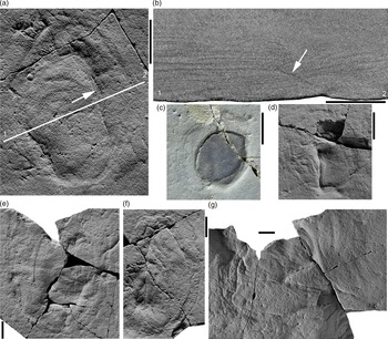
The studied samples show that the stretching of various parts of the Dickinsonia body occurred along their own, sometimes curvilinear, trajectories and over different distances. The complex of multidirectional pulling and tearing structures could form a separate compact fossil. Its cast, preserved at the base of the layer, looks like a shallow depression and is oval in shape as a whole, corresponding to the original position of the Dickinsonia body on the surface of the microbial mat (Fig. 9). This completed, fully formed structure is not accompanied by an imprint of the body, at least in the part, located at the bottom of the layer. It consists of fragments of the pulling and tearing structures that are separated by more or less deep grooves. One of these grooves extends into the overlying sandstone, where it develops into a joint plane, intersecting the bedding (Fig. 9b) and fixed by a thin coat of clay. It seems that the groove and the joint plane continuing it marks a place where Dickinsonia enters the sediment and trace the movement through it. In general, this entire complex of fossil structures can be classified as an escape trace (fugichnia) of Dickinsonia.
Escape traces of Dickinsonia: (a, b, d–g) fully formed; and (c) incomplete. (a, b) Specimen no. 4716/5266 (a, plan view; b, cross-section along the line 1–2); arrows point to the furrow and the jointing surface, tracing the place of entry and passage of Dickinsonia through the sediment. (c) Specimen no. 4716/5268; one of the stages of the formation of an escape trace, when the body imprint is surrounded on all sides by pulling structures, and its peripheral part is lifted into a sandstone layer. (d) Specimen no. 4716/5267; (e) specimen no. 4716/5264; (f) specimen no. 4716/5067; and (g) specimen no. 4716/5006. All photographs, except for (c), were made with the coating with ammonium chloride; all scale bars represent 1 cm.

The completed escape traces were found in the L2(XII) burial event horizon only in the area with a minimum thickness of the sand layer. Imprints of medium and large bodies are extremely rare there (Table 1). It can be assumed that Dickinsonia from Lyamtsa with a body length of more than 18 mm, when buried under a sediment layer of about 3 cm (excluding diagenetic shrinkage), retained the ability to move and could get out to the seafloor surface, but it lost this ability with a layer thickness of about 15 cm or more. Dickinsonia with a body length of less than 14 mm was unable to move even when the sediment layer was 3 cm in thickness.
5. Discussion
In the L2(XII) burial event horizon, the sharp inconsistency in the orientation of long axes of the gutters and the direction of the transfer of sandy material that filled these gutters indicates that, at the initial moments of the layer formation, two events separated in time happened: (1) the disruption of the continuity of the microbial mat and erosion of the underlying silty sediment; and (2) the sand precipitation, the filling of the gutters formed during the first event and the formation of a cross-bedded lower part of the layer. It can be assumed that the first event led to the mobilization of fine mineral grains. Due to their adhesion to organic materials, the structures described here were able to be preserved. The second event led to the burial of the living Dickinsonia. In the places where the layer of the precipitated sediment was rather thin, large individuals were able to get out from under it, leaving traces of movement and escape.
The flow force during the formation of the L2(XII) burial event horizon was strong. In the beginning, it was sufficient for the local rupture of the microbial mat with the formation of gutters and later for the transfer of large clay pebbles; this is not a single observation. During the formation of the Z2(III) burial event horizon of the Zimnie Gory locality, a flow tore off large pieces of the microbial mat with an area of tens of square metres and, at the remaining areas, the mat was lifted up with rolled-up edges (Ivantsov, Reference Ivantsov2011 a). However, even in this case, Dickinsonia, along with a number of other organisms, remained on the surface of the mat. There are cases when the flow, without displacing Dickinsonia specimens themselves, lifted up parts of their bodies and deposited sediment under the raised areas (Evans et al. Reference Evans, Droser and Gehling2015). Such resistance to transfer is hardly possible for organic bodies lying freely on the sea bottom, the density of which approaches the density of the water (Schopf & Baumiller, Reference Schopf and Baumiller1998; Retallack, Reference Retallack2007). The distribution of large objects along the fossiliferous bed L2(XII) is characteristic. There were imprints of soft-bodied organisms on the bottom of the layer, and above them there were clay pellets (the initial density of which was obviously higher than that of the organic bodies). Above all these, there were redeposited fragments of the microbial mat (the density of the substance of which was probably of the same order as in the organisms). The pellets and pieces of microbial mat are positioned according to the expected difference in their density, while the imprints are located where we would expect to find quartz pebbles, rather than the bodies of non-skeletal organisms. The ability of Dickinsonia and other macroorganisms of the Flinders–Belomorian fossil community to resist the transfer and upwards movement of suspended matter cannot be explained by anything other than the adhesion. A very rare case is described (two specimens per several thousand known from South Australia and the White Sea region) when the body of Dickinsonia was torn into two parts, and both parts remained lying side by side (Gehling et al. Reference Gehling, Droser, Jensen, Runnegar and Briggs2005; Evans et al. Reference Evans, Huang, Gehling, Kisailus and Droser2019 b; Droser et al. Reference Droser, Tarhan, Evans, Surprenant and Gehling2020). The reason why the torn-off fragment was not carried away by the flow of water or sandy suspension remains unknown.
A notable feature of the L2(XII) burial event horizon is depressions from clay pellets visible at the base of the fossiliferous layer (Fig. 2b). The pellets themselves (they are mudstone lenses at the moment) lie within the layer near its base. The depressions were formed during the dehydration and contraction of the pellets. At the same time, the volume reduction was accompanied by the upwards rise of mainly the lower surface of the pellet (Bobrovskiy et al. Reference Bobrovskiy, Krasnova, Ivantsov, Luzhnaya (Serezhnikova) and Brocks2019). The rise phenomenon was also responsible for the unusually deep imprints of the soft bodies of Ediacaran organisms. The upwards movement, against the force of gravity, can be explained by the ‘floating’ of relatively less dense organic materials compared with the sandy sediment (the Dickinsonia bodies and the mass of the microbial mat located under it).
On each medium and large sample we studied from the L2(XII) burial event horizon, in addition to the body imprint there is an imprint of some thin structure that rose above the substrate as a constituent element. It is difficult to dispute that it and the structures associated with it belong to some specific Dickinsonia specimen. In our conclusion, all of them were formed due to the mucous secretions of Dickinsonia itself (Fig. 4). It is difficult to imagine that such an ‘ephemeral’ unstable and flexible material as mucus could be a source of the formation of sufficiently distinct imprints with deep and well-defined details. That is why one of the authors of this article suggested that, in the case of the formation of fossil traces of the movement of various Ediacaran organisms, mineral particles were added to the mucus (Ivantsov, Reference Ivantsov2009, Reference Ivantsov2010; Ivantsov et al. Reference Ivantsov, Nagovitsyn and Zakrevskaya2019 b). The presence of such mineral particles, namely, grains of silt and sandy size, is now revealed for the Lyamtsa imprints, which became possible due to the application of the method of gentle washing of samples. The authors are not aware of any other hypotheses about the primary composition of the material from which these structures could be created.
Mucus is used by many modern invertebrates, including those with a rather simple structure, as an intermediary material in contact with the substrate during movement and adhesion (Smith, Reference Smith1991, Reference Smith1992; Davies & Case, Reference Davies and Case1997; Whittington & Cribb, Reference Whittington and Cribb2001). It would therefore not be a long stretch to assume that Dickinsonia was able to secrete mucus and used it for the same purposes. Fossil structures similar to imprints of a mucous film and stretching structures were described in the literature for Dickinsonia and related organisms as a trace of contraction, and as the last feeding trace, respectively (Wade, Reference Wade1968; Seilacher, Reference Seilacher1989; Ivantsov, Reference Ivantsov2011 a, Reference Ivantsov2013; Evans et al. Reference Evans, Gehling and Droser2019 a, b). Both structures were formed by the body of Dickinsonia, which was in a position slightly different from the final imprint, or had a larger area. It has been shown that the feeding trace was created by a living organism that expanded over the surface of the microbial mat (Ivantsov, Reference Ivantsov2011 a, Reference Ivantsov2013; Evans et al. Reference Evans, Gehling and Droser2019 a), while the trace of contraction was presumably left by a dead body compressed during post-mortem dehydration (Wade, Reference Wade1968; Seilacher, Reference Seilacher1989; Gehling et al. Reference Gehling, Droser, Jensen, Runnegar and Briggs2005). Each of these two types of trace fossils is a cast of the imprint of the ventral surface of the organism on the microbial mat. Both of them are represented on the bottom of the fossil-bearing layer in positive and slightly convex relief. The grooves on them are casts of the grooves of the Dickinsonia body, along which the isomer boundaries pass and they extend these boundaries outwards. In contrast to these two types of trace fossils, the imprint of the supposed mucus film with a halo of grooves is pressed into the surface of the fossil-bearing layer, that is, it has a concave and negative relief. The halo is often developed only at one margin of the body imprint, while the feeding traces and the traces of contraction surround a body from all sides. In the case of a fully developed escape trace there is no body imprint at all near a group of stretching and lift-off structures, inconsistent with a contractional mechanism of formation. The orientation of the grooves of the halo and of the boundaries separating the isomers on the body imprint is often markedly different (Fig. 5d, e). This implies that the body changed its position after the formation of the structures. The number of furrows on the stretching structures can be significantly higher than the number of the corresponding isomers of the adjacent body area. Moreover, only a few of the linear depressions of the halo can be correlated with isomer boundaries. Loop-shaped, Y-shaped and more complex grooves and depressions, often obliquely arranged to one another, curved and intersecting, do not find a direct correspondence with the known morphology of the ventral side of Dickinsonia. But such grooves (recall that the initial structures were ridges on the surface of the mat) could represent threads of mucus stretching behind various relief elements of the margin of the Dickinsonia body, as well as of its ventral and even dorsal sides.
Fedonkin (Reference Fedonkin2002) interprets the contraction trace as an imprint of a soft body extending beyond the carapace, representing the main part of the Dickinsonia fossil remain. The pulling and tear-off structures resemble the body imprint even more since, in contrast to the contraction trace, they are preserved in the same negative relief. We cannot offer a criterion for reliably distinguishing a mucous membrane from a thin outgrowth of a soft body using an imprint. Indirect evidence of difference, such as a predominant arrangement of the structures at one of the body margins, a variety of surface relief, a clear correspondence between the external contour of the deformed body part and the contour of the pulling structure, can be disputed. Even the escape trace can be interpreted as a body imprint from which the carapace was ripped off. However, according to our data, the dissected structure of Dickinsonia did not represent a shield or carapace. It had a shape of a thin-walled sack and was externally covered with an integument layer (Ivantsov et al. Reference Ivantsov, Zakrevskaya and Nagovitsyn2019 c). With such structure, the presence of extensive outgrowths capable of being pulled under the body is unlikely.
We would prefer not to enter into extensive polemics with Retallack here. Dickinsonia, as it appears in our works, is not comparable with any lichen either in structure (a three-dimensional body with a basement membrane and ectodermal tissue; Ivantsov et al. Reference Ivantsov, Zakrevskaya and Nagovitsyn2019 c) or in the growth mode (which is based on the addition of elements at one end of the body; Ivantsov et al. Reference Ivantsov, Zakrevskaya, Nagovitsyn, Krasnova, Bobrovskiy and Luzhnaya (Serezhnikova)2020). An affinity with lichens is also made unlikely from the presumed composition of the substance (high cholesteroid content; Bobrovskiy et al. Reference Bobrovskiy, Hope, Ivantsov, Nettersheim, Hallman and Brocks2018) as well as the ability to move (chains of various traces; Ivantsov et al. Reference Ivantsov, Nagovitsyn and Zakrevskaya2019 b; this study). Nevertheless, the furrows of the pulling and tear-off structures may resemble rhizoids of lichens or liverworts. However, if we assume that these are the imprints of the torn-off rhizoids themselves, then the absence of the ends with which they attached to the thallus will remain unclear. These furrows cannot be traces of rhizoids pulled out of the substrate since they are preserved in negative relief, the same as the body remains.
Evans et al. (Reference Evans, Droser and Gehling2015) explained the lift of the body edge of Australian Dickinsonia by a mechanical effect of the lateral pressure of the water flow on it. In the study of the Lyamtsa locality, we did not measure the orientation of the raised areas relative to the direction of the palaeoflow. However, the lift of the two opposite body edges, as demonstrated for example in specimen no. 4716/5165 (Fig. 6a), the lift of the body edge in one direction accompanied by pulling of the other edge in the opposite direction, as in specimen no. 4716/5209 (Fig. 5d), and the slit-like shape of the bend of the same specimen is difficult to explain by the impact of the flow alone. On the material of L2(XII) burial event horizon, we did not find evidence confirming an extraneous effect on the body of Dickinsonia. However, we do not state that the lift of the body edge could not be done by the flow. After all, the force of its pressure was sufficient to rupture sections of the mat, form gutters and move muddy pebbles. For example, the fact that Dickinsonia continued to move with one lifted edge may be an indication of external influence on that edge. However, according to our hypothesis, most of the stretching and lift-off structures recorded for the samples from L2(XII) burial event horizon were formed as a result of the independent movements of living Dickinsonia. These movements could be performed by the animals in the course of attempts to avoid burial. In the same way, we previously interpreted rough folds covering the imprints of bodies specifically as a consequence of the attempts to avoid burial (Ivantsov, Reference Ivantsov2011 a).
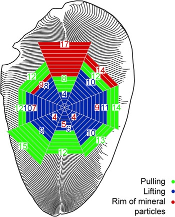
The lifted areas are most characteristic of the anterior sectors of the Dickinsonia body (Fig. 10). The stretching structures are more evenly distributed, with a slight predominance of the posterior sectors over the anterior ones. A number of specimens clearly show the opposite of the arrangement of the arcs of the mineral rim and wide stripes of the stretching structures (Fig. 7d). Apparently, the anterior part of the Dickinsonia body was more mobile, capable of making movements in various directions and planes, while the posterior part was more inclined to passive movement along the surface of the substrate. This is consistent with the visible anteroposterior body asymmetry of Dickinsonia and related organisms, and with data on the preferred orientation of these animals during movement (Gehling et al. Reference Gehling, Droser, Jensen, Runnegar and Briggs2005; Ivantsov, Reference Ivantsov2011 a, Reference Ivantsov2013; Evans et al. Reference Evans, Gehling and Droser2019 a).
Distribution of the structures by sectors of the Dickinsonia body. The numbers correspond to the number of detected cases.

The formation of the rim of silty-sand particles around the body of Dickinsonia was probably a by-product of the adhesion of the organism to its substrate, the microbial mat. Moreover, this process was also due to mucus. In the process of adhesion, modern organisms use mucus as a glue to increase adhesion to the substrate, or as a sealer to maintain negative pressure under the foot (Smith, Reference Smith1991, Reference Smith1992; Davies & Case, Reference Davies and Case1997; Whittington & Cribb, Reference Whittington and Cribb2001). In this case, the negative pressure is created by the muscles that rise the centre of the foot from the substrate (Smith, Reference Smith1991; Davies & Case, Reference Davies and Case1997). We assume that Dickinsonia also used mucus as a sealer. However, it created the underpressure in a peculiar way via a centrifugal dispersion of the mucus film. Mineral particles moved along the way with the mucus and accumulated at the margins of the body. The rhythmic alternation of concentric stripes can be seen on sample no. 4716/5252 (Fig. 8c). They are represented by wide light-coloured stripes that are enriched in silty particles, and narrow dark stripes that are depleted in silty particles. This alternation possibly reflects the wavelike nature of the radial movement of the material under the body of Dickinsonia. The mineral rim is the most pronounced at the lateral margins of the body (Fig. 10). This can be explained by the greater length of the isomers in the middle part of the body and, accordingly, the larger area from which the mineral particles were collected.
The mineral rings and arcs, as well as the imprints of a hypothetical mucous membrane, are therefore traces of Dickinsonia’s adhesion to the substrate. Apparently, they were able to fossilize only in the exceptional case of the L2(XII) burial event horizon. Specimen no. 4716/5252 demonstrates at least four unfinished adhesion attempts, the first of which were made while the surface of the microbial mat was covered with the silty particles. The presence of a small amount of sandy material in the latest rims probably indicates that, by the time of their formation, there were changes during the taphonomic event, and sandy grains were beginning to be added to the precipitated silt material, which subsequently resulted in mass precipitation of the sand and the final burial of Dickinsonia. A visual representation of the efficiency of the adhesion of Dickinsonia to the substrate is specimen no. 4716/5268 (Fig. 9c), in which all marginal areas were lifted into the overlying sediment and the central part of the body still remained on the bedding plane.
The ridges, described as the locomotion traces of intravitally damaged Dickinsonia specimens, are similar to the ridges of the stretching structure formed behind the non-deformed specimens, but are higher and wider due to the concentration of the material that composed them in the deformed parts of the body. The lift-off structure often develops into the pulling structure, which is equally often limited to the lifted margin of the body. These structures can record one process of the Dickinsonia movement. Many Dickinsonia imprints are asymmetric to some extent. One or more of their sectors turns out to be wider or longer than the sectors opposite to them across the longitudinal axis. For example, the front right side of specimen no. 4716/5252 is stretched compared with the front left side. Moreover, the direction in which the stretching occurred coincides with the direction of movement of this individual (Fig. 8a, b). It can be assumed that, when moving along the surface of the mat, Dickinsonia carried forward a certain part of the body (mainly from the anterolateral sectors), somehow fixed it in a new place and then pulled the rest of the body to it. A series of closely located mineral arcs at one end of the body and an associated series of pulling structures at the opposite end, both visible on specimen no. 4716/5290 (Fig. 7d), may reflect the intermittent nature of the movement. Although the end bearing a sub-triangular lobe was at the forefront of the movement, each specific impulse could not be performed exclusively by the anterior sectors, but this was possible by any of the anterolateral sectors. There is one extremely deformed specimen in our collection, in which a significant part of the body was lost during the lifetime. Based on the trajectory of the traces left by this specimen, the anterior end in the direction of movement was located in the left anterolateral sector of its body (Fig. 3d).
The lifting into the overlying unconsolidated sediment was probably carried out in the same way. The covered Dickinsonia contracted some part of its body, lifted and pushed it up, where it was fixed and pulled up the rest of the body. It is possible that the area raised by the stream could give rise to this movement. This hypothesis connects all the described structures.
6. Conclusion
Only a few of the researchers of the Ediacaran biota currently doubt the mobility of Dickinsonia. Perhaps only G. Retallack remains faithful to the hypothesis of their attachment, still considering these organisms as terrestrial fungi or lichens, overgrowing hypothetical Ediacaran palaeosols (Retallack & Broz, Reference Retallack and Broz2020), despite the growing criticism of these views (Waggoner, Reference Waggoner1995; Sperling & Vinther, Reference Sperling and Vinther2010; Callow et al. Reference Callow, Brasier and McIlroy2013; Evans et al. Reference Evans, Droser and Gehling2015, Reference Evans, Gehling and Droser2019 a; Tarhan et al. Reference Tarhan, Droser, Gehling and Dzaugis2015; Lücking & Nelsen, Reference Lücking and Nelsen2018). However, Dickinsonia individuals were clearly adhered at the time of their burial, as evidenced by the location of their fossil remains on the bottom of the layer and their ability to resist flow (Retallack, Reference Retallack2007; Ivantsov, Reference Ivantsov2011 a; Evans et al. Reference Evans, Droser and Gehling2015). At the same time, Dickinsonia was a mobile animal. The dataset on the feeding traces (Ivantsov & Malakhovskaya, Reference Ivantsov and Malakhovskaya2002; Gehling et al. Reference Gehling, Droser, Jensen, Runnegar and Briggs2005; Ivantsov, Reference Ivantsov2011 a; Evans et al. Reference Evans, Gehling and Droser2019 a), extended by the structures interpreted as locomotion traces (Ivantsov et al. Reference Ivantsov, Nagovitsyn and Zakrevskaya2019 b and this study), the structures of pulling the body over the surface of the substrate and the escape traces, as well as the recorded successive attempts of adhesion, provide convincing evidence for this. It turns out that Dickinsonia and the organisms close to it were sometimes mobile and sometimes adhered. This double modality of behaviour is characteristic of animals living in an environment of frequent changes in hydrodynamic activity. It is unlikely that this change was associated with relatively rare catastrophic storms. It is obvious that we must reconsider our understanding of the preferred depth of the Dickinsonia habitats, with their transfer to shallower environments above the fair-weather wave base.
The movement intermittency and the ability of Dickinsonia to push its body through unconsolidated sediment may serve as a new argument in favour of the assumption that these animals had musculature. Previously, this assumption was made after an analysis of compression wrinkles, sometimes observed on the imprints, and extended chains of feeding traces formed by Dickinsonia (Gehling, Reference Gehling1991; Gehling et al. Reference Gehling, Droser, Jensen, Runnegar and Briggs2005; Evans et al. Reference Evans, Gehling and Droser2019 a). However, this musculature was apparently rather weak and, with the thickness of the burial sediment of > 15 cm, even relatively large specimens of Dickinsonia lost the opportunity to free themselves and escape to the surface.
Acknowledgments
The authors are very grateful to AL Nagovitsyn (Arkhangelsk Regional Lore Museum, Arkhangelsk, Russia). Thanks to his extremely careful attitude to fossil remains and thoughtfulness it became possible to discover the structures described here. The authors are grateful to S Jensen (Universidad de Extremadura) and GE Budd (Uppsala University) for their valuable and constructive comments that improved the quality of the paper. Fieldwork and preparation of the fossil material were carried out with the support of the grant for the fundamental research on the topic of the state assignment of Paleontological Institute, Russian Academy of Sciences, ‘The Emergence of Life, the Formation of the Biosphere, and the Development of Ancient Biotas’. Laboratory and theoretical studies were supported by the Russian Science Foundation (RSF) (MZ, grant no. 19-14-00346).