Implications
The identification of G protein-coupled receptors (GPR) 41 and GPR43 in this experiment indicate that the rabbit genome contains GPR41 and GPR43 genes. A better understanding of their expression pattern could be useful to study the short-chain fatty acid (SCFA)-GPR41 and -GPR43 interactions and their modulation of host physiology in response to nutrition in rabbit. They might also represent a novel link between gut microbiota and physiological processes.
Introduction
Short-chain fatty acids, primarily including acetate, propionate and butyrate, are not only an important energy source for animals, but also regulate rumen development, insulin and glucagon secretion, gastrointestinal motility and blood flow, and cell proliferation, apoptosis, and differentiation (Scheppach and Weiler, Reference Stoddart, Smith and Milligan2004; Bourdu et al., Reference Bourdu, Dapoigny, Chapuy, Artigue, Vasson, Eechelotte, Bommelaer, Eschalier and Ardid2005; Wong et al., Reference Wong, de Souza, Kendall, Emam and Jenkins2006; Al-Lahham et al., Reference Al-Lahham, Peppelenbosch, Roelofsen, Vonk and Venema2010). The mechanism underlying the regulatory effects of volatile fatty acid is unknown. Recent studies showed that two orphan GPR41 and GPR43, were discovered to be receptors for SCFAs (Le Poul et al., Reference Livak and Schmittgen2003). So GPR41 and GPR43 are also called free fatty acid receptor 3 (FFAR3) and 2 (FFAR2), respectively. Both receptors couple to Gα i /Gα o but GPR43 also couples to Gα q (Covington et al., Reference Covington, Briscoe, Brown and Jayawickreme2006; Nøhr et al., Reference Nilsson, Kotarsky, Owman and Olde2013). During the past decade, research interest in FFARs has been growing with an aim to better understand the modulation of host physiology in response to nutrition.
Rabbits are true non-ruminant herbivores and are considered hind gut ferenters. Their large cecum has a capacity 10 times that of their stomach. It contains up to 40% of the intestinal contents and enables efficient digestion of a high-fiber diet (Kohles, Reference Kimura, Ozawa, Inoue, Imamura, Kimura, Maeda, Terasawa, Kashihara, Hirano, Tani, Takahashi, Miyauchi, Shioi, Inoue and Tsujimoto2014). There is a rich and diverse bacterial ecosystem in cecum, which can ferment the dietary fiber and produce much SCFAs (Dorota et al., Reference Dorota, Bożena, Milan, Ewa, Wojciech and Józef2015). The composition of SCFAs in the cecum of rabbits is relatively clear (60 to 80 mM of acetate, 8 to 20 mM of butyrate and 3 to 10 mM of propionate), which is different with the rumen of cattle and sheep (60% acetate, 25% propionate and 15% butyrate). The potency orders of each SCFA for GPR41 is propionate>butyrate>acetate, whereas GPR43 is equally sensitive to each SCFA. Acetate is more selective for GPR43 than GPR41. So we infer that the expression pattern of rabbit GPR43 and GPR41 is also different with other animal. In this study, we determined whether the rabbit genome contains GPR41 and GPR43 genes, and investigated the expression pattern of these two genes in different tissues and developmental stages.
Material and methods
Animals
Eight nests of New Zealand Rabbits were individually housed in self-made cages (60×40×40 cm). Temperature (20±2°C), humidity (60.1±4.5%) and lighting (12 h light and 12 h dark) were maintained according to commercial conditions. Diets were formulated according to the recommendations from the NRC (1977) and de Blas and Mateos (1998) and pelleted by using pressure. The diameter of the pellets was 4 mm. Lactating female rabbit received a diet containing 17.5% CP, 13.5% crude fiber and 11 MJ/kg of digestible energy. The rabbits from weaning to slaughter in present experiment received a starter diet containing 16% CP, 14% crude fiber and 10.50 MJ/kg of digestible energy. All rabbits had free access to food and water during the rearing period.
Tissue collection
Various rabbit tissues (spleen, thymus, pancreas, lung, duodenum, jejunum, ileum, colon, cecum and adipose tissue) were collected from eight New Zealand rabbits (with four males and four females) at 15 (newborn, 248.76±13.48 g), 30 (weaning, 643.47±27.69 g), 60 (growing, 1287.75±24.64 g) or 90 (finishing, 2359.65±18.69 g) days after birth. The samples of the whole intestinal tract were removed, and segments were taken from the midpoint of duodenum, (5 cm from the pylorus), jejunum (50% along the small intestine) and ileum (5 cm from the ileal–cecal junction) (Freeman et al., Reference Freeman, Bentsen, Thwaites and Simmons1995). After being snap-frozen in liquid nitrogen, the tissue samples were stored at −80°C.
Ribonucleic acid isolation and reverse transcription
Total RNA was isolated using the TRIzol method (Invitrogen, San Diego, CA, USA) according to the manufacturer’s protocol. All samples were treated with DNase I (Invitrogen) before reverse transcription. The quality and the quantity of the RNA was determined by agarose gel electrophoresis and using a biophotometer (Eppendorf, Hamburg, Germany). Real-time (RT) reactions (10 µl) contained 500 ng total RNA, 5 mmol/l MgCl2, 1 µl RT buffer, 1 mmol/l deoxy-ribonucleoside triphosphate, 2.5 U avian myeloblastosis virus, 0.7 nmol/l oligodeoxythymidylate and 10 U ribonuclease inhibitor (TaKaRa, Dalian, China).
The primers for the GPR41, GPR43 and glyceraldehydes-3-phosphate dehydrogenase (GAPDH) genes (reference gene) were designed using Primer 5 and were based on the predicted sequences of rabbit GPR41 (Accession number: XM_002722237.2) and GPR43 (Accession number: XM_002722218.2), as well as GAPDH (Accession number: NM_001082253.1). All the primers used in this study were synthesized by Sangon (Shanghai, China) and are listed in Supplementary Table S1.
Polymerase chain reaction
To amplify the protein-coding regions of rabbit GPR41 and GPR43 messenger RNAs (mRNA), equal complimentary DNA (cDNA) amounts from different tissues were pooled, and 2 μl of mixed cDNA was incubated with 12.5 μl of PCR Master Mix (CWBIO, Beijing, China) and 1 mM of rGPR41-1-F/R primers or rGPR43-1-F/R primers, respectively, in a total volume of 20 μl. The conditions of the PCR reaction were 94°C for 2 min, 35 cycles of 94°C for 30 s, 60°C for 30 s and 72°C for 30 s, followed by extension at 72°C for 2 min. In total, 2 μl of the PCR products were re-amplified after diluting them 50 times with ddH2O, using 1 mM of rGPR41-2-F/R primers or rGPR43-2-F/R primers under the same conditions. The quality of the products of the second amplification was determined by 1% agarose gel electrophoresis. The amplified PCR products were sequenced by Sangon.
Rapid amplification of cDNA ends
Rapid amplification of cDNA ends (RACE) was used to amplify the 3’ and 5’ end regions of the rabbit GPR41 and GPR43 cDNAs. In this study, we used the SMARTer RACE cDNA Amplification Kit (Clontech, Beijing, China) for the 3’ end regions and the Rapid Amplification of cDNA Ends Version 2.0 Kit for the 5’ end regions. Total pancreas and adipose tissue RNA from eight 90-day-old rabbits was used for RACE of rabbit GPR41 and GPR43. The RACE primers were designed based on the identified PCR sequences using Primer 5.
For 3’ RACE of GPR41 and GPR43, first-strand cDNA was transcribed from 1 μg of total RNA using 3’ coding sequence primer A. cDNAs were used as templates in subsequent nested PCRs to amplify the 3’ end of rabbit GPR41 and GPR43 cDNAs with parallel primer sets of universal prime (UPM) with 3’RACE GPR41-1 or 3’RACE GPR43-1 and UPM with 3’RACE GPR41-2 and 3’RACE GPR43-2 (Supplementary Table S1). For 5’RACE, 1 μg of total RNA was pooled for first-strand cDNA synthesis using 5’RACE GPR41-1 or 5’RACE GPR43-1 primers and Superscript II RT enzyme. RNase Mix was used to remove any remaining RNA and terminal deoxynucleotidyl transferase enzyme and deoxycytidine triphosphate were used to obtain dT-tailed cDNAs after purification. These cDNAs were used as templates in subsequent nested PCRs to amplify the 5’RACE sequences of rabbit GPR41 and GPR43 cDNAs using parallel primer sets of abridged anchor primer with 5’RACE GPR41-2 or 5’RACE GPR43-2 and abridged universal amplification primer with 5’RACE GPR41-3 or 5’RACE GPR43-3 (Supplementary Table S1). The PCR products of the second amplification were determined by 1% agarose gel electrophoresis and sequenced by Sangon.
Real-time polymerase chain reaction
To determine the expression of GPR41 and GPR43 messenger RNA in rabbit tissues in the different developmental stages, Real-time PCR (RT-PCR) analysis was conducted using the Applied Biosystems 7500 Real-Time PCR System (Applied Biosystems, Foster, CA, USA). Each RT reaction served as a template in a 20 µl PCR reaction containing 0.2 μmol/l of each primer and SYBR green master mix (TaKaRa). The primer sequences are listed in Supplementary Table S2. Primers were designed for exon–intron junctions using Primer 5.0 software. The RT-PCR reactions were performed at 95°C for 10 s (pre-denaturation), followed by 40 cycles consisting of denaturation at 95°C for 5 s and annealing and extension at 60°C for 40 s. A standard curve was plotted to calculate the efficiency of the RT-PCR primers. Glyceraldehydes-3-phosphate dehydrogenase (GAPDH) was used as the housekeeping gene, and the results of the relative mRNA quantification were verified using β-actin levels (ΔCT). On the basis of the CT values, β-actin and GAPDH mRNA expression were stable across the treatments in this study (P>0.1). The comparative CT method (2−ΔΔCT) was used to quantitate mRNA expression (Livak and Schmittgen, Reference Li, Su, Zhou and Yao2001). The specificity of the amplification products was verified by melting curve analysis and cDNA sequencing.
Statistical analysis
The data are presented as the means±SEM. The homogeneity of variances among the treatments was confirmed using Bartlett’s test. All data were subjected to one-way ANOVA analysis to test the effect of the treatment. For gene expression analysis, n=8. When the effect of treatment was significant, differences between means were assessed by Duncan’s multiple range analysis. P<0.05 was considered statistically significant.
Results
Rabbit genome contains G protein-coupled receptor 41 and G protein-coupled receptor 43 genes
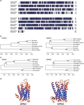
A comparison of the rabbit genome in the GenBank database using the Basic Local Alignment Search Tool program revealed that rabbit GPR41 and GPR43 genes are highly similar to human, bovine, rat and mouse sequences. Similarities between GPR41 in the rabbit and human (NM_005304.3), bovine (NM_001145233.1), Capra hircus (XM_005692314.1), porcine (XM_003127053.2), mouse (NM_001033316.2) and rat (NM_001108912.1) were 84%, 84%, 84%, 83%, 81% and 80%, respectively. Similarities between GPR43 in the rabbit and human (NM_005306.2), porcine (NM_001278758.1), bovine (NM_001163784.1), rat (NM_001005877.1), mouse (NM_146187.4) and C. hircus (NM_001285655.1) were 80%, 82%, 79%, 84%, 84% and 80%, respectively. These data in Figure 1b showed that the mRNA sequence of rabbit GPR41 and GPR43 were highly conserved throughout vertebrate evolution.
Alignments of rabbit G protein-coupled receptor 41 (GPR41)/G protein-coupled receptor 43 (GPR43) and rat GPR41/GPR43 amino acid sequences (a) (The rabbit GPR41 and GPR43 amino acid sequences are deduced from their messenger RNA (mRNA) sequences in this study. The GenBank accession numbers for the rat GPR41 and GPR43 amino acid sequences are NP_001102382.1 and NP_001005877.1. The identical amino acid residues between the rat and rabbit sequences are shaded). Phylogenetic analysis of mRNA sequence (b) and amino acid sequences (c) of rabbit GPR41/GPR43 from Homo sapiens, Mus musculus, Sus scrofa, Bos taurus, Rattus norvegicus and Capra hircus. The phylogenetic analysis was conducted in Molecular Evolutionary Genetics Analysis 4.0. The three-dimensional structures of rabbit GPR41/43 amino acid sequences (d). This diagram was generated by SWISS-MODEL online software (https://swissmodel.expasy.org/). The α-helix residues are colored with red, and loop and unassigned residues with other colors.

Full-length amplification and tissue expression of rabbit G protein-coupled receptor 41 and G protein-coupled receptor 43
Full-length rabbit GPR41 was cloned from pancreas cDNA, whereas GPR43 was cloned from adipose cDNA by RACE. A nucleotide sequence of 957 bp (Accession number: KU585902), representing the open reading frame (ORF) cDNA sequence of rabbit GPR41, was obtained. Open reading frame finder (http://www.ncbi.nlm.nih.gov/gorf/orfig.cgi) was used to derive the amino acid (AA) sequence. Rabbit GPR41 mRNA was predicted to encode a 318-AA protein. Multiple AA alignment results among human, bovine, rat, mice and porcine GPR41 were processed using ClustalW2 multiple sequence alignment (http://www.ebi.ac.uk/Tools/msa/clustalw2/).
Similarities between rabbit and human, bovine, rat, mouse, porcine and C. hircus GPR41 were 77%, 85%, 87%, 78%, 87% and 78%, respectively. A nucleotide sequence of 1793 bp (Accession number: KU585903), representing the full-length cDNA sequence of rabbit GPR43, was obtained. The ORF of rabbit GPR43 was 125 to 1117 bp. Rabbit GPR43 mRNA was predicted to encode a 330-AA protein. Similarities between rabbit and human, bovine, rat, mouse, porcine and C. hircus GPR43 were 81%, 75%, 83%, 84%, 81% and 75%, respectively. Alignments of GPR41 and GPR43 AA sequences of rabbit and rat were shown in Figure 1a. These data in Figure 1c showed that the AA sequence of GPR41 and GPR43 were highly conserved throughout vertebrate evolution. The phylogenetic tree was processed using Molecular Evolutionary Genetics Analysis 4.0 (https://swissmodel.expasy.org/).
The analysis results also indicated that these two proteins were membrane proteins containing seven transmembrane domains. Using the SWISS-MODEL online software, we predicted the three-dimensional structures of both GPR41 and GPR43 (Figure 1d).
Pooled RNA was used in this section. The rabbits sampled in this study were of the same breed, from different families, of identical age and comparable body weight, and were raised in the same housing facility using the same feed, so that variations in gene expression would be negligible, and it would be acceptable to use pooled RNA to describe the distribution of GPR41 and GPR43 in various tissues. Real-time PCR analysis indicated that both GPR41 and GPR43 mRNAs were expressed in the tested tissues, including the spleen, thymus, pancreas, lung, duodenum, jejunum, ileum, colon, cecum and adipose tissue.
Gene expression of rabbit G protein-coupled receptor 41 and G protein-coupled receptor 43 in different tissues and developmental stages
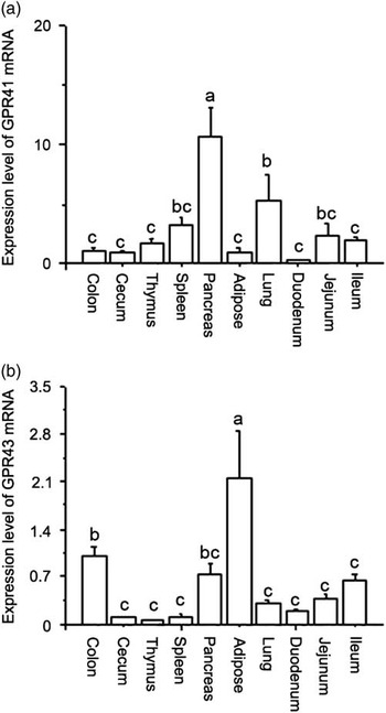
G protein-coupled receptor 41 and GPR43 were expressed in a tissue-specific and time-associated manner. G protein-coupled receptor 41 was most abundantly expressed in the pancreas (Figure 2a), which had significantly higher expression levels than any other tested tissue (P<0.05). Its expression level was higher in the lung than in the thymus (P<0.05), and comparable in the spleen, duodenum, jejunum, ileum, colon, cecum and adipose tissue (P>0.05). The highest mRNA levels of GPR43 were detected in the adipose tissue (Figure 2b), which showed more robust expression than any of the other tissues (P<0.05). GPR43 mRNA levels were higher in the colon than in the ileum (P<0.05), and there were no differences among the other tissues (P>0.05).
The messenger RNA (mRNA) levels of rabbit G protein-coupled receptor 41 (GPR41) (a) and G protein-coupled receptor 43 (GPR43) (b) in different tissues. Values are shown as the mean±SEM (n=8). a,b,cSignificantly different mean value (P<0.05).

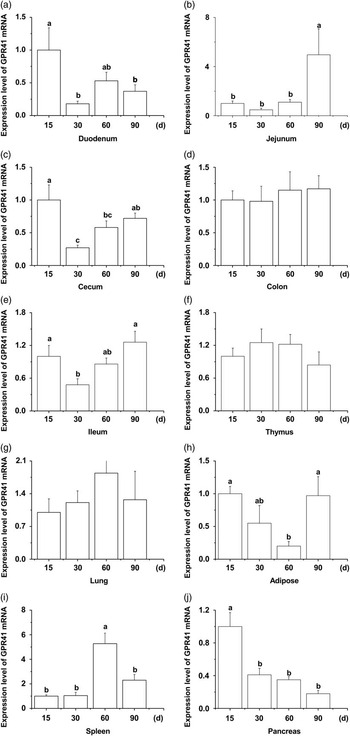
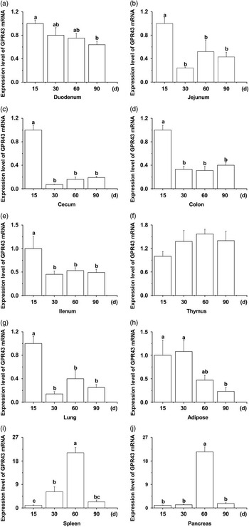
Rabbit GPR41 and GPR43 were also differentially expressed at different developmental stages in several tissues (Figures 3 and 4). The highest expression levels of GPR41 in duodenum, cecum (P<0.05) and pancreas (P<0.05) were at 15th day, when compared with the other developmental stages. Later on, the expression levels of GPR41 in the duodenum, cecum and pancreas were down-regulated. The expression levels of GPR41 in jejunum, ileum, adipose tissue and spleen were up-regulated during growth, with peaks at 90th day in the jejunum, ileum and adipose tissue, and 60th day in the spleen. However, rabbit GPR41 was not differentially expressed in different developmental stages in the colon, thymus and lung. The expression pattern of GPR43 at different developmental stages was different from that of GPR41. G protein-coupled receptor 43 was highly expressed in the duodenum, jejunum, ileum, colon, cecum and lung at 15th day, whereas the expression levels in the pancreas and spleen increased later after birth, with the highest expression at 60th day. However, GPR43 expression in the thymus and in adipose tissue was comparable at all developmental stages.
The rabbit G protein-coupled receptor 41 (GPR41) messenger RNA (mRNA) levels in the duodenum (a), jejunum (b), cecum (c), colon (d), ileum (e), thymus (f), lung (g), adipose tissue (h), spleen (i) and pancreas (j) at different developmental stages (15, 30, 60 and 90 days). The values are shown as the mean±SEM (n=8). a,b,cSignificantly different mean value (P<0.05).

The rabbit G protein-coupled receptor 43 (GPR43) messenger RNA (mRNA) levels in the duodenum (a), jejunum (b), cecum (c), colon (d), ileum (e), thymus (f), lung (g), adipose tissue (h), spleen (i) and pancreas (j) at different developmental stages (15, 30, 60 and 90 days). The values are shown as the mean±SEM (n=8). a,b,cSignificantly different mean value (P<0.05).

Discussion
Previous researchers have identified SCFAs as endogenous ligands for GPR41 and GPR43 (Brown et al., Reference Brown, Goldsworthy, Barnes, Eilert, Tcheang, Daniels, Muir, Wigglesworth, Kinghorn, Fraser, Pike, Strum, Steplewski, Murdock, Holder, Marshall, Szekeres, Wilson, Ignar, Foord, Wise and Dowell2003; Le Poul et al., Reference Livak and Schmittgen2003; Nilsson et al., Reference Nøhr, Pedersen, Gille, Egerod, Engelstoft, Husted, Sichlau, Grunddal, Poulsen, Han, Jones, Offermanns and Schwartz2003). The identification of SCFA receptors, GPR41 and GPR43, might clarify the mechanism of action of SCFAs in various physiological processes. Subsequent studies identified GPR41 and GPR43 in various tissues in several species, including human (Karaki et al., Reference Kim, Cho, Kim, Lim, Kim, Lee and Kwon2008; Tazoe et al., Reference Tazoe, Otomo, Karaki, Kato, Fukami, Terasaki and Kuwahara2009), rat (Karaki et al., Reference Karaki, Tazoe, Hayashi, Kashiwabara, Tooyama, Suzuki and Kuwahara2006), porcine (Li et al., Reference Le Poul, Loison, Struyf, Springael, Lannoy, Decobecq, Brezillon, Dupriez, Vassart, Van Damme, Parmentier and Detheux2014) and bovine (Wang et al., Reference Wang, Gu, Heid, Akers and Jiang2009). Our study focused on the identification of rabbit GPR41 and GPR43 and their tissue expression pattern.
In the present study, the GenBank search revealed that the rabbit genome contains GPR41 (XM_002722237.2) and GPR43 (XM_002722218.2) genes, located in tandem on chromosome 6. They are highly similar to GPR41 and GPR43 genes in the human, bovine, rat and mouse genomes. In several instances, these genes present over 80% similarity across different species. However, most of the GPR41 and GPR43 mRNA sequences available in GenBank are derived from computational prediction, and the organization of the GPR41 and GPR43 genes has not been characterized in most species. The AA sequences of rabbit GPR41 and GPR43 were deduced based on the nucleotide sequences and predicted a 318-AA and a 330-AA protein, respectively. Multiple alignments showed that the rabbit GPR41 and GPR43 proteins are highly similar to the human, bovine, rat and mouse proteins. The structural prediction indicated that these GPR41 and GPR43 were membrane proteins containing seven putative transmembrane domains.
Real-time PCR results indicated that GPR41 and GPR43 were expressed in a variety of rabbit tissues, including the spleen, thymus, pancreas, lung, duodenum, jejunum, ileum, colon, cecum and adipose tissue. However, GPR41 expression in adipose tissue is controversial. Studies detected GPR41 expression in human adipose tissue (Le Poul et al., Reference Livak and Schmittgen2003), mouse white adipose tissue and undifferentiated Ob-luc cells (Xiong et al., Reference Xiong, Miyamoto, Shibata, Valasek, Motoike, Kedzierski and Yanagisawa2004). Conversely, it was not detected in adipose tissue in mice (Hong et al., Reference Hong, Nishimura, Hishikawa, Tsuzuki, Miyahara, Gotoh, Choi, Feng, Chen, Lee, Katoh, Roh and Sasaki2005; Wang et al., Reference Wang, Gu, Heid, Akers and Jiang2009). The reason for this discrepancy remains unclear; the inconsistent results might be due to differences in sample origins and experimental techniques.
Our studies showed highest level of rabbit GPR41 in the pancreas, which is consistent with the finding in bovine pancreas. G protein-coupled receptor 41 was expressed in mouse and human pancreatic β-cells and mediated an inhibition of insulin secretion by coupling to Gi-type G proteins (Stoddart et al., Reference Scheppach and Weiler2008; Tang et al., Reference Tang, Ahmed, Gille, Lu, Gröne, Tunaru and Offermanns2015). Further research is required to determine the precise mechanisms of action of GPR41 in the pancreas. We found that GPR43 mRNA levels were highest in the adipose tissue than in other tested tissues, which implies that GPR43 may be associated with lipid metabolism (Ulven, Reference Ulven2012). The result is in consistent with the previous study that GPR43 could be directly involved in adipocyte differentiation via increasing PPARγ2 and leptin genes expression (Hong et al., Reference Hong, Nishimura, Hishikawa, Tsuzuki, Miyahara, Gotoh, Choi, Feng, Chen, Lee, Katoh, Roh and Sasaki2005) Further, GPR43 mediated the stimulatory effect of SCFAs on the inhibition of lipolysis (Ge et al., Reference Ge, Li, Weiszmann, Wang, Baribault, Chen and Li2008). G protein-coupled receptor 43-deficient mice are obese on a normal diet, whereas mice over-expressing GPR43 specifically in adipose tissue remain lean even when fed a high-fat diet (Kimura et al., Reference Kohles2013).
Our results also revealed that the expression levels of GPR41 and GPR43 varied at different developmental stages in the tested tissues. This time-associated expression pattern might be caused by the development of rabbit gut microbiota and the SCFA-producing capacity at different ages. Due to the low density of gut microbiota, the concentration of SCFAs is quite low in the gut lumen at birth. Afterwards, the gastrointestinal tract is colonized by a variety of bacteria, including Lactobacilli, Streptococci and Enterobacteria (Mackie et al., 1999; Kim et al., Reference Karaki, Mitsui, Hayashi, Kato, Sugiya, Iwanaga, Furness and Kuwahara2013). The abundant diversity and high population of the gut microbiota induces a great increase in SCFA generation, which may up-regulate the expression of their receptor. However, in most of the tested tissues, the expression levels of GPR41 and GPR43 were down-regulated in the growing and finishing phases. This finding might indicate that rabbit GPR41 and GPR43 mainly exert their functions in the early developmental stages, especially before 60 days.
The high expression level of GPR41 (in duodenum, cecum and ileum) and GPR43 (in whole intestinal tract) at 15th day (newborn) may be related to high SCFA levels in the milk. Guilloteau et al. (Reference Guilloteau, Martin, Eeckhaut and Ducatelle2010) found that mammalian milk contain high level of SCFAs. Besides those SCFAs produced in the intestine by the intestinal microbiota, the SCFAs could also be produced by the hydrolysis of milk lipids by salivary and gastric lipases and may be utilized in the upper the gastrointestinal tract (Stark and Lee, Reference Stark and Lee1982). In addition, very high levels of butyrate β-oxidation are detected in colonic epithelial cells in newborn rats (Ogawa et al., Reference Ogawa, Ben, Pons, Paolo and Fernández1992).
In conclusion, our study has shown that the rabbit genome encodes GPR41 and GPR43 genes, and these two genes are expressed in a variety of rabbit tissues, including the spleen, thymus, pancreas, lung, duodenum, jejunum, ileum, colon, cecum and adipose tissue. The expression of GPR41 and GPR43 mRNA occurs in a tissue-specific and time-associated manner, suggesting that these two receptors may have different functions in different tissues and at different developmental stages. The SCFA-GPR41 and -GPR43 interactions might also represent a novel link between gut microbiota and physiological processes in rabbit. However, further research is required to determine the precise mechanisms of action of GPR41 and GPR43 in various physiological pathways and their functions exerted in the rabbit development.
Ethics statement
All study procedures were approved by the Shandong Agricultural University Animal Care and Use Committee (SDAUA-2015-011) and were in accordance with the Guidelines for Experimental Animals established by the Ministry of Science and Technology (Beijing, China).
Acknowledgments
This work was supported by the Modern Agro-industry Technology Research system (CARS-44-B-1), Agricultural Major Application Technology Innovation Project of Shandong Province (2015-2016), Project Funded by the China Postdoctoral Science Foundation (2015M580601) and the Youth Science and Technology Innovation Fund of Shandong Agricultural University (2015–16).
Supplementary material
To view supplementary material for this article, please visit https://doi.org/10.1017/S175173111700043X