Case report
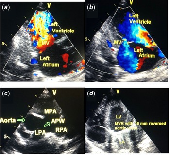
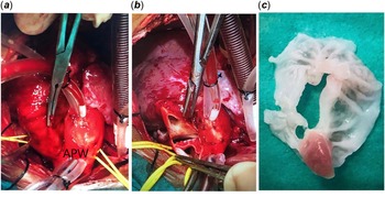
A 5-month-old boy was admitted to our centre for rapid breathing, poor feeding, and failure to thrive. His body weight and height were 4750 gr and 57 cm, respectively. Physical examination revealed a resting oxygen saturation of 95%. There was no sign of desaturation either at rest or on exertion. Cardiac auscultation revealed a significant diastolic murmur at the apex and a continuous precordial murmur. Chest X-ray showed cardiomegaly and plethoric lung fields. Two-dimensional echocardiography revealed a severe mitral stenosis with a maximum of peak 28 mmHg and mean 14 mmHg gradients measured with coloured-wave doppler (Fig 1a). There were two separate papillary muscles within the mitral valve. The posterior papillary muscle was originating high on the posterior wall of the left ventricle. Both leaflets were found to adhere to the same papillary muscle. Left ventricular inflow was seen turbulent through the fenestrations of the hammock mitral valve, and moderate insufficiency was observed (Fig 1b). Mitral annulus was 18 mm, and the left atrium was measured 35 × 35 mm. Mild insufficiency of the tricuspid valve and mild dilatation of both the right and the left ventricles were detected. Biventricular systolic function was good, and there was no pericardial effusion. There was a 20 × 25 mm defect between the main pulmonary artery and the ascending aorta (Fig 1c). The aortic arch was left-sided with no coarctation. The diagnosis was confirmed as a hammock mitral valve accompanying aortopulmonary window. Depending on the clinical and echocardiographic evaluations, cardiac catheterisation was not deemed mandatory. Following median sternotomy, total excision of the thymus was performed. Pericard was opened wide enough to provide exposure of the aorta and the pulmonary arteries. Aortic and main pulmonary arterial pressure was measured directly. Aortic pressure was 75/38 (57) mmHg, while the main pulmonary arterial pressure was 74/34 (55) mmHg. A typical external fusion line was detected between the proximal ascending aorta and the main pulmonary artery (Fig 2a). Aorto-bicaval cannulation was performed. A vent was placed in the right superior pulmonary vein. Under deep hypothermic (24°C) cardiopulmonary bypass, antegrade cold blood hyperkalaemic cardioplegic arrest and an antegrade cerebral perfusion support (20 minutes), main pulmonary arteriotomy was performed and 25 × 25 mm type 1 aortopulmonary window was exposed before the pulmonary artery bifurcation. Aortopulmonary window was repaired with a transpulmonary approach by using a Dacron patch (Fig 2b). Pulmonary arteriotomy was closed primarily. Left atrial access was obtained via a right atriotomy and creation of an atrial septal defect. The mitral leaflets were developed like a membranous structure. The surface of this structure was smooth, and there were no traces of commissures. There were two separate papillary muscles. Rudimentary and short chordae arose and connected to the medial side leaflets which were originating from the posterior papillary muscle (Fig 2c). The native mitral valve was excised. We preferred to use interrupted 4/0 Ti-Cron sutures with pledgets which were directly placed into the annulus to implant the 16-mm mechanical aortic valve (Medtronic AP360) in the reverse position. The interatrial septum was closed primarily with creating a 5-mm septal defect. The right atriotomy was closed primarily. The patient was favourably weaned from the cardiopulmonary bypass without any inotropic support. Finally, the aortic pressure measurement was 102/65 (79) mmHg, whilst the main pulmonary arterial pressure measurement was 43/13 (22) mmHg after weaning from the cardiopulmonary bypass. The post-operative course was uneventful. Post-operative echocardiographic evaluations revealed neither insufficiency nor stenosis of the replaced mechanical mitral valve with a normal biventricular function (Fig 1d). Oral warfarin was started at a dose of 0.5 mg/day and uptitrated to a targeted goal international normalised ratio of 3. Heparin was continued until optimal dosing for Warfarin was obtained in an inpatient setting. Oral aspirin was administered with 5 mg/kg/day at beginning from the first post-operative day. The patient discharged on the ninth post-operative day. Follow-up echocardiogram within the first- and sixth-month outpatient clinic control revealed no evidence of mitral insufficiency or stenosis.
( a ) Two-dimensional transthoracic echocardiography parasternal short-axis view showing a severe mitral stenosis with coloured-wave doppler. ( b ) Two-dimensional transthoracic echocardiography parasternal short-axis view showing left ventricular inflow with a turbulent flow through the fenestrations of the hammock mitral valve, and moderate insufficiency was observed with coloured-wave doppler. ( c ) Two-dimensional transthoracic echocardiography parasternal short-axis view showing type 1 aortopulmonary window between the proximal ascending aorta and the main pulmonary artery. ( d ) Two-dimensional transthoracic echocardiography parasternal short-axis view showing the replaced mechanical mitral valve with a 16-mm mechanical aortic valve (Medtronic AP360) in the reverse position. APW: aortopulmonary window, LA: left atrium, LPA: left pulmonary artery, LV: left ventricle, MPA: main pulmonary artery, MV: mitral valve, RPA: right pulmonary artery.

( a ) Intraoperative view of the type 1 aortopulmonary window. A typical external fusion line was detected between the proximal ascending aorta and the main pulmonary artery. ( b ) Intraoperative view of a 25 × 25 mm type 1 aortopulmonary window was exposed before the pulmonary artery bifurcation. Aortopulmonary window was repaired with a transpulmonary approach by using a Dacron patch. ( c ) Intraoperative view of the excised native mitral valve. The mitral leaflets were developed like a membranous structure. The surface of this structure was smooth, and there were no traces of commissures. APW: aortopulmonary window.

Discussion
Aortopulmonary window occurs in about 0.1–0.2% of all CHDs either in isolation or associated with other various complex cardiac malformations. Reference Talwar, Agarwal and Choudhary1 Progressive pulmonary arterial hypertension and its consequences are more common in aortopulmonary window. Initial diagnosis can generally be achieved by transthoracic echocardiography. Aortopulmonary window may be classified into three types: type 1 between the proximal ascending aorta and the main pulmonary artery; type 2 between the aorta and the proximal right pulmonary artery; type 3 with a combination of types 1 and 2 defects. Reference Mori, Ando, Takao, Ishikawa and Imai2 If untreated, patients may rapidly develop pulmonary vascular obstructive disease. Surgical repair should be offered as soon as the diagnosis is confirmed. Transaortic, transpulmonary, and transwindow patch closure are proposed approaches for a complete separation of systemic and pulmonary circulations. Kumar et.al, stated that surgical outcomes after aortopulmonary window repair is excellent even if the surgery was performed beyond the age of 3 months. Reference Kumar, Singh, Thingnam, Mishra and Jaswal3
Congenital mitral stenosis results in both functional and anatomic obstruction of inflow into the left ventricle with an estimated incidence of 0.4%. Reference Remenyi and Gentles4 The hammock mitral valve is a rare congenital cardiac anomaly with dysplastic and shortened chordae inserting in a muscular mass of the posterior left ventricular wall resulting in stenosis, regurgitation, or both. Reference Remenyi and Gentles4,Reference Zegdi, Khabbaz, Chauvaud, Garcon, Carpentier and Deloche5 This pathology occurs due to developmental deficiency during the differentiation of muscles into chordal tissue. The intermixed and abnormal papillary muscles which were implanted below the posterior leaflet moderately obstruct the valvar orifice. The posteriorly implanted papillary muscles which were crossed over towards the orifice by the chordae tendinea of the anterior leaflet produces the hammock appearance. Surgery for mitral valve repair in cases of congenital mitral stenosis remains limited and particularly challenging especially in infants and children due to the size and immature fragile leaflet tissues. Reference Zegdi, Khabbaz, Chauvaud, Garcon, Carpentier and Deloche5 Hammock mitral valve is the most difficult malformation to correct by reconstructive techniques as the orifice of the mitral valve is partially obstructed by abnormal papillary muscles and intermixed chordae. Reference Delmo Walter and Hetzer6 The chordae tendinea also may not be established easily with a risk of transection and creating a flail segment after a reconstructive surgical procedure. Reference del Nido and Baird7 We speculate that mitral valve replacement in patients with hammock mitral valve is a reliable strategy for palliating the disease in infants. Though, there are conflicting data regarding the outcomes after mitral valve replacement in infants. Ibezim et al. stated that the odds ratio for early mortality after mitral valve replacement under the age of 2 years was 7.8. Reference Ibezim, Sarvestani and Knight8 In contrast, Rafii et al. declared that mitral valve replacement is not a risk factor for mortality under the age of 2 years. Reference Rafii, Davies, Carroll, Quaegebeur and Chen9 Another concern for mechanical mitral valve replacement in infants is placement of a prosthetic valve which is larger than the annulus with a risk of complete heart block, compression of the circumflex coronary artery, and potential obstruction of the left ventricular outflow tract. However, annulus growth continues after mitral valve replacement, to allow for a larger valve placement at reoperation. Reference Durham, Burkhart, Dearani, Puga, O’Leary and Schaff10 To the best of our knowledge, successful management of severe congenital mitral stenosis is challenging in infants. However, valve replacement should be considered when the predominant characteristic is stenosis.
Acknowledgements
None.
Financial support
This research received no specific grant from any funding agency, commercial, or not-for-profit sectors.
Conflicts of interest
None.
Ethical standards
The authors assert that all procedures contributing to this work comply with the ethical standards of the Helsinki Declaration of 1975, as revised in 2008.