Introduction
Schizophrenia is a serious psychotic disorder characterized by abnormal perception, thought processes and behaviors. Although the pathophysiological mechanism of schizophrenia is unclear, it has been hypothesized that schizophrenia arises from dysfunctional integration of a distributed network of brain regions (Friston & Frith, Reference Friston and Frith1995) or misconnection of neural circuitries, leading to impaired brain function (Venkatasubramanian, Reference Venkatasubramanian2007; Liu et al. Reference Liu, Liang, Zhou, He, Hao, Song, Yu, Liu, Liu and Jiang2008; Broyd et al. Reference Broyd, Demanuele, Debener, Helps, James and Sonuga-Barke2009). Therefore, elucidating functional connections between multiple brain regions is important for understanding this disease (Broyd et al. Reference Broyd, Demanuele, Debener, Helps, James and Sonuga-Barke2009; Goghari et al. Reference Goghari, Sponheim and MacDonald2010; Rotarska-Jagiela et al. Reference Rotarska-Jagiela, van de Ven, Oertel-Knochel, Uhlhaas, Vogeley and Linden2010).
Abnormal functional connectivity of schizophrenia has been repeatedly reported within a group of brain regions known as the default mode network (DMN) (Liang et al. Reference Liang, Zhou, Jiang, Liu, Tian, Liu and Hao2006; Liu et al. Reference Liu, Liu, Liang, Hao, Tan, Kuang, Yi, Xu and Jiang2006; Bluhm et al. Reference Bluhm, Miller, Lanius, Osuch, Boksman, Neufeld, Theberge, Schaefer and Williamson2007; Williamson, Reference Williamson2007; Zhou et al. Reference Zhou, Liang, Tian, Wang, Hao, Liu, Liu and Jiang2007; Calhoun et al. Reference Calhoun, Kiehl and Pearlson2008; Jafri et al. Reference Jafri, Pearlson, Stevens and Calhoun2008; Pomarol-Clotet et al. Reference Pomarol-Clotet, Salvador, Sarro, Gomar, Vila, Martinez, Guerrero, Ortiz-Gil, Sans-Sansa, Capdevila, Cebamanos and McKenna2008; Whitfield-Gabrieli et al. Reference Whitfield-Gabrieli, Thermenos, Milanovic, Tsuang, Faraone, McCarley, Shenton, Green, Nieto-Castanon and LaViolette2009). This network includes the medial prefrontal cortex (MPFC), anterior and posterior cingulate cortex, precuneus, inferior temporal cortex and lateral parietal cortex. The DMN is characterized by elevated coherent spontaneous activation when subjects are at rest, and deactivation when subjects perform cognition-related tasks (Raichle et al. Reference Raichle, MacLeod, Snyder, Powers, Gusnard and Shulman2001). Although the exact role of the DMN remains to be confirmed, it has been implicated in endogenously generated thought, self-referential activities, monitoring and inner speech (Sonuga-Barke & Castellanos, Reference Sonuga-Barke and Castellanos2007; Williamson, Reference Williamson2007; Broyd et al. Reference Broyd, Demanuele, Debener, Helps, James and Sonuga-Barke2009).
Similar alternating patterns between hyper- and hypo-activities has also been observed in another group of brain regions, including the dorsolateral PFC (DLPFC), inferior parietal cortex (IPC) and supplementary motor area (SMA). However, unlike the DMN, these brain regions are more active during cognitive tasks and less active at rest, and thus are collectively called the task-positive network (TPN) (Fox et al. Reference Fox, Snyder, Vincent, Corbetta, Van Essen and Raichle2005). Of note, activities in the DMN and the TPN are reported to be temporally anticorrelated not only during cognitive tasks but also possibly at rest (Fox et al. Reference Fox, Snyder, Vincent, Corbetta, Van Essen and Raichle2005).
This inverse temporal correlation was interpreted as competition or functional segregation for opposite goals between neural networks. Furthermore, the strength of this anticorrelation was associated with response time in cognitive functions (Kelly et al. Reference Kelly, Uddin, Biswal, Castellanos and Milham2008) and performance in working memory tasks (Hampson et al. Reference Hampson, Driesen, Roth, Gore and Constable2010). Taken together, the DMN and the TPN, and the competition (i.e. anticorrelated relationship) between them, are crucial to neurocognitive functions.
Increased functional connectivity within the DMN and increased anticorrelation between the DMN and TPN have been reported in patients with schizophrenia (Liang et al. Reference Liang, Zhou, Jiang, Liu, Tian, Liu and Hao2006; Garrity et al. Reference Garrity, Pearlson, McKiernan, Lloyd, Kiehl and Calhoun2007; Zhou et al. Reference Zhou, Liang, Tian, Wang, Hao, Liu, Liu and Jiang2007; Whitfield-Gabrieli et al. Reference Whitfield-Gabrieli, Thermenos, Milanovic, Tsuang, Faraone, McCarley, Shenton, Green, Nieto-Castanon and LaViolette2009; Wang et al. Reference Wang, Metzak and Woodward2010a). Increased connectivity within the DMN may reflect excessive attentional orientation to introspective thought and may contribute to disturbances of thought in schizophrenia (Whitfield-Gabrieli et al. Reference Whitfield-Gabrieli, Thermenos, Milanovic, Tsuang, Faraone, McCarley, Shenton, Green, Nieto-Castanon and LaViolette2009). By contrast, increased anticorrelation between the DMN and TPN suggests excessive antagonism between intro- and extrospective psychological processing, and may interfere with the cognitive function of the patient with schizophrenia (Garrity et al. Reference Garrity, Pearlson, McKiernan, Lloyd, Kiehl and Calhoun2007). Consistent with this notion, Garrity et al. (Reference Garrity, Pearlson, McKiernan, Lloyd, Kiehl and Calhoun2007) found that the DMN showed greater deactivation when performing cognitive tasks in patients with schizophrenia, and greater deactivation in the medial frontal gyrus and precuneus were correlated with positive symptoms and attentional deficits.
Neurocognitive dysfunction, especially in the domains of memory, attention and executive functions, has been widely reported in schizophrenia and is considered a core feature of the disease (Mesholam-Gately et al. Reference Mesholam-Gately, Giuliano, Goff, Faraone and Seidman2009). However, the neural mechanism underlying the cognitive deficit in schizophrenia is still elusive. Given the tight linkage between the DMN and neurocognitive functions and the well-known cognitive deficit in the disease, it is important to examine the relationship between the intrinsic activity/connectivity of the DMN and deficient neurocognitive performance in schizophrenia. Challenges in addressing this issue may exist in two aspects. First, medications used by patients may be a confounding factor. Indeed, atypical antipsychotic drugs have been shown to have significant effects on the brain function of patients with schizophrenia (Snitz et al. Reference Snitz, MacDonald, Cohen, Cho, Becker and Carter2005). Therefore, it is important to elucidate this relationship in first-episode, treatment-naive schizophrenia (FES) patients to avoid the influence of the chronicity of the illness and the effects of treatment. Second, given the heterogeneity and large inter-patient variance often observed in both imaging and behavioral studies of schizophrenia, a relatively large sample size is necessary. In the present study we have overcome these two challenges by assessing intrinsic activity/connectivity of the DMN and the performance of a battery of neurocognitive tests in a large group of patients with FES. Based on these measurements, we have explored the relationship between aberrant brain activity/connectivity and neurocognitive function in patients. We have also examined the association between abnormal brain activity/connectivity and the severity of psychotic symptoms.
Method
Subjects
In total 228 subjects including 115 FES patients (53 males, 62 females) and 113 healthy controls (57 males, 56 females) participated in this study. All patients were recruited at the Mental Health Center of West China Hospital, Sichuan University from August 2005 to October 2010. Healthy controls were recruited from the local area through poster advertisement. Both groups were matched for sex, age and education level. All participants were right-handed based on the Annett Handedness Scale (Annett, Reference Annett1970). The study was approved by the ethical committee of the West China Hospital of Sichuan University. All patients and controls provided written informed consent.
Clinical assessment
The patients were interviewed by an experienced psychiatrist using the Structured Clinical Interview for DSM-IV (SCID-P; First et al. Reference First, Spitzer and Williams1997). DSM-IV criteria for schizophrenia or schizophreniform psychosis were used for diagnosis (APA, 1994). Diagnostic results showed 94 patients with schizophrenia and 21 patients with schizophreniform psychosis. Patients diagnosed with schizophreniform psychosis were followed up for at least 6 months and all met the DSM-IV criteria for schizophrenia. Psychopathology associated with schizophrenia was evaluated using the positive and negative syndrome scales (PANSS), which provided the total score and six factor scores including Negative, Positive, Excited/Activation, Anxious/Depressed, Disorganized and Withdrawn (Van den Oord et al. Reference Van den Oord, Rujescu, Robles, Giegling, Birrell, Bukszár, Murrelle, Möller, Middleton and Muglia2006). All controls were screened with the SCID non-patient version (SCID-NP;First et al. Reference First, Spitzer and Williams1997) for the lifetime absence of psychiatric illnesses. Subjects with organic brain disorders, alcohol and/or drug abuse, pregnancy or any other severe physical illness such as brain tumor or epilepsy were excluded from the study.
Magnetic resonance imaging (MRI) data acquisition
All participants were scanned using a 3-T MRI system (EXCITE, General Electric, USA). Functional MRI (fMRI) images were obtained using a gradient–echo echo-planar imaging (EPI) sequence [repetition time (TR)/echo time (TE) = 2000/30 ms, flip angle = 90°, slice thickness = 5 mm, no slice gap, in-plane resolution = 3.75 × 3.75 mm2, matrix size = 64 × 64, field of view (FOV) = 240 × 240 mm2, slice number = 30]. Each resting-state fMRI scan contained 200 image volumes. During fMRI scanning, participants were instructed to lie still with eyes closed, remain relaxed and refrain from focusing on any particular thought. Data from 20 subjects (11 patients and nine controls) were discarded because of excessive head movement (translational movement >1.5 mm or rotation >1.5°), resulting in a total of 104 patients and 104 controls for further analysis.
Neurocognitive assessment
A battery of neurocognitive tests was applied in the present study. The immediate and delayed Logical Memory subtest of the Wechsler Memory Scale (Wechsler, Reference Wechsler1987) was used to evaluate verbal memory and paragraph recall abilities. The immediate and delayed Pattern Recognition Memory (PRM) test and the Rapid Visual Information Processing (RVP) test from the Cambridge Neuropsychological Test Automated Battery (CANTAB) were used to assess visual memory and sustained attention respectively. The Trail Making Test (TMT, parts A and B; Reitan, Reference Reitan1992) and the Digit Symbol Test (DST) from the Wechsler Intelligence Scales (Wechsler, Reference Wechsler1981) were used to assess processing speed (Mesholam-Gately et al. Reference Mesholam-Gately, Giuliano, Goff, Faraone and Seidman2009). Eighty patients and 72 healthy controls completed all of the neurocognitive tests whereas the other 24 patients and 32 controls only participated in the imaging session.
Data processing
Preprocessing and statistical analysis of functional images were carried out using the Statistical Parametric Mapping package (SPM5, Wellcome Department for Imaging Neuroscience, London, UK). For each subject, the first 10 image volumes were discarded to allow the fMRI signal to reach a steady state. Initial analysis included slice time correction and realignment. The resulting images were then spatially normalized to the Montreal Neurological Institute (MNI) EPI template in SPM5, resampled to 3 × 3×3 mm3, and subsequently smoothed with an isotropic Gaussian kernel (full-width at half-maximum = 6 mm). Finally, all preprocessed fMRI data were linearly detrended and band-pass filtered (0.01 <f < 0.08 Hz) using the Resting-State fMRI Data Analysis Toolkit (REST) software package (http://resting-fmri.sourceforge.net). The default brain mask in REST was used. Nuisance covariates including six motion parameters and the global signal were regressed out.
Fractional amplitude of spontaneous low-frequency fluctuations (fALFF)
fALFF calculated based on the method described by Zou et al. (Reference Zou, Zhu, Yang, Zuo, Long, Cao, Wang and Zang2008) provide a quantitative measure of spontaneous fluctuations of neural activities (Fox et al. Reference Fox and Raichle2007). In brief, fALFF were calculated as the ratio of the power spectrum of low frequency (0.01–0.08 Hz) to that of the entire frequency range. For the purpose of standardization, the fALFF value of each voxel was divided by the global mean fALFF value for each individual subject, calculated from voxels within the brain mask, with the background and other non-brain tissue signals removed. For each group, significant fALFF values were assessed by using a one-sample t test, thresholded at p < 0.05, corrected for multiple comparisons using the false discovery rate (FDR) criterion.
Functional connectivity
Resting-state functional connectivity was quantified by using the seed-based correlational analysis (Fransson, Reference Fransson2005) implemented in the REST software package (http://resting-fmri.sourceforge.net). Considering the pivotal role of the posterior cingulate cortex (PCC) in the DMN (Fransson & Marrelec, Reference Fransson and Marrelec2008), a spherical region of interest (ROI, radius = 10 mm) centered at the PCC (0, −50, 30) was chosen as the seed (Fransson, Reference Fransson2005). The seed position showed local peak intensity of the fALFF value in both groups, and was also close to the seed used by Fransson (Reference Fransson2005). The regionally averaged time series of the seed ROI was first extracted and used as the reference. The correlational analysis was then performed on a voxel-by-voxel basis by correlating the time series of each voxel to the reference. The correlation coefficient (r value) of each voxel was transformed to the z value using the Fisher r-to-z transformation. The strength of resting-state functional was quantified by using z values. Significant connectivity for each subject group was evaluated by using one-sample t tests voxel by voxel, thresholded at p < 0.05, corrected for multiple comparisons using the FDR criterion, and cluster size >10 voxels.
Statistical analysis
Demographic differences between the patient and control groups were analyzed using independent-sample t tests and χ2 tests, and p < 0.05 was deemed statistically significant. The neurocognitive function difference between the two groups was examined using ANOVA with age and educational level as covariates.
Alterations in fALFF and functional connectivity in patients relative to controls were evaluated by using two-sample t tests in a voxel-by-voxel manner. T-value maps were thresholded at p < 0.05, corrected for multiple comparisons using the FDR criterion and cluster size >10 voxels.
Relationships between aberrant brain activity/connectivity and neurocognitive performance and also clinical symptoms in patients were evaluated separately by using partial correlation analysis, with age, educational year and gender as covariates. First, we identified local peaks of each cluster in the T-value maps resulting from comparing fALFF and functional connectivity strength (in z values) between patients and controls. A spherical ROI was then constructed with one of these loci as the center and a radius of 10 mm. Subsequently, regionally averaged values of fALFF and functional connectivity strength were extracted separately from these ROIs. Finally, these values were correlated to neurocognitive performance and the severity of clinical symptoms in patients respectively. The statistical significance level for this analysis was set at p < 0.05.
Results
Demographics
Age (patients: mean = 25.36 years, s.d. = 8.260; controls: mean = 26.61 years, s.d. = 8.904; p = 0.296), educational level (patients: mean = 12.14, s.d. = 3.061; controls: mean = 12.74, s.d. = 3.532; p = 0.191) and gender ratio (patients: 49 males, 55 females; controls: 50 males, 54 females; p = 0.890) were not significantly different between the patient and control groups (p > 0.05). The illness duration of patients was 39.54 ± 31.88 weeks. The average of the total PANSS score of the patient group was 92.14 ± 17.48, suggesting that patients were in the acute phase of the disease.
Neurocognitive function in patients with FES and healthy controls
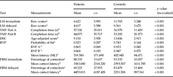
There was a significant difference in the performance of neurocognitive tests between the two groups (Table 1). Patients' performance was considerably worse than healthy controls in all neurocognitive tests.
Neurocognitive performance in patients with FES and healthy controls

FES, First-episode, treatment-naive schizophrenia; LM, logical memory; TMT, Trail Making Test; DSC, digit symbol coding; RVP, rapid visual information processing; PRM, pattern recognition memory; Probability of hit, the probability of making a correct response; RVP A′, sensitivity to the correct target; RVP B′′, the tendency to respond; ML, the mean time taken to respond; Mean correct latency, the mean time taken to respond correctly; s.d., standard deviation.
a Lower scores represent poorer neuropsychological performance.
b Higher scores represent poorer neuropsychological performance.
Resting-state functional connectivity
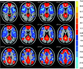
Figure 1 shows the spatial pattern of the DMN and TPN revealed by correlational analysis with the seed of the PCC. The MPFC, PCC, precuneus and lateral parietal cortex showed significant positive connectivity with the PCC, together constituting the DMN. The bilateral DLPFC, bilateral inferior parietal lobe and bilateral SMA showed significant negative connectivity with the PCC, together constituting the TPN. These results were consistent with the spatial pattern of the DMN and TPN in the literature (Fox et al. Reference Fox, Snyder, Vincent, Corbetta, Van Essen and Raichle2005; Fransson, Reference Fransson2005).
One-sample t tests of the seed-based [posterior cingulate cortex (PCC), coordinates: (0, −50, 30)] correlational analysis for healthy controls. The threshold was p < 0.001, corrected for multiple comparison using the false discovery rate (FDR) criterion, extent threshold 10 voxels. The left side of the image represents the right side of the brain. Color bar indicates the T score. A, PCC/precuneus; B, medial prefrontal cortex (MPFC); C, lateral parietal cortex; D, dorsolateral PFC (DLPFC); E, inferior parietal lobe; F, supplementary motor area (SMA).

Altered fALFF and DMN functional connectivity in patients with FES
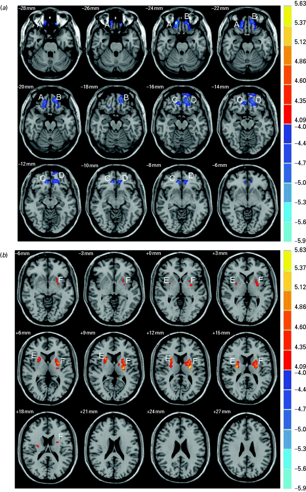
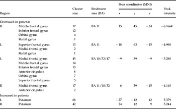
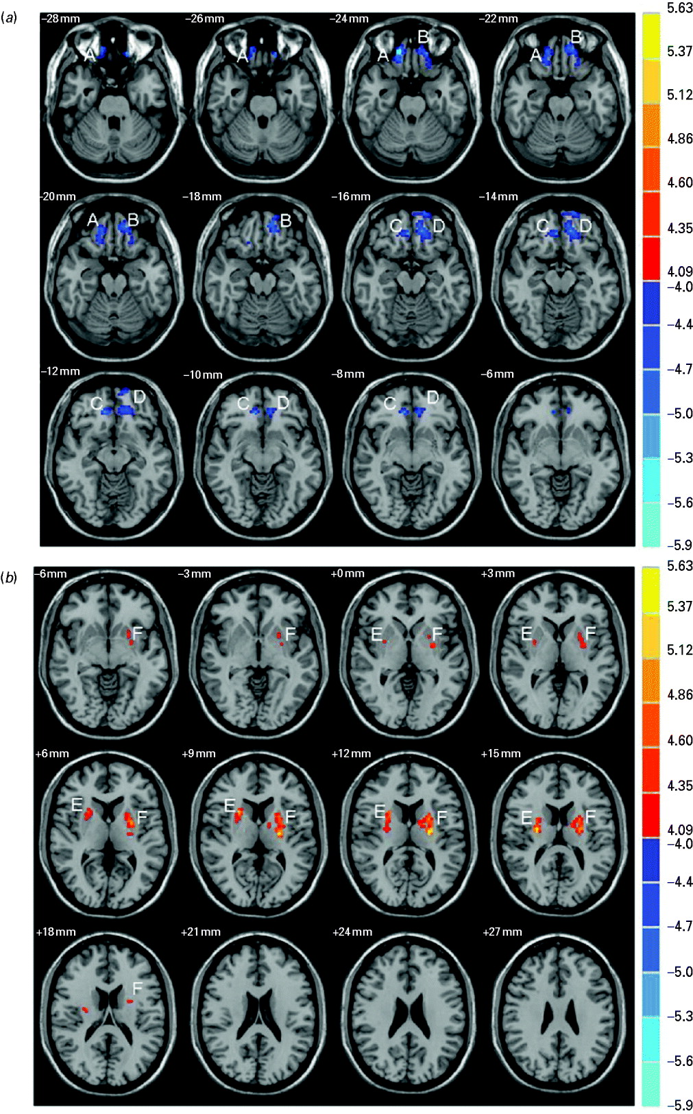
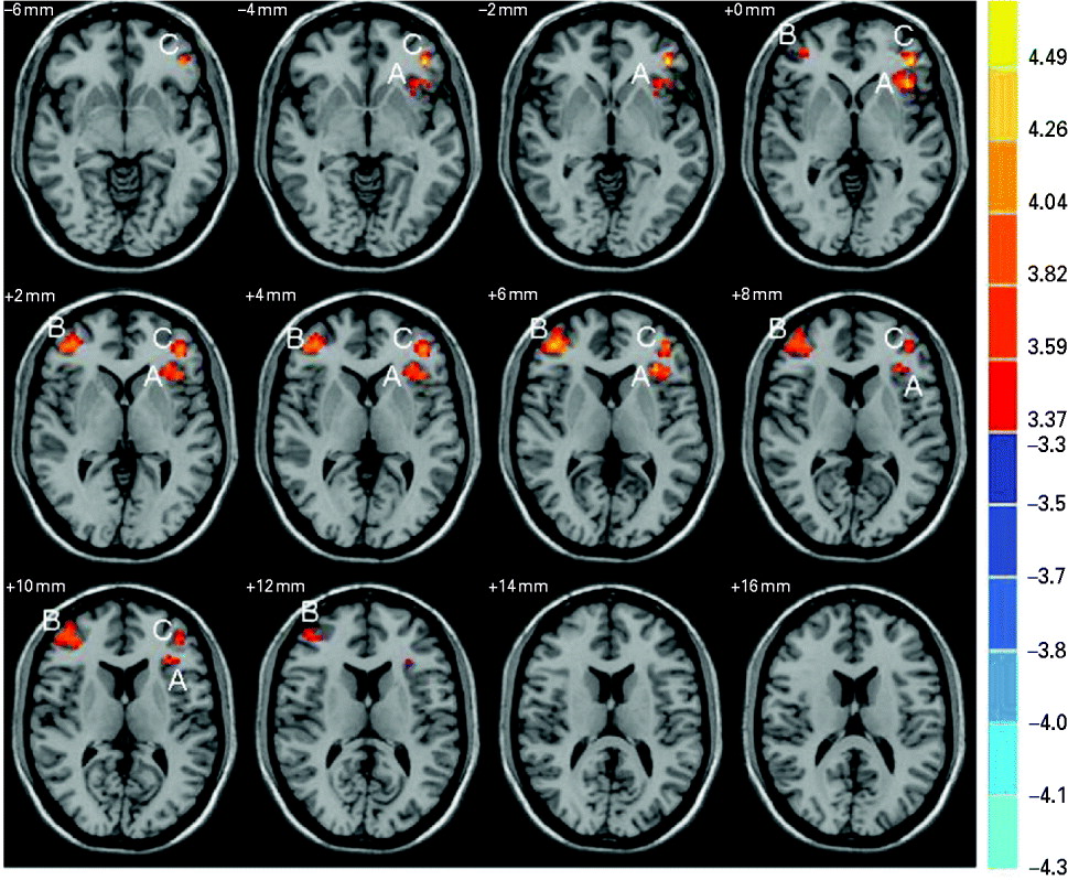
Relative to the healthy controls, patients with FES showed significantly decreased fALFF in the bilateral orbitofrontal cortex (OFC) and the MPFC (Fig. 2a and Table 2). In addition, significantly increased fALFF were found in the bilateral putamen in the patient group (Fig. 2b and Table 2). Furthermore, significantly increased anticorrelated functional connectivity with the PCC (i.e. DMN) was observed in the TPN including the left middle frontal gyrus, right inferior frontal gyrus (located in the DLPFC) and left insula (Fig. 3) in the patient group.
Regions showing (a) decreased and (b) increased fragment amplitude of low-frequency fluctuations (fALFF) in first-episode, treatment-naive schizophrenia (FES) patients. The left side of the image represents the right side of the brain. Color bar indicates the T score. A, Right middle frontal gyrus, extending to the right inferior frontal gyrus and orbital gyrus; B, left superior frontal gyrus, extending to the left medial frontal gyrus and rectus; C, right medial frontal gyrus, extending to the right anterior cingulated; D, left medial frontal gyrus, extending to the left middle frontal gyrus, inferior frontal gyrus, anterior cingulate, orbital gyrus and superior frontal gyrus; E, right putamen; F, left putamen.
Regions showing increased connectivity with the seed region of the posterior cingulate cortex (PCC)/precuneus in patients with first-episode, treatment-naive schizophrenia (FES). The left side of the image represents the right side of the brain. The color bar indicates the z score. A, Left insula (peak coordinates: −36 21 0; peak z score: 5.080), extending to the left inferior frontal gyrus; B, right inferior frontal gyrus (peak coordinates: 45 45 6; peak z score: 4.705), extending to the right middle frontal gyrus; C, left middle frontal gyrus (peak coordinates: −42 39 0; peak z score: 4.902), extending to the left inferior frontal gyrus.
Difference in fALFF between patients with FES and healthy controls

fALFF, Fragment amplitude of low-frequency fluctuations; FES, first-episode, treatment-naive schizophrenia; MNI, Montreal Neurological Institute; R, right; L, left.
Relationships between aberrant brain activity/connectivity and neurocognitive performance and clinical symptoms
fALFF
In the patient group, the fALFF of the left putamen were positively associated with the factors excited/activation (r = 0.319, p = 0.039) and disorganization (r = 0.269, p = 0.049). The fALFF of the right middle frontal gyrus were inversely correlated with the factors excited/activation (r = − 0.201, p = 0.033) and disorganization (r = − 0.232, p = 0.018), and also with TMT-B (r = − 0.289, p = 0.008). The fALFF of the left superior frontal gyrus were negatively correlated with the mean executive time of TMT-A (r = − 0.248, p = 0.031) and positively correlated with the score of Digit Symbol Coding (DSC) (r = 0.250, p = 0.029).
DMN functional connectivity
In the patient group, the functional connectivity between the left insula and the PCC was positively correlated with the factors positive (r = 0.376, p = 0.025) and excited/activation (r = 0.333, p = 0.022), and also positively associated with mean latency of the RVP (r = 0.302, p = 0.020).
To examine the possibility that the correlations between imaging indices and cognitive performance were mediated by clinical symptoms, we further analyzed the partial correlation between the imaging data (functional connectivity and fALFF) and neurocognitive performance, controlling for clinical symptoms. We found that the correlation previously identified between fALFF of the right middle frontal gyrus and TMT-B disappeared (r = − 0.102, p = 0.383). This result suggests that this correlation is more likely to reflect psychosis than cognition. In addition, we identified an inverse correlation between fALFF of the right putamen and the RVP after controlling for clinical symptoms (r = − 0.338, p = 0.008).
Discussion
In the present study we found that resting-state brain activity and DMN connectivity in patients with FES differed significantly from those of healthy controls. In the patient group, compromised fALFF were observed in the bilateral OFC and MPFC whereas increased fALFF were found in the bilateral putamen. Importantly, the changes in fALFF in the left putamen and right OFC were correlated with the severity of symptoms including excited/activation and disorganization. In addition, aberrant anticorrelated functional connectivity between the DMN and TPN was observed in patients. Greater anticorrelated functional connectivity between the PCC and the left insula was correlated with the severity of clinical symptoms including positive, excited/activation and disorganization.
We also found that the neurocognitive performance in FES patients, mainly in the domains of memory, attention and processing speed, was significantly worse than that in healthy controls. This result is consistent with previous studies suggesting that neurocognitive dysfunction starts to appear in early phases of the illness (Townsend & Norman, Reference Townsend and Norman2004; Wang et al. Reference Wang, Vassos, Deng, Ma, Hu, Murray, Collier and Li2010b). Of note, the performance of the DST and the executive time of TMT-A were associated with fALFF in the left OFC; the executive time of TMT-B was associated with fALFF in the right OFC in patients; and the mean latency of RVP was correlated with fALFF in the right putamen in patients. In addition, the magnitude of hyperconnectivity between the PCC and the left insula was also correlated with the mean latency of RVP in patients. Notably, the latter is a good indicator of sustained attention (Cognition, 2004).
Consistent with previous reports (Snitz et al. Reference Snitz, MacDonald, Cohen, Cho, Becker and Carter2005; Huang et al. Reference Huang, Lui, Deng, Chan, Wu, Jiang, Zhang, Jia, Li, Li, Chen, Li and Gong2010), smaller amplitudes of spontaneous neural activities were found in the OFC and MPFC in patients with FES. These functional alterations were also in line with lower cerebral blood flow and reduced cortical thickness in the same brain areas in schizophrenia found by dynamic susceptibility contrast (DSC) MRI (Peruzzo et al. Reference Peruzzo, Rambaldelli, Bertoldo, Bellani, Cerini, Silvia, Pozzi Mucelli, Tansella and Brambilla2011) and structural neuroimaging techniques (Witthaus et al. Reference Witthaus, Kaufmann, Bohner, Ögüdal, Gudlowski, Gallinat, Ruhrmann, Brüe, Heinz, Klingebiel and Juckel2009) respectively. A resting-state fMRI study previously reported increased fALFF in the MPFC (Hoptman et al. Reference Hoptman, Zuo, Butler, Javitt, D'Angelo, Mauro and Milham2009), but this study was performed in chronic schizophrenia patients undergoing medical treatment, and thus the contradiction could result from medications used by patients before MRI scanning. Indeed, a recent study reported that fALFF increased after a short-term treatment with second-generation antipsychotic medications (Lui et al. Reference Lui, Li, Deng, Jiang, Wu, Tang, Yue, Huang, Chan, Collier, Meda, Pearlson, Mechelli, Sweeney and Gong2010).
Aberrant spontaneous activity in the OFC in FES patients can be implicated in multiple functions. The OFC is defined as the ventral surface of the prefrontal lobe. It receives inputs from the temporal association cortex, amygdala and hypothalamus, making it one of the highest integrated centers for emotional processing. The OFC is related to the appreciation of emotions of either self or others and the acquisition of appropriate behaviors or inhibition of inappropriate behaviors (Clark et al. Reference Clark, Boutros and Mendez2010). Most patients with OFC lesions are emotionally labile, irritable and impulsive (Bramham et al. Reference Bramham, Morris, Hornak, Bullock and Polkey2009). In addition, the OFC plays an important role in mediating attention during cognitive tasks and suppressing distraction (Clark et al. Reference Clark, Boutros and Mendez2010). In the present study, we found that the neural activity of the bilateral OFC was weakened, and the degree of alteration in the right OFC was correlated with psychopathological severity of the illness, especially with symptoms of excited/activation and disorganization. Importantly, a correlation between aberrant neural activity in OFC and the processing speed of cognitive functions was also found in the present study. The processing speed refers to the speed at which different cognitive operations can be executed. It represents a very general cognitive capability and many high cognitive domains, including executive function, involve speed-dependent internal processes (Dickinson et al. Reference Dickinson, Ramsey and Gold2007). In summary, these results suggest that the OFC plays an important role in psychotic symptoms, especially those of excited/activation and disorganization. In addition, dysfunction of the OFC was associated with abnormal processing speed, and this linkage may be responsible for the failure to execute cognitive tasks by patients.
In the current study we have found that compromised intrinsic neural activity in the MPFC has occurred during early stages of the illness. As part of the DMN, the MPFC is involved in self-monitoring processing (Fransson, Reference Fransson2005) and has been suggested to be associated with decreased illness insight in schizophrenia (Lee et al. Reference Lee, Brown, Egleston, Green, Farrow, Hunter, Parks, Wilkinson, Spence and Woodruff2006). Consistent with this view, the improvement in illness insight was accompanied by increased intrinsic activity of the left MPFC in patients with schizophrenia after recovering from the acute episode (Lee et al. Reference Lee, Brown, Egleston, Green, Farrow, Hunter, Parks, Wilkinson, Spence and Woodruff2006). Taken together, our data implied that decreased illness insight and impaired source monitoring might also occur during early stages of schizophrenia.
The bilateral putamen also showed altered intrinsic neural activity in patients with FES. The putamen is involved in dopamine metabolism and has been implicated in the pathophysiology of schizophrenia (Brandt & Bonelli, Reference Brandt and Bonelli2008). One study has reported increased striatal dopamine transmission in FES patients (Schmitt et al. Reference Schmitt, Meisenzahl, Frodl, La Fougere, Hahn, Moller and Dresel2009), which has been associated with the pathology of the disease (Kegeles et al. Reference Kegeles, Abi-Dargham, Frankle, Gil, Cooper, Slifstein, Hwang, Huang, Haber and Laruelle2010). In the present study, hyperactivity of the left putamen was correlated with the excited/activation and disorganization symptoms, which reflect irritable, impulsive and disorganized behaviors in schizophrenia (Van den Oord et al. Reference Van den Oord, Rujescu, Robles, Giegling, Birrell, Bukszár, Murrelle, Möller, Middleton and Muglia2006). Taken together, the dysfunction of the putamen in early stages of the illness supports the hypothesis that schizophrenia is associated with the hyperdopaminergic state in the striatum (Howes & Kapur, Reference Howes and Kapur2009).
Enhanced connectivity between the DMN and left insula has also been observed in patients with FES in the present study. The insular cortex is a highly developed mesocortical brain region that is a major component of the limbic system. As a sensory integration region through its connections with cortical and subcortical regions, the insular cortex plays an important role in human emotion and cognition (Wylie & Tregellas, Reference Wylie and Tregellas2010). Previous studies have revealed decreased cerebral blood flow and gray matter volume in the insular cortex in FES patients (Crespo-Facorro et al. Reference Crespo-Facorro, Kim, Andreasen, O'Leary, Bockholt and Magnotta2000). The functional disconnection of the insula has also been reported in schizophrenia (Liang et al. Reference Liang, Zhou, Jiang, Liu, Tian, Liu and Hao2006; Bora et al. Reference Bora, Fornito, Radua, Walterfang, Seal, Wood, Yücel, Velakoulis and Pantelis2010). Abnormalities in paralimbic regions (e.g. the insular cortex) may lead to perceptual disturbances that account for psychotic symptoms in schizophrenia (Crespo-Facorro et al. Reference Crespo-Facorro, Kim, Andreasen, O'Leary, Bockholt and Magnotta2000; Clark et al. Reference Clark, Boutros and Mendez2010). The present study has shown that enhanced connectivity between the DMN and the left insula was related to the severity of psychiatric symptoms and sustained attention deficit. These results are consistent with the previous finding that humans with lesions in the insula demonstrated sensory deficit similar to patients with schizophrenia (Wylie & Tregellas, Reference Wylie and Tregellas2010). Therefore, we speculate that abnormalities in the insular cortex and its connections with the DMN may be related to the difficulty to differentiate internal and external sensory events in patients with schizophrenia.
In the current study, head motion in both groups was strictly controlled (translational movement <1.5 mm and rotation <1.5°). These stringent criteria were adopted because recent evidence suggests that even very subtle head motion may introduce spurious correlations in functional connectivity (Power et al. Reference Power, Barnes, Snyder, Schlaggar and Petersen2012; Satterthwaite et al. Reference Satterthwaite, Wolf, Loughead, Ruparel, Elliott, Hakonarson, Gur and Gur2012; Van Dijk et al. Reference Van Dijk, Sabuncu and Buckner2012). In addition, we estimated the level of head movement in patients [translational movement (mean±s.d.) = 1.17 ± 0.28 mm; rotation (mean ± s.d.) = 1.10 ± 0.33°] and controls [translational movement (mean±s.d.) = 1.16 ± 0.27 mm; rotation (mean ± s.d.) = 1.10 ± 0.26°]. The data suggest that there is no significant difference in head motion between the control and patient groups (translational movement: p value = 0.073; rotation: p value = 0.078). Therefore, it is unlikely that our results were biased by head motion difference between the control and patient groups.
There are several limitations in the present study. First, we used a case–control design, which cannot explore progressive changes at different stages of the disease. Further studies are needed to address this issue. Second, functional connectivity was examined by using the seed-based correlational approach with the PCC selected as the seed, under the assumption that the PCC is one of the core regions of the DMN (Fransson & Marrelec, Reference Fransson and Marrelec2008). However, other brain regions involved in the DMN should also be considered. In addition, abnormal functional connectivity at the whole-brain network level can be explored by using other strategies such as independent component analysis. Other cognitive domains such as social cognition and executive function should be assessed and analyzed in future studies. Of note, it is not possible to know whether the differences we observed are part of the neural mechanisms underlying the disorder, or an epiphenomenon. Symptoms and cognitive function that were correlated with intrinsic activity/connectivity could be secondary to symptoms rather than intrinsic to the neural mechanisms of the disorder. Finally, anticorrelated functional connectivity is complicated by the preprocessing procedure of global signal regression (Murphy et al. Reference Murphy, Birn, Handwerker, Jones and Bandettini2009). However, in the present study we focused on the difference in anticorrelated functional connectivity between the healthy control and patient groups, and thus our result should be less susceptible to this limitation (Kelly et al. Reference Kelly, Uddin, Biswal, Castellanos and Milham2008; Hampson et al. Reference Hampson, Driesen, Roth, Gore and Constable2010).
Nevertheless, the results of our study have provided important evidence suggesting that altered spontaneous activities and DMN connectivity start to appear in the early stages of schizophrenia. These changes are linked to neurocognitive dysfunction and the severity of the symptoms. This link should provide important insights into understanding the neural mechanisms underlying the neurocognitive deficits of the disease.
Acknowledgments
This work was partly funded by the National Nature Science Foundation of China (81130024, 30530300 and 30125014, T.L.), the National Basic Research Program of China (973 Program 2007CB512301, T.L.), the Ph.D. Programs Foundation of the Ministry of Education of China (20110181110014, T.L.), the National Key Technology R&D Program of the Ministry of Science and Technology of China during the 12th Five-Year Plan (2011BAZ02530, T.L.), a National Alliance for Research on Schizophrenia and Depression (NARSAD) Independent Investigator Award (T.L.), the Wellcome Trust (International Collaborative award to T.L. and D.A.C.), and the China Postdoctoral Science Foundation (20090461337 and 201003699, W.D.).
Declaration of Interest
None.