Introduction
The anatomy of the membranous labyrinth and its effect on disease processes is extremely difficult to study given the limited access to the otic capsule. Advances in imaging tools, such as micro computed tomography (CT) technologyReference Uzun, Curthoys and Jones1, Reference Uzun-Coruhlu, Curthoys and Jones2 and high-resolution magnetic resonance imaging,Reference Thylur, Jacobs, Go, Toga and Niparko3, Reference Monsanto, Pauna, Kwon, Schachern, Tsuprun and Paparella4 allow new perspectives of this anatomy, with relevance to current and future interventions.
Interventions on the oval window are not limited to stapes surgery (which in itself is evolving in terms of endoscopic ear surgery techniques),Reference Sproat, Yiannakis and Iyer5 but the oval window is currently also used for drug delivery.Reference Salt, Hartsock, Gill, King, Kraus and Plontke6, Reference King, Salt, Kel, Eastwood and O'Leary7 A detailed understanding of the deep relations of the footplate is crucial to assess the risks and outcomes of such interventions.Reference Mukherjee, Uzun-Coruhlu, Curthoys, Jones, Bradshaw and Pohl8 In addition to the utricle and saccule, these deep relations include the periotic connective tissue and the membrana limitans. The membrana limitans is a membrane-like structure that connects the otoliths to the bony margins of the vestibule, and in some cases to the deep surface of the stapes footplate itself. These structures have been described in the past,Reference Nomura9, Reference Hara and Kimura10 though the bulk of the literature is focused on guinea pig rather than human data.
A detailed three-dimensional (3D) study of utricular and saccular maculae in relation to the footplate has been reported previously.Reference Uzun-Coruhlu, Curthoys and Jones2, Reference Mukherjee, Uzun-Coruhlu, Curthoys, Jones, Bradshaw and Pohl8 However, the present study describes the anatomical variations of the membrana limitans in human subjects, its relation to the stapes footplate and its potential impact on stapes surgery. Furthermore, this study demonstrates the visualisation of these structures using the 3D visualisation tool of augmented reality. To our knowledge, this is the first demonstration of this anatomy in such a view.
Materials and methods
Human cadaveric study
All human temporal bones were handled in accordance with anatomy licensing rules. Eighteen cadaveric temporal bones were drilled to isolate the otic capsule. The first eight bones (three paired and two unpaired) were fixed with Karnovsky's fixative (3 per cent paraformaldehyde, 0.5 per cent glutaraldehyde in phosphate buffer),Reference Mukherjee, Uzun-Coruhlu, Curthoys, Jones, Bradshaw and Pohl8 which is known to minimise shrinkage. They were then soaked in 2 per cent osmium tetroxide, and scanned in a micro-CT scanner with scan width ranging from 11.89 µm to 55 µm. The otic capsule was intact to exclude sectioning artefacts, and no alcohol was used in order to avoid tissue shrinkage.
The next 10 temporal bones (4 paired and 2 unpaired) were fresh and drilled within 48 hours post-mortem. The preservation technique was identical to that in the first set. Only minimal osmium was used in these bones. Unlike the previous sample of temporal bones, osmium was trickled into the perilymphatic space through a fenestra drilled into the superior canal and drained through a cochleostomy to minimise sedimentation artefact. Average scan time was 8 hours, and scan resolution in this second batch of fresh temporal bones ranged from 1 µm to 13 μm. Specimens were placed in a 64-bit micro-CT scanner (MicroXCT-400; Xradia, Pleasanton, California, USA).
The methods, including details about the scanning parameters, have been described in detail in earlier publications.Reference Uzun-Coruhlu, Curthoys and Jones2, Reference Thylur, Jacobs, Go, Toga and Niparko3 Data analysis was conducted with VGStudio Max CT software, versions 1 and 2. This allowed axial, sagittal and coronal plane views of each structure, and enabled 3D reconstruction.
The micro-CT data attained in tagged image file format (‘TIFF’) files were segmented to focus on the vestibule. They were then converted to Digital Imaging and Communications in Medicine (‘DICOM’) file format, and rendered into a 3D file. The data were published using augmented reality technology. This enabled visualisation of these structures from outside of the vestibule, to help orientate the structures and view them as a 3D-rendered ‘stack’. It also allowed further visualisation of the nerve supply and the anatomy of the vestibule from inside and out. These methods of creating 3D files have been described previously.Reference Chiarovano, Cheng and Mukherjee11, Reference Cheng, Mukherjee and Curthoys12 The augmented reality digital content can be visualised with a smart device such as an iPhone or iPad, using a free software application called Hyperspaces (Inglobe Technologies, Ceccano, Italy).
Guinea pig temporal bone study
All procedures were approved by the Animal Ethics Committee of Sydney University (protocol numbers L29/4-2010/3/5266 and K22/3-2013/2/5964), and conformed to the National Health and Medical Research Council Principles of Laboratory Animal Care.
A guinea pig weighing between 400 g and 600 g was deeply anaesthetised with pentobarbitone sodium (Nembutal® (100 mg/kg), the ilium). Fixation was achieved via intracardiac perfusion, first with phosphate buffer (300 ml containing heparin), followed by 3 per cent paraformaldehyde (in phosphate buffer). The temporal bone was removed and stored in paraformaldehyde. It was then stained en bloc with osmium tetroxide (2 per cent weight/volume in phosphate buffer) by being placed in plastic tubes and undergoing gentle rotation by a standard laboratory rotator for 3 days.
The radiographic datasets were imported into hardware-based back-projection reconstruction software supplied by Xradia, producing an image series of 1024Reference Uzun-Coruhlu, Curthoys and Jones2 pixels in 16-bit axial slices, and reconstructed as per the human cadaver scanning protocol.
Results
Human temporal bone results
One temporal bone was excluded because of scarring or abnormality seen in the saccule, which showed several adhesions to the stapes footplate and concurrent cochlear hydrops. The patient's family did not declare any ear diseases, but, because of ethical constraints, further medical information about the patient could not be attained. The other ear of this patient showed no abnormality and was included as an unpaired bone in the dataset.
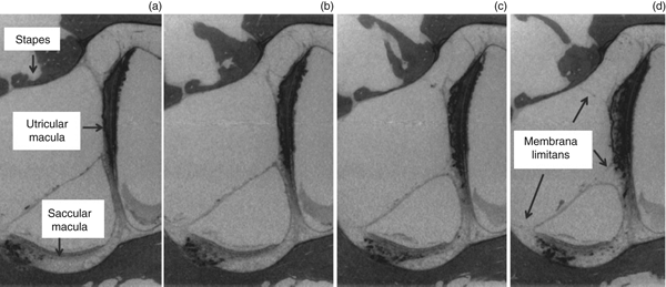
Thus, in total, 17 ears (of 10 patients) were studied, of which 7 sets were paired. It was noted that 9 of the 17 ears (53 per cent) displayed an attachment of the membrana limitans to the stapes footplate (Figures 1 and 2). These nine ears were from five patients (one unilateral and four bilateral ears). Therefore, 5 out of 10 patients had abnormalities where the utricular macula was directly attached to the stapes footplate via thin fibrillary attachments of the membrana limitans. Of the 17 bones, in all of the 7 paired sets (14 bones), it was noted that if attachments were present on one side (4 out of 7 sets), then such attachments were present bilaterally in every case (100 per cent of this sample). In this sample, all attachments were to the posterior one-third of the stapes footplate only; the membrana limitans did not have any attachments to the mid- or anterior-third of the footplate.
Images of several sections on the stapes footplate where the membrana limitans attached to the footplate in the posterior-third of the footplate. Note that the thickness of the stapes footplate (seen in parts a–d) is due to the view of the section through the posterior arch and not due to the presence of pathology, as this finding was not present in other views.

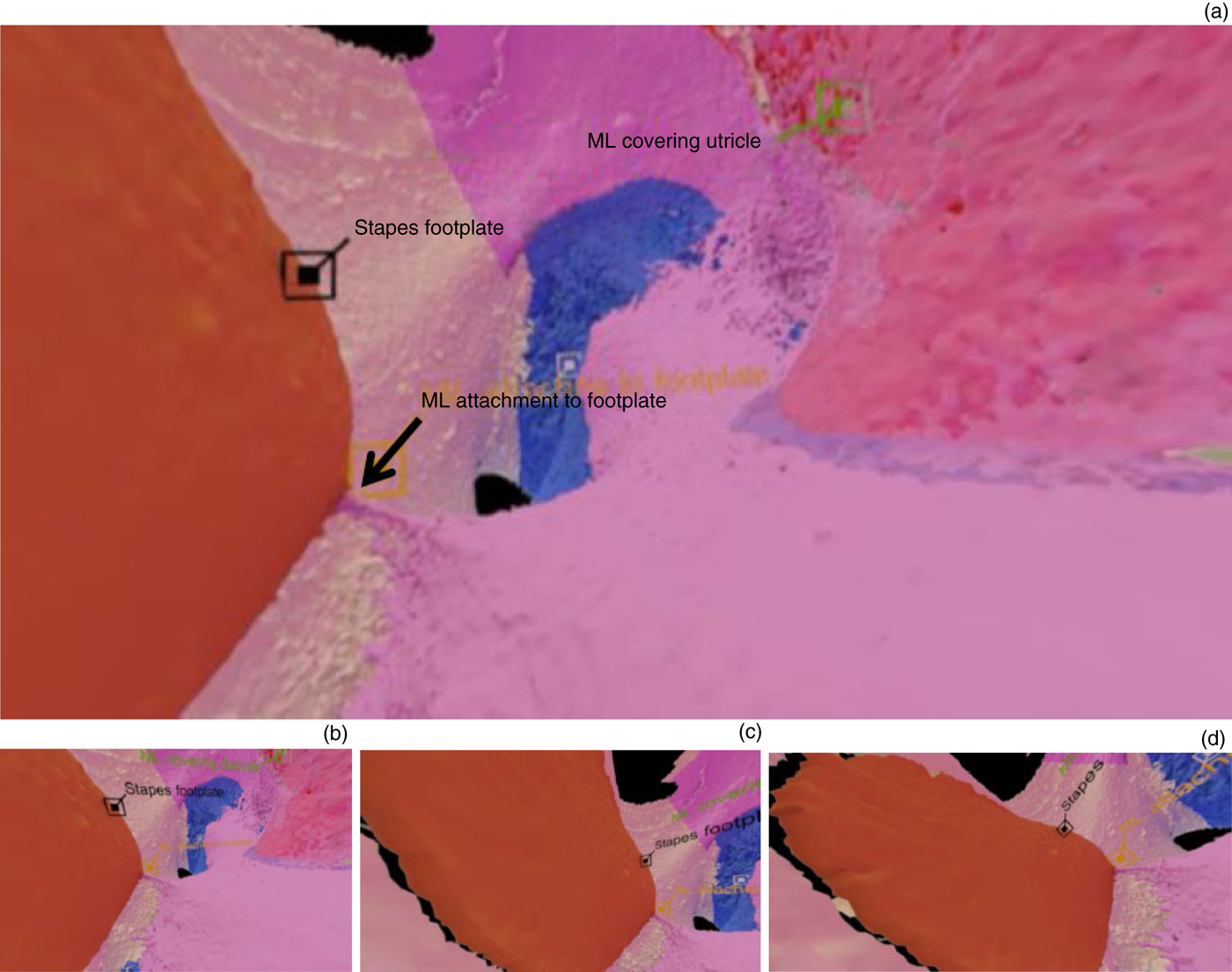
A three-dimensional view from within the anterior part of the vestibule looking posteriorly, with different camera angles shown in parts (a) to (d). Part (a) shows: the stapes footplate (brown); the membrana limitans (‘ML’; shown in pink), with attachments to the footplate; the saccule (red), beneath the membrana limitans; and the utricle (blue), most of which is also covered by the membrana limitans.

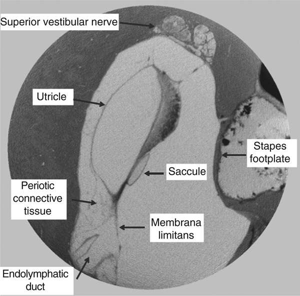
The attachment patterns of the membrana limitans in humans were similar to those reported in guinea pigs (Figure 1). It extended across the utricular macula to the cochlea anteriorly, posteriorly to the ampullated and non-ampullated ends of the lateral canal and to the ampullated end of the posterior canal. Superiorly, the ampullated end of the superior canal was spared, with the membrana limitans instead curving laterally to attach superiorly to the oval window (Figure 3). It also did not form a continuous membrane through the vestibule. It was more a sieve-like structure, acting as a support rather than a barrier. Where the membrana limitans did not attach to the footplate, it attached to the superior part of the vestibule, immediately above the stapes footplate (Figure 3). Figure 3 further serves as a demonstration of how the same information can be viewed as augmented reality content on a smart device (see Figure 3 legend for viewing instructions).
A coronal micro computed tomography section through the vestibule of a human, showing a thin fibrillary network of mesh-like connective tissue medial to the utricle, encasing the endolymphatic duct. There is no such tissue present lateral to the membrana limitans, facing towards the stapes footplate. The most superior part of the membrana limitans attaching anterior to the superior vestibular nerve fascicles can sometimes attach onto the superior part of the stapes footplate. This figure also serves to demonstrate the augmented reality content. Viewers need to download the free Hyperspaces application onto their iPhone, open the application, and type in the password ‘Vestibule’. The smartphone needs to be aimed at this figure. After downloading, a three-dimensional (3D) image will appear; application users should move the mobile telephone closer to dissect through the 3D otic capsule.

Guinea pig temporal bone results
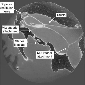
One guinea pig temporal bone was used to supplement the description of the membrana limitans in humans in light of the superior visualisation of this structure in the guinea pig. The membrana limitans in the guinea pig is distinctly different from humans, forming a continuous membrane (Figure 4). The appearance of this is consistent with the description of the membrana limitans in guinea pigs in the literature.Reference Nomura9, Reference Hara and Kimura10 In comparison, the human membrana limitans appeared to be a mesh-like structure, with non-continuous bands strapping the saccular membrane and utricular macula to the superior, medial and inferior part of the vestibule (Figure 1). Periotic connective tissue is lacking lateral to this membrane in both species, but is abundant medially.
A coronal micro computed tomography section through the vestibule of the guinea pig, showing the utricle, membrana limitans (‘ML’) and stapes footplate. The membrana limitans is present as a continuous and distinct membrane; it attaches superiorly to the stapes footplate, and inferiorly reflects from the utricular membrane to the lateral bony limit of the vestibule.

Discussion
The membranous labyrinth is supported in the perilymphatic space by a rich fibrillary network of tissue called the periotic connective tissue. This tissue holds the membranous semicircular canals in the outer circumference of the bony canal, and tethers the utricle and saccule to the superior and medial parts of the vestibule respectively (Figure 3). There is a clear absence of periotic connective tissue lateral to the utricle and saccule, though it is present in abundance on the medial side (Figure 3). This is likely attributed to the membrana limitans, which is well described in guinea pigsReference Hara and Kimura10 but poorly described in human ears. In the guinea pig, the membrana limitans is reported to structurally separate parts of the membranous labyrinth,Reference Nomura9 but this was not found in the human ears in this study (Figures 1 and 3).
Compartmentalisation in guinea pigs is reported to have implications on the variability of drug absorption in the inner ear, particularly as the ultrastructure of the membrana limitans has been shown to consist of fibrocytes, and tight and gap junctions.Reference Hara and Kimura10 Hence, in guinea pigs, it is possible that there is some degree of active transport of medications. However, given the otic capsule bone differences, such morphological information is difficult to attain from human specimens, and further study is needed. Nevertheless, the differences noted in the structural appearance of the membrana limitans in the two species are important, as the guinea pig is frequently used as an animal model to understand neurotological interventions in humans.
Implications of surgical interventions
Backous et al.Reference Backous, Aboujaoude, Minor and Nages13 reported the presence of such membrana limitans attachments to the stapes footplate; however, in their sample, this only occurred in 26 per cent of temporal bones (34 of 130 ears). The majority of connections were in the posterior-third of the footplate (18 out of 34). It occurred with less frequency in the posterior and middle parts together (15 out of 34), and occurred only in one bone in the middle section alone. There were no attachments in the anterior-third.
The sample size in the current study is much smaller, but the incidence of these attachments was a lot higher and the attachments were only found in the posterior-third of the footplate. The reasons for this difference may not be limited just to sample error. Specimen preparation and assessment techniques may play a significant role. Backous et al.Reference Backous, Aboujaoude, Minor and Nages13 reported section slice width on histology of 24 µm, with only every 10th section being committed to histological analysis. Thus, because of technological limitations of data analysis, the sections studied were 0.24 mm apart, which may underestimate the incidence of these attachments. One advantage of micro-CT data is that every slice is examined. In this study, slice width ranged from 1 µm to 55 µm. Thus, newer technologies, which allow more detailed data examination, may reveal that the true incidence of such fine attachments is higher than previously reported. It is imperative for surgeons operating in this area to be aware, irrespective of whether a laser or a trephine is being used to manipulate the footplate, that the posterior-third of the footplate and, less frequently, the middle-third of the footplateReference Backous, Aboujaoude, Minor and Nages13 may have attachments that could induce vestibular damage and sensorineural hearing loss.
The findings support the safety of stapedotomy surgery over stapedectomy surgery,Reference Fisch14–Reference Sedwick, Louden and Shelton17 and raise concern that footplate manipulation alone may cause vertigo in cases where otosclerotic focus on the footplate has not fixed the posterior part as much as the anterior part of the footplate. Though stapes surgery is performed under local and general anaesthesia, local anaesthetic surgery would make it easier to detect any physiological effect of footplate manipulation if such adhesions were present. Furthermore, if problems are encountered in the first side, there is a high risk that such findings are present on the contralateral side too, given that these attachments were present bilaterally in all cases in this study. Backous et al.Reference Backous, Aboujaoude, Minor and Nages13 also predicted that footplate manipulation may play a role in patients who experience pressure or sound-induced vertigo, without the presence of a canal dehiscence or obvious fistula, if the footplate alone was hypermobile, or could be responsible for hyperacusis or hypersensitivity in the presence of normal audiovestibular test and imaging findings.
Implications of three-dimensional assessment
The value of generating 3D tools to understand such anatomy is vital in pushing the frontiers of understanding the inner ear. The role of augmented and virtual reality has been discussed in relation to the teaching of technical skills in otological and rhinological surgery,Reference Chiarovano, Cheng and Mukherjee11, Reference Cheng, Mukherjee and Curthoys12, Reference Ruthenbeck, Hobson, Carney, Sloan, Sacks and Reynolds18–Reference Wiet, Stredney and Wan21 but the role of augmented reality as an educational tool for understanding inner-ear anatomy is still evolving. This is the first such study of inner-ear anatomy, with the resolution of structures demonstrated in micrometres. An added advantage of 3D-rendered digital data is that these can be viewed in multiple ways, such as in virtual reality or 360-degree video format.
• The periotic connective tissue (membrana limitans) that supports the utricle and saccule has not been well described in human subjects
• Knowledge of this anatomy is important for surgical interventions involving the stapes
• In over 50 per cent of temporal bones, membrana limitans ran from utricular macula directly to the stapes footplate, and was likely to occur bilaterally
• Guinea pig and human anatomy of this region may vary, which may influence findings when using the guinea pig as an animal model in experiments
• Three-dimensional understanding of this anatomy is important for training surgeons and developing future technological inner-ear interventions
The challenges encountered in this process include the segmentation of large data files acquired through micro-CT and the compression of that data for use in augmented reality and virtual reality, which require relatively low-resolution files for optimal performance. Thus, the challenge involves selectively preserving the detail of the structures of interest during segmentation. In this study, this was overcome by sub-selecting a small area for assessment (only the vestibule and stapes from the entire otic capsule). This is an important challenge to recognise in clinically translating micro-technology in a communicable format widely usable by clinicians. Furthermore, it highlights the challenges of future technologies, particularly in the realms of nanoscience and nanorobotic surgery.Reference Yamaan and Vyas22 There is also the need to communicate this difficult anatomy to collaborating scientists in a quick and efficient manner, to enable the design of interventions and access into the membranous labyrinth, which is a highly complex 3D structure. It is therefore important to push our current frontiers of data visualisation and 3D anatomy, so we are better able to understand current therapeutic interventions and design future innovations to treat and interact with the inner ear.
Conclusion
The attachment of the membrana limitans varies in individuals; it may attach the utricle directly to the stapes footplate in some patients, which has important consequences for surgical interventions. The posterior part of the footplate is most vulnerable. Surgeons should also be aware that if problems of vertigo are encountered on one side with stapes surgery, extra caution should be exercised with surgery on the contralateral ear, as it may be due to the membrana limitans attachments to the stapes footplate bilaterally. It is likely that these structures also play a role in drug delivery, though the exact physiological effect of the membrana limitans and periotic tissue on drug diffusion needs further study on human tissue. Three-dimensional visualisation and interaction with these structures is important to further our understanding of current interventions, while also allowing us to design future innovations, in terms of both diagnostic and treatment strategies for the inner ear.
Acknowledgements
The authors wish to acknowledge the Garnett Passe and Rodney Williams Memorial Foundation, and the RPA Institute of Academic Surgery, for providing grant support to fund this study.
Competing interests
None declared