INTRODUCTION
Polydorid species of the family Spionidae belong to nine genera: Polydora Bosc, 1802; Dipolydora Verrill, 1879; Pseudopolydora Czerniavsky, 1881; Boccardia Carazzie, 1895; Polydorella Augener, 1914; Tripolydora Woodwick, 1964; Boccardiella Blake & Kudenov, Reference Blake and Kudenov1978; Caraziella Blake & Kudenov, Reference Blake and Kudenov1978; and Amphipolydora Paterson & Gibson, 2003 (Benham, Reference Benham, Harmer and Shipley1896; Walker, Reference Walker2011). Polydorid species possess special major spines in an enlarged or modified 5th chaetigerous segment. These species occupy a diversity of habitat niches, and this has undoubtedly contributed to their evolutionary success (Blake, Reference Blake, Blake, Hilbig and Scott1996).
Polydorids contain a group that is well known for its boring activities in calcareous substrates such as coralline algae, corals, mollusc shells and barnacles (Blake, Reference Blake, Blake, Hilbig and Scott1996). In this regard, certain polydorid species have been the subject of considerable study due to their impact on commercially important molluscs produced in aquaculture. These impacts include reduced commercial value, growth rate, meat yield and heavy mortality (Mori et al., Reference Mori, Sato, Nomura and Imajima1985; Handley & Bergquist, Reference Handley and Bergquist1997; Lleonart et al., Reference Lleonart, Handlinger and Powell2003; Simon et al., Reference Simon, Ludford and Wynne2006; Sato-Okoshi et al., Reference Sato-Okoshi, Okoshi and Shaw2008, Reference Sato-Okoshi, Okoshi, Koh, Kim and Hong2012). Polydorid species that associate with mollusc shells are achieving global distribution due to commercial transport of host mollusc shells (Radashevsky & Olivares, Reference Radashevsky and Olivares2005; Simon et al., Reference Simon, Ludford and Wynne2006). Because of their invasiveness, these polydorid species are a source of concern from both economic and ecological perspectives (Cohen & Carlton, Reference Cohen and Carlton1998).
There are several species complexes within the Polydora group which include different species that are morphologically indistinguishable from each other. At the same time, some Polydora species show a high degree of intraspecific variation, particularly with respect to pigmentation patterns. Although molecular biological approaches are considered effective for the identification of species within various taxa, to date, little molecular information has been obtained for the Spionidae, particularly polydorid species. Consequently, there are few GenBank sequences available for Polydora complex species particularly for material sourced from Asia and the Pacific Rim. Obtaining molecular information on polydorid species is of both scientific and economic importance, as is characterizing morphological variation at the species level.
This study represents a first attempt to determine the molecular sequences of the 18S rRNA gene of three morphologically similar species of the genus Polydora, which are commonly known as oyster shell borers, using materials sourced from Japan and Australia. Here, we not only determine the molecular sequences of individuals from a single population of these species but also of individuals from several populations of different waters and multiple individuals with or without particular pigmentation in each species to confirm the gene sequences of the species and to assess the morphological divergence in these species.
MATERIALS AND METHODS
Morphological observations
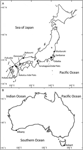
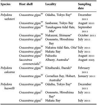
Individuals of three Polydora species, Polydora websteri, P. calcarea and P. haswelli, were collected in 2011 from the shells of wild and cultured oysters Crassostrea gigas along the coasts of the Pacific Ocean (Kitaibaraki; Odaiba, Sanbanse, Tokyo Bay; Tanakagawa tidal flats, Mie), the Inland Sea (Oonoseto, Hiroshima Bay; Nakatsu tidal flats, Oita), and the Sea of Japan (Hakata Bay; Fukuoka; Nakaumi, Shimane) (Figure 1; Table 1). Polydora individuals were also extracted from the shells of cultured oysters Saccostrea commercialis from Albany, south-western Australia, in 2005, and from wild oysters Crassostrea gigas from Cornelian Bay, Hobart, Australia, in 2011 (Figure 1; Table 1). The shells were collected from intertidal to subtidal elevations. The polychaete worms, which inhabit self-excavated burrows inside the shells, were extracted by fracturing the shells with pliers and a hammer.
Location of sampling sites in (A) Japan and (B) Australia.

Boring Polydora species collected for morphological observation and genetic analysis from oysters in Japan and Australia.

C, cultured; W, wild; *, sample for rRNA analysis.
After the worms were removed from the shells, morphological characteristics were observed under a stereo microscope (Olympus SZX 16) in both live and preserved conditions in 10% neutral formalin in seawater. Pictures were taken by the camera (Olympus DP 73) fixed to the microscope.
Specimens were deposited in the National Museum of Nature and Science, Tokyo (NSMT).
DNA extraction, PCR amplification, sequencing and phylogenetic analysis
Genetic analysis was performed on 11 specimens of P. websteri, two specimens of P. calcarea and two specimens of P. haswelli, obtained from the locations detailed in Table 1 and Figure 1. Genomic DNA was extracted from live or ethanol-preserved tissues. All animal tissues were washed by several transfers in filtered (pore size 0.2 µm) seawater and distilled water to remove as much extraneous matter as possible. Polymerase chain reaction (PCR) tubes (0.2 ml) containing 50 µl of 10% Chelex® suspension (Bio-Rad Laboratories Inc., Richmond, CA) and animal tissues were heated at 95°C for 20 minutes to extract genomic DNA according to the method of Richlen & Barber (Reference Richlen and Barber2005). Extracted DNAs were used as templates to amplify the target regions. All PCRs were performed in a thermal cycler in a reaction mixture (25.0 µl) containing 1.0 µl of template DNA, 0.2 mM of each deoxynucleoside triphosphate (dNTP), 1× PCR buffer, 2.0 mM MgSO4, 0.4 U of KOD-Plus- ver. 2 DNA polymerase (Toyobo, Osaka, Japan; with intensive 3′ → 5′ exonuclease activity), and 0.2 nM of each primer. Three primer pairs (18S-1F1/18S-1R632, 18S-2F576/18S-2R1209 and 18S-3F1129/18S-R1772: Nishitani et al., Reference Nishitani, Nagai, Hayakawa, Kosaka, Sakurada, Kamiyama and Gojobori2012) were used to amplify the nuclear 18S rRNA gene of the three Polydora species. The PCR cycling conditions were as follows: initial denaturation at 94°C for 2 minutes; 38 cycles at 94°C for 15 seconds, 54°C for 30 seconds, and 68°C for 45 seconds. PCR amplification products were checked on 1.0% agarose gels, with ethidium bromide staining. To remove unincorporated PCR primers and dNTPs, 1.5 µl of the PCR products were treated with 0.6 µl exonuclease I and shrimp alkaline phosphatase (Exo SAP-IT) (USB Corp., Cleveland, OH, USA) at 37°C for 15 minutes, followed by incubation at 80°C for 15 minutes to inactivate the enzymes. The PCR products were sequenced directly using an automated DNA sequencer (ABI 3100; Applied Biosystems). The forward and reverse complementary sequences were combined and the three nucleotide sequences obtained with the three primer pairs were aligned using GENETYX software (Genetyx Corp., Tokyo, Japan). Partial sequences of the nuclear 18S rRNA gene were aligned with the sequences of related species obtained from GenBank using the Clustal W algorithm (Thompson et al., Reference Thompson, Higgins and Gibson1994), and neighbour-joining (NJ) trees were generated using Molecular Evolutionary Genetics Analysis (MEGA) software version 4.0 (Tamura et al., Reference Tamura, Dudley, Nei and Kumar2007) with the default setting. Bootstrapping with 1000 replications was used to estimate the reliability of the phylogenetic trees.
RESULTS
Morphological characteristics of Polydora websteri, P. calcarea and P. haswelli
Family SPIONIDAE Grube, 1850
Genus Polydora Bosc, 1802
Polydora websteri Hartman in Loosanoff and Engle, 1943
(Figure 2)
Polydora websteri: Sato-Okoshi, Reference Sato-Okoshi1999: 832, 834; Read, Reference Read2010: 1–18.
Polydora haswelli: Sato-Okoshi et al., Reference Sato-Okoshi, Okoshi and Shaw2008: 495, 497.
MATERIALS
Extracted from the shells of wild Crassostrea gigas, from Odaiba and Sanbanse, Tokyo Bay; Tanakagawa tidal flats, Mie; Nakaumi, Shimane; Nakatsu tidal flats, Oita; shells of cultured C. gigas from Oonoseto, Hiroshima Bay; Hakata Bay; Fukuoka, Japan, in 2011. Extracted from the shells of cultured Saccostrea commercialis from Albany, Australia, in 2005. More than 10 individuals were examined per site.
MORPHOLOGY OF NEW MATERIALS
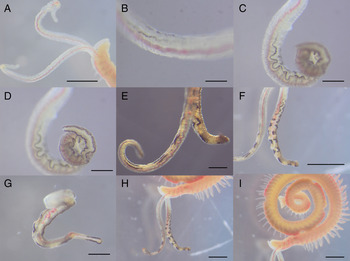
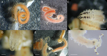
Medium-sized worms up to 18 mm long and 1.0 mm in width at chaetiger 5, with up to 120 chaetigers. Colour in life, light tan with variable amounts of black pigmentation only on palps. Palps with weak (Figure 2A, B) to conspicuous black pigmentation (Figure 2C, D) along margin: some continuously black (Figure 2A–D); others discontinuously black along margin of palps (Figure 2E–G), the latter showing black dot-like or black bar-like pattern instead. Black pigments fade after fixation in formalin.
Polydora websteri. Pictures of live individuals from Odaiba, Tokyo Bay, showing pigment variation particularly on the palps: (A, B) inconspicuous and narrow black pigmentation along the margin of the palps, lateral views; (C, D) C and D show the same palp, but in different fields of focus. Slight black pigmentation on the palps with conspicuous black along the margins; (E–H) discontinuously black along the margins of the palps, showing black dot-like or black bar-like pattern, (I) fairly long branchiae continuing almost to the end of the body. Pygidium flaring disc, white in color. Scale bars: A, F, H & I, 1 mm; B, D, 0.5 mm; E,G, 0.25 mm.

Prostomium strongly to weakly bilobed, curved downward anteriorly; caruncle extension size-dependent, reaching posterior end of chaetiger 2 to end of chaetiger 3. No occipital tentacle. Eyes absent or present, though present in most specimens; if present, up to four, trapezoidal in arrangement. Chaetiger 1 with short notopodial lobe, notochaetae absent, with neurochaetae on neuropodia only. Winged capillary notochaetae of chaetigers 2–4, 6, and subsequent segments arranged in two rows. No special notochaetae present on posterior chaetigers. Chaetigers 2–4 and 6 with winged capillary neurochaetae arranged in two rows. Bidentate hooded hooks, main fang with right angle to shaft, and acute angle with apical tooth, constriction on shaft, replace fascicle of neurochaetae from chaetiger 7, numbering from four to eight per chaetiger up to mid-body, then decreasing to one in posterior chaetigers. Branchiae from chaetiger 7, fairly long throughout with bright branchiate blood vessels in middle, reaching full size on chaetiger 10 or posteriorly, overlapping mid-dorsal, then decreasing in length and continuing to last 1/4 of body or near the posterior end (Figure 2C).
Chaetiger 5 modified and enlarged, major spines falcate with lateral tooth or sheath, four to seven spines emergent per row, plus approximately two spines buried inside, alternating with slender, pennoned accompanying chaetae. Winged capillary noto- and neurochaetae present on chaetiger 5.
Pygidium small disc-like to largely flared, with dorsal gap; white to semi-transparent white in colour (Figure 2C).
Polydora calcarea (Templeton, 1836)
(Figure 3)
Polydora calcarea: Radashevsky & Pankova, Reference Radashevsky and Pankova2006: 245–252.
MATERIALS
Extracted from the shells of wild Crassostrea gigas attached to concrete slope in Kitaibaraki, Ibaraki Prefecture, Japan, in 2011, and from the shells of wild C. gigas in Cornelian Bay, Hobart, Australia, in 2011. More than 50 individuals were examined per site.
MORPHOLOGY OF NEW MATERIALS
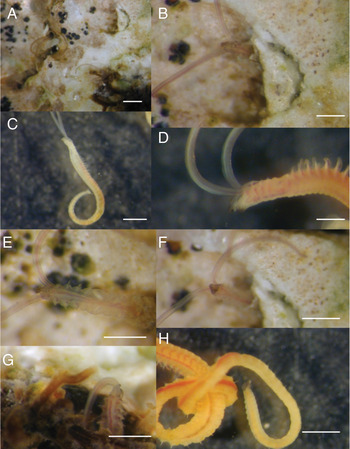
Small to medium-sized worms up to 12 mm long and 0.7 mm in width at chaetiger 5, with up to 85 chaetigers. Colour in life, light tan with variable black pigmentation on palps, prostomium, peristomium, chaetigers 1–4, and pygidium. Palps transparent, or black pigmentation on and along margin of palps (Figure 3A, B). Black pigment or no pigment on both sides of anterior end of prostomium (Figure 3B–E); anteroventral side of peristomium (Figure 3F), with pair of black pigment or no pigment on peristomium (Figure 3B, D, E & G) and dorsal side of chaetigers 1–4 (Figure 3E, G). Pygidium conspicuously black all over (Figure 3H) to slightly browned, or totally transparent (Figure 3C).
Polydora calcarea. Pictures of live individuals from Kitaibaraki showing pigment variation: (A, B, E–G) anterior chaetigers and palps protruding from their burrows. Black pigmentation can be seen on the palps, and on both the dorsal and ventral sides of the prostomium and peristomium; (C) entire individual extracted from its burrow, lateral view; (D) black pigmentation on the anterior body, lateral view; (H) middle to posterior body with black pygidium. Scale bars: A–C & E–H, 1 mm; D, 0.5 mm.

Prostomium weakly incised or rounded and slightly curved downward anteriorly, caruncle extension reaching posterior end of chaetiger 2. No occipital tentacle. Eyes absent or present; if present, up to four, trapezoidal in arrangement. Chaetiger 1 with short notopodial lobe, notochaetae absent, and neurochaetae on neuropodia only. Winged capillary notochaetae arranged in two rows on chaetigers 2–4, 6, and succeeding chaetigers. No special notochaetae on posterior chaetigers. Chaetigers 2–4 and 6 displayed winged capillary neurochaetae arranged in two rows. Bidentate hooded hooks of main fang with right angle to shaft, and acute angle with apical tooth, constriction on shaft, replace fascicle of neurochaetae from chaetiger 7, numbering from six to eight per chaetiger up to mid-body, then decreasing to one in posterior chaetigers. Branchiae from chaetiger 7 continuing only to 1/3 or 1/2 of body (Figure 3C).
Chaetiger 5 modified and larger than chaetigers 4 and 6; thickened spines of chaetiger 5 falcate, with lateral tooth or sheath, alternating with slender, pennoned accompanying chaetae; from five to six spines emergent per row, plus approximately two spines buried inside. Winged capillary noto- and neurochaetae present on chaetiger 5, one to three anterodorsal and two to five posteroventral, respectively.
Pygidium scoop- to flare-like with dorsal gap, with or without black pigmentation (Figure 3C, H).
Polydora haswelli Blake & Kudenov, Reference Blake and Kudenov1978
(Figure 4)
Polydora haswelli Blake & Kudenov, Reference Blake and Kudenov1978: 259–260.
Polydora sp.: Sato-Okoshi, Reference Sato-Okoshi1999: 836.
Polydora haswelli: Read, Reference Read2010: 1–18.
MATERIALS
Extracted from the shells of wild Crassostrea gigas in Odaiba, Tokyo Bay; cultured C. gigas in Oonoseto, Hiroshima Bay; and Hakata Bay, in 2011. More than five individuals were examined per site.
MORPHOLOGY OF NEW MATERIALS
Medium-sized worms up to 25 mm long and 1.1 mm in width at chaetiger 5, with up to 130 chaetigers. Colour of living specimens light tan with variable black pigmentation on palps, prostomium, peristomium, and anterior chaetigers 1–6 (Figure 4A–F). Palps without marginal black but with two to 11 distinct to inconspicuous black bars (Figure 4A, B, E, F). Black pigmentation on anterior end and along both sides of prostomium (Figure 4A–F), with a pair of black pigment or no pigment on peristomium, and black pigmentation on dorsal side of chaetigers 1–6, usually 1–4 (Figure 4A–D) and ventral side of chaetigers 1–4 (Figure 4E). Some without conspicuous pigmentation on ventral side.
Polydora haswelli. Live individuals showing pigment variation: (A) entire individual from Hakata Bay, Eateral view. Black pigmentation along the prostomium and anterior chaetigers, and black bars on the palps; (B–E) individuals from Hiroshima. Distinct black pigmentation along the prostomium, peristomium and anterior chaetigers 1–6, and black bars on the palps. Dorsal (B–D) and ventral (E) views; (F) anterior end of an individual from Tokyo Bay, lateral view. Palps with distinct black bars. Scale bars: A, B & E, 1 mm; C, D & F, 0.5 mm.

Prostomium weakly incised anteriorly, caruncle extending to middle of chaetiger 4. No occipital tentacle. Eyes absent or present; if present, up to four, trapezoidal in arrangement. Chaetiger 1 without notochaetae, only neurochaetae present. Winged capillary notochaetae arranged in two rows on chaetigers 2–4, 6, and succeeding segments. No special notochaetae on posterior chaetigers. Chaetigers 2–4 and 6 with winged capillary neurochaetae arranged in two rows. Bidentate hooded hooks, main fang with right angle to shaft, and acute angle with apical tooth, constriction on shaft, replace fascicle of neurochaetae from chaetiger 7, six to 10 per chaetiger up to mid-body, then decreasing in number in posterior chaetigers. Branchiae from chaetiger 7, long, touching mid-dorsal, then decreasing in length and continuing nearly to end of body (Figure 4A, B).
Chaetiger 5 largely modified, major spines thickened and falcate with large lateral flange and delicate smaller flange, up to eight in a row accompanying pennoned slender chaetae. Both winged capillary noto- and neurochaetae present on chaetiger 5.
Pygidium disc- to flare-like, with dorsal gap (Figure 4A).
Phylogenetic analysis of 18S rRNA gene sequences
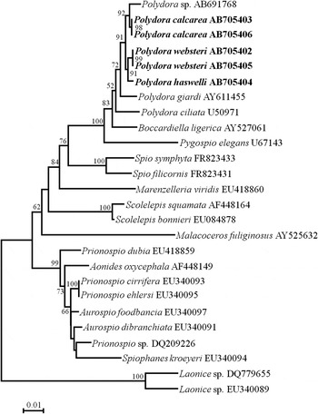
Nuclear 18S rRNA gene sequences (1771 base pairs) were determined for the 11 individuals of P. websteri, two individuals of P. calcarea and two individuals of P. haswelli. DNA sequence analyses revealed that the nucleotide sequence of the 18S rRNA gene was identical within a species and distinct between species (GenBank accession number: Polydora websteri from Japan AB705402, P. websteri from Australia AB705405; Polydora calcarea from Japan AB705403, P. calcarea from Australia AB705406; and Polydora haswelli AB705404). The phylogenetic tree, constructed using available 18S rRNA gene sequences, and the position of the three Polydora species with respect to other spionid taxa, are shown in Figure 5. Polydora formed a generic group. Three boring species examined in this study formed one group. Polydora sp. (Teramoto et al., unpublished data) and P. calcarea were resolved as sister species, as were P. websteri and P. haswelli.
A neighbour-joining (NJ) tree inferred from the nuclear 18S rRNA gene sequences of spionid polychaetes. The gene sequences of the three Polydora species obtained in this study are highlighted in boldface type. Bootstrap values of >50% are given as percentage of 1000 bootstrap replicates given at respective nodes. The scale bar represents the number of substitutions per site.

DISCUSSION
The three species described here, namely, P. websteri, P. calcarea and P. haswelli, belong to the Polydora ciliata/websteri group, members of which are characterized by a lateral subdistal flange or tooth on the falcate spines of chaetiger 5, as defined by Blake (Reference Blake, Blake, Hilbig and Scott1996). These species resemble each other very closely, which often leads to confusion in identification. The present study documents a diverse range of intraspecific morphology, particularly regarding pigmentation, among specimens of the three species collected from Japan and Australia.
Polydora websteri is a well-known, presumptively cosmopolitan species that has been described from the shells of various mollusc species worldwide (Blake, Reference Blake, Blake, Hilbig and Scott1996; Read, Reference Read2010). This species has been the subject of taxonomic confusion, because it possesses the typical, simple morphological characteristics of the Polydora ciliata/websteri group without other conspicuous features (Radashevsky, Reference Radashevsky1999). Insufficient species accounts and poorly preserved specimens have exacerbated this confusion (Radashevsky & Williams, Reference Radashevsky and Williams1998). To date, P. websteri has been reported to morphologically resemble P. aggassizi, P. aggregata, P. brevipalpa, P. ciliata, P. curiosa, P. limicola, P. neocaeca and P. haswelli (Blake, Reference Blake, Blake, Hilbig and Scott1996; Williams & Radashevsky, Reference Williams and Radashevsky1999). Therefore, it is possible that these related species have been misidentified and improperly recorded. According to our molecular analysis, reports of P. haswelli from shells of the cultured oyster Saccostrea commercialis collected in Albany, Australia (Sato-Okoshi et al., Reference Sato-Okoshi, Okoshi and Shaw2008), should be revised as P. websteri.
In comparison with other Polydora species inhabiting Japanese and Australian waters, P. websteri possesses fairly long branchiae continuing to close to the end of the body with conspicuous red blood vessels in each branchia. This characteristic holds promise as a useful key for species determination. In addition to the distinct longer branchiae, shell infestation by P. websteri often forms mud blisters. This species also excavates a shallow burrow, and thus shows both boring activity and somewhat non-boring morphological and ecological characteristics (i.e. morphological and ecological characteristics of non-boring species are: conspicuous thicker and wider branchiate blood vessels; strong and vigorous palp movement and high crawling activity; forming mud blisters instead of self-excavating). Polydora websteri, which inhabits hosts associated with muddy environments particularly oyster beds, may thus represent an intermediate stage between non-boring and boring strategies, as previously discussed by Sato-Okoshi (Reference Sato-Okoshi2000).
On the other hand, divergence in palp pigmentation and pygidium morphology make positive identification difficult. Careful attention is required, particularly with regard to the pattern of palp pigmentation: P. websteri exhibits either (1) continuous black pigmentation along the margins of the palps (Figure 2B–D) or (2) partially discontinuous black pigmentation along the palp margins, resembling black bars (Figure 2E–H). Our results suggest that it is necessary to distinguish palps with discontinuous black along the margins from palps with apparently independent black bars. Palps with distinct and independent black bars (Figure 4A–C, E, F) are considered to be one of the most important morphological characteristics of P. haswelli in terms of species identification.
The other two species examined here have also been reported to resemble several Polydora species. Polydora calcarea is morphologically similar to P. manchenkoi (Radashevsky & Pankova, Reference Radashevsky and Pankova2006), whereas P. haswelli resembles P. neocaeca (Read, Reference Read2010). We found the pigmentation patterns of these two species, P. haswelli and P. calcarea, to be variable and to resemble each other, which may lead to misidentification of the species. The most distinct characters differentiating the two species are: (1) distinct black bars on the palps only in P. haswelli; (2) a difference in the number of branchiate chaetigers: branchiae continue only to 1/3 or 1/2 of the body in P. calcarea, whereas they continue almost to the end of the body in P. haswelli; and (3) black pigmentation on the pygidium only in P. calcarea.
Morphological, biological, and ecological approaches have traditionally been used to identify polydorid species, in addition to differences in methyl green staining (Read, Reference Read2010) and allozyme analyses (Mustaquim, Reference Mustaquim1988; Manchenko & Radashevsky, Reference Manchenko and Radashevsky1993, Reference Manchenko and Radashevsky1998; Radashevsky & Pankova, Reference Radashevsky and Pankova2006). Our work demonstrates that a molecular biological approach can provide a key to differentiating between species with a similar morphology. There is, however, a scarcity of molecular information on Spionidae, particularly for polydorid species. To date, molecular sequences for several genes are available only for eight of the 147 known polydorid species: Polydora sp. (Teramoto et al., unpublished data), P. ciliata, P. cornuta, P. triglanda, P. giardi, Dipolydora carunculata, Boccardia proboscidea and Boccardiella ligerica from GenBank (Walker, Reference Walker2011; Teramoto et al., unpublished data). Among the polydorids, sequence data for the 18S rRNA gene are available only for Polydora sp. from Japan (Teramoto et al., unpublished data), P. ciliata (source locality unstated) (unpublished) and P. triglanda from Taiwan (unpublished), P. giardi from France (Rousset et al., Reference Rousset, Rouse, Siddal, Tillier and Pleijel2004), Dipolydora carunculata from Russia (unpublished) and Boccardiella ligerica from Germany (Struck & Purschke, Reference Struck and Purschke2005). Owing to the short length of the partial sequence data, Polydora triglanda and D. carunculata were not included in the phylogenetic tree (Figure 5). As we could not confirm whether the species P. giardi (in Rousset et al., Reference Rousset, Rouse, Siddal, Tillier and Pleijel2004) is a Dipolydora giardi (Polydora giardi is revised to Dipolydora giardi by Blake, Reference Blake, Blake, Hilbig and Scott1996) or some another Polydora species, we left the result as it is and expected to discuss and solve this matter in a future study.
This study examined the 18S rRNA gene sequence of the presumed cosmopolitan species Polydora websteri, and two other oyster-boring species, P. calcarea and P. haswelli, all of which belong to the Polydora ciliata/websteri group. Our results provide genetic evidence of independent species, and morphological characteristics for distinguishing them. Pigmentation characterized by distinct independent black bars on the palps is the morphologically invariant characteristic of P. haswelli, and can be a key to distinguishing this species from others. Intraspecific morphological variation in caruncle extension, number of branchiate chaetigers, shape of pygidium, and other body pigmentation is common. This intraspecific variation may be due to differences in size and environmental factors. Further molecular approaches are expected to be applied in polydorid groups not only for the accurate discrimination of these species but for elucidating genealogical relationships among the taxa.
ACKNOWLEDGEMENTS
We express our sincere thanks to Drs M. Hamaguchi, K. Okoshi, J. Shaw, P. Hutchings, C. Macleod, T. Kimura and K. Yamaguchi for their kind collaboration in collecting oysters. Thanks are due to Drs S. Keable and L.M. Walker, Australian Museum, who loaned the type materials of Polydora haswelli. This study was partially supported by Grants-in-Aids from the Ministry of Education, Science, Sports and Culture of Japan to Waka Sato-Okoshi (grant number 21580216) and from the Research Institute of Marine Invertebrates Foundation to Hirokazu Abe.