Introduction
Metastatic cutaneous squamous cell carcinoma (SCC) is the most common malignancy of the parotid gland in Australia and New Zealand.Reference O'Hara, Ferlito, Takes, Rinaldo, Strojan and Shaha 1 Cutaneous SCC of the head and neck region has a 2–5 per cent metastasis rate, with intra-parotid lymph nodes being the most common site for metastatic spread.Reference O'Hara, Ferlito, Takes, Rinaldo, Strojan and Shaha 1 , Reference O'Brien, McNeil, McMahon, Pathak, Lauer and Jackson 2 Clinical and pathological factors of cutaneous SCC that have been shown to correlate with a greater risk of developing metastases to lymph nodes include: thick lesions (over 4–5 mm), large size (over 2 cm), high-grade, perineural invasion, lymphovascular invasion, location on or around the pinna, local recurrence, and immunosuppression.Reference Veness, Palme and Morgan 3 The incidence of locoregional metastases in patients with cutaneous SCC with these adverse factors is increased to 16–35 per cent.Reference O'Hara, Ferlito, Takes, Rinaldo, Strojan and Shaha 1 Although only a minority of cutaneous SCC lesions metastasise, given the high incidence of cutaneous SCC in Australasia, the absolute number of patients affected is of clinical significance.
The published evidence suggests that the optimum treatment of metastatic cutaneous SCC of the parotid is surgical resection with adjuvant radiotherapy.Reference O'Hara, Ferlito, Takes, Rinaldo, Strojan and Shaha 1 , Reference Veness, Morgan, Palme and Gebski 4 Surgery alone may be appropriate for selected patients who have early disease with low-risk histopathological features. There is controversy surrounding the extent of the parotid surgery, both regarding the extent of parotid resection (whether the deep lobe should be cleared) and facial nerve resection. Clinically positive neck nodes should be treated with neck dissection. The treatment of the clinically and radiologically node-negative (N0) neck is debatable. The incidence of occult cervical nodal disease in these cases can be as high as 35–50 per cent,Reference Veness, Morgan, Palme and Gebski 4 , Reference Moore, Weber, Prieto, El-Naggar, Holsinger and Zhou 5 and therefore many advocate selective neck dissection or radiation treatment to the neck. Lateral temporal bone resection may have a role in the treatment of selected tumours.Reference Shao, Wong, McIvor, Mylnarek, Chaplin and Izzard 6 – Reference O'Connor, Behan, Toner, Kinsella, Beausang and Timon 9
Various patient, tumour and treatment factors have been shown to be prognostic indicators in metastatic cutaneous SCC of the parotid, but discrepancies exist between studies. Patient characteristics that may affect outcomes include immune status and age.Reference Shao, Wong, McIvor, Mylnarek, Chaplin and Izzard 6 , Reference Chen, Roman, Sosa and Judson 10 – Reference Oddone, Morgan, Palme, Perera, Shannon and Wong 14 Increasing size of the parotid tumour, close or positive surgical margins, facial nerve involvement, cervical node involvement, and extra-parenchymal spread have been associated with poorer outcomes in some studies.Reference Chen, Roman, Sosa and Judson 10 , Reference Oddone, Morgan, Palme, Perera, Shannon and Wong 14 – Reference Hinerman, Indelicato, Amdur, Morris, Werning and Vaysberg 16
We reviewed patients with parotid metastases from cutaneous SCC who were treated surgically at a tertiary hospital over a 10-year period. We aimed to compare our outcomes to those published in the literature, and to identify prognostic factors associated with reduced disease-specific survival and locoregional control. We hypothesised that more extensive surgery (such as lateral temporal bone resection or facial nerve resection) would improve locoregional control and survival by providing greater margins.
Materials and methods
The Christchurch Hospital coding system was used to identify all patients who had undergone parotidectomy or lateral temporal bone resection between 1 January 2003 and 31 December 2012. Patients with histology showing metastatic cutaneous SCC of the parotid were included in the study. Cases of lateral temporal bone resections performed for disease other than metastatic cutaneous SCC of the parotid were excluded. Patients with direct invasion of the parotid from overlying cutaneous SCC were excluded. All patients had been discussed at a multidisciplinary meeting prior to treatment.
Facial nerve resection (complete sacrifice or removal of selected branches) was planned when there was pre-operative facial weakness. In the remaining cases, the decision to resect branches of the nerve was made intra-operatively if the nerve could not be dissected with a clear margin from the tumour. Lateral temporal bone resection was performed when there was clinical or radiological suspicion that the tumour was adherent to or invading the temporal bone or external auditory canal. The lateral portion of the temporal bone was resected en bloc in continuity with the parotid and neck specimen.
With regard to management of the cervical lymph nodes, if these were clinically and radiologically negative, the patient received a level II and III neck dissection in continuity with the parotid surgery. If the primary skin lesion arose from the posterior scalp, a level Va neck dissection was also included. If there was clinical or radiological evidence of neck node involvement, the patient underwent comprehensive neck dissection in continuity with the parotid surgery.
Primary outcomes were disease-specific survival and locoregional control. Locoregional recurrence was defined as recurrence of disease in the neck or parotid bed. The variables analysed included: patient age, gender, ethnicity, site of cutaneous primary lesion, side of surgery, pre-operative facial nerve function, extent of surgery, facial nerve resection and post-operative radiotherapy.
The Kaplan–Meier method was used to calculate overall survival, disease-specific survival and locoregional control. The Cox proportional hazard model was used to analyse the strength of association between the various prognostic factors and the two primary outcomes: disease-specific survival and locoregional control. A p value of less than 0.05 was considered statistically significant. All statistical tests were performed using SAS software, version 9.3 (SAS Institute, Cary, North Carolina, USA).
This retrospective review was approved by the Canterbury District Health Board audit committee.
Results
Over the 10-year period, 64 parotidectomies were performed in 63 patients. There were 53 men (84 per cent) and 10 women (16 per cent), with a mean age of 72 years at the time of surgery (range, 44–92 years). Fifty-three patients (84 per cent) were of ‘New Zealand European’ or ‘other European’ ethnicity. There were seven (11 per cent) immunocompromised patients (two renal transplant patients, two patients on azathioprine for Crohn's disease, two on long-term prednisone and one patient with chronic lymphoid leukaemia).
The pinna was the most common site of the primary lesion (19 cases; 30 per cent), followed by the temple (14 cases; 22 per cent). The right side was more commonly affected (39 cases; 61 per cent). There were eight patients (13 per cent) with pre-operative facial nerve palsy; of these, five underwent lateral temporal bone resection with facial nerve resection, and three had a parotidectomy with facial nerve resection.
Forty-seven patients (75 per cent) underwent superficial or total parotidectomy without facial nerve sacrifice (with one renal transplant patient having bilateral superficial parotidectomies for bilateral disease). Eight patients (13 per cent) underwent superficial or total parotidectomy with resection of one or more branches of the facial nerve. Eight patients (13 per cent) underwent lateral temporal bone resection with parotidectomy (with or without facial nerve resection). All patients underwent simultaneous ipsilateral neck dissection.
The majority of patients (n = 51; 81 per cent) had adjuvant radiotherapy. Based on favourable histopathological findings, four patients were not offered post-operative radiotherapy. Adjuvant radiotherapy was planned in two patients but they did not receive it (one developed distant metastatic disease and one died of an unrelated cause prior to the start of radiotherapy). Six patients declined radiotherapy, two of whom were immunocompromised. Of these patients, two developed local and then subsequent distant recurrence and died of this disease, two developed distant recurrence from which they died, and two died of unrelated causes. The four patients who were not offered adjuvant radiotherapy (in light of favourable histopathological features) did not develop locoregional or distant recurrence in the follow-up period.
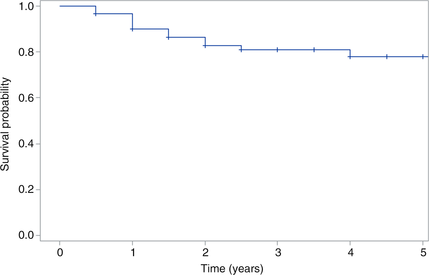
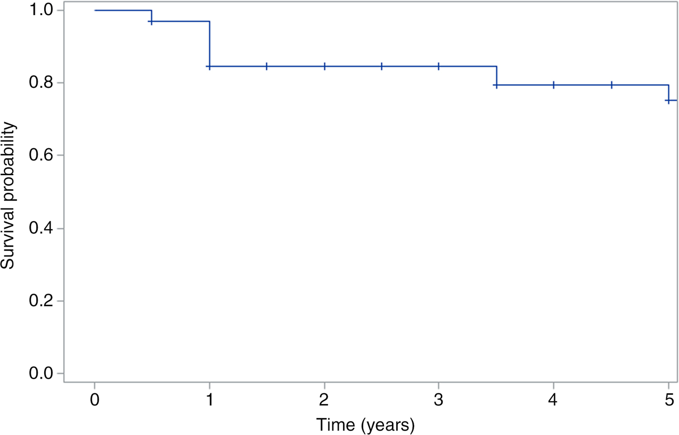
Median follow-up duration was 38 months. The overall survival rate was 72 per cent at two years and 53 per cent at five years. The disease-specific survival rate was 83 per cent at two years and 78 per cent at five years (Figure 1). The locoregional control rate was 85 per cent at two years and 72 per cent at five years (Figure 2).
Kaplan–Meier curve of disease-specific survival (all patients). ‘+’ = censored

Kaplan–Meier curve of locoregional control (all patients). ‘+’ = censored

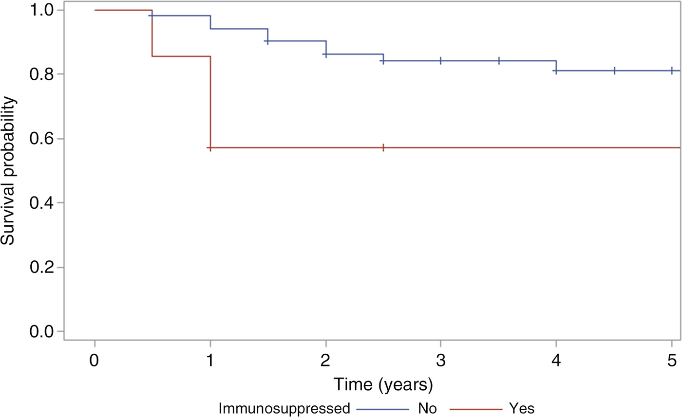
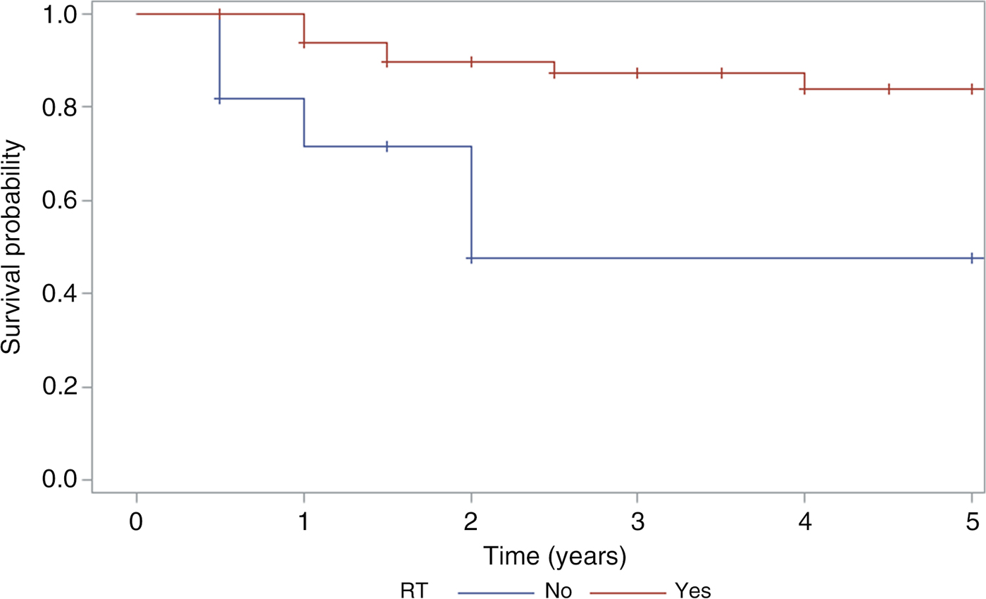
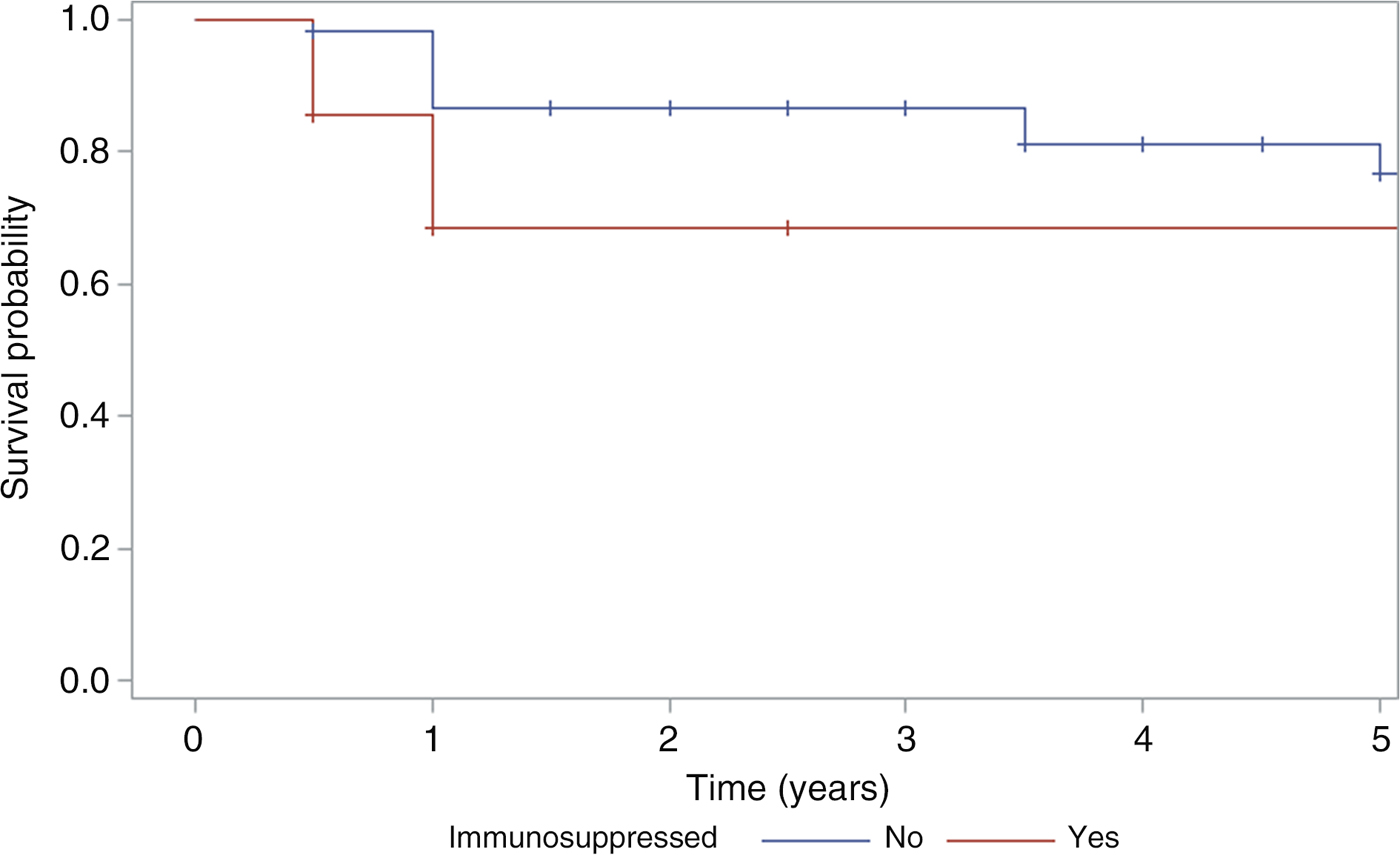
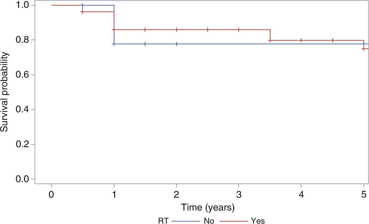
Immune state and adjuvant radiotherapy were significant prognostic factors for disease-specific survival. There was a statistically significant decrease in five-year disease-specific survival in immunosuppressed patients compared to immunocompetent patients (57 per cent vs 81 per cent; p = 0.039, Cox proportional hazard model) and in patients who did not receive post-operative radiotherapy (48 per cent vs 84 per cent; p = 0.0076, Cox proportional hazard model) (Figures 3 and 4). Age, gender, presence of pre-operative facial nerve weakness, facial nerve resection and type of surgical resection were not statistically significant prognostic factors for disease-specific survival.
Kaplan–Meier curve comparing disease-specific survival in immunosuppressed and non-immunosuppressed patients. ‘+’ = censored

Kaplan–Meier curve comparing disease-specific survival in patients who received adjuvant radiotherapy (RT) with those who did not. ‘+’ = censored

There was slightly poorer locoregional control in immunocompromised patients (69 per cent vs 77 per cent at five years), but this did not reach statistical significance (p = 0.25). Neither radiotherapy nor any of the other variables were significant prognostic factors for locoregional recurrence (Figures 5 and 6).
Kaplan–Meier curve comparing locoregional control in immunosuppressed and non-immunosuppressed patients. ‘+’ = censored

Kaplan–Meier curve comparing locoregional control in patients who received adjuvant radiotherapy (RT) with those who did not. ‘+’ = censored

Discussion
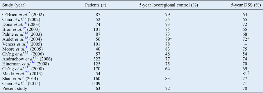
Our five-year disease-specific survival rate of 78 per cent and locoregional control rate of 72 per cent compare favourably with other published results. Previous series have shown five-year disease-specific survival rates ranging from 54 per cent to 77 per cent, and five-year locoregional rates between 48 per cent and 83 per cent (Table I).Reference O'Brien, McNeil, McMahon, Pathak, Lauer and Jackson 2 , Reference Veness, Morgan, Palme and Gebski 4 – Reference Shao, Wong, McIvor, Mylnarek, Chaplin and Izzard 6 , Reference Chen, Roman, Sosa and Judson 10 , Reference Ch'ng, Maitra, Lea, Brasch and Tan 12 , Reference Palme, O'Brien, Veness, McNeil, Bron and Morgan 13 , Reference Audet, Palme, Gullane, Gilbert, Brown and Irish 15 – Reference Makki, Mendez, Taylor, Trites, Bullock and Flowerdew 22
Locoregional control and disease-specific survival rates for metastatic cutaneous scc of parotid gland

*Three-year locoregional control and disease-specific survival rates. †Studies include patients with metastatic cutaneous squamous cell carcinoma to cervical nodes alone. ‡Two-year disease-specific survival rate. SCC = squamous cell carcinoma; DSS = disease-specific survival
In this study, adjuvant radiotherapy and immune status were the only factors that had a significant association with disease-specific survival. None of the variables analysed had a significant association with locoregional control.
Immune status has been identified as a prognostic factor in metastatic cutaneous SCC of the parotid in other studies, with survival and recurrence rates comparable to this series.Reference Shao, Wong, McIvor, Mylnarek, Chaplin and Izzard 6 , Reference Khurana, Mentis, O'Brien, Hurst, Stevens and Packham 11 , Reference Ch'ng, Maitra, Lea, Brasch and Tan 12 , Reference Oddone, Morgan, Palme, Perera, Shannon and Wong 14 , Reference Southwell, Chaplin, Eisenberg, McIvor and Morton 23 In a recent study, Shao et al. found that immune status was the only factor significantly associated with survival or locoregional control in their series of 160 patients undergoing surgery for metastatic cutaneous SCC to the parotid.Reference Shao, Wong, McIvor, Mylnarek, Chaplin and Izzard 6 In our study, immune status did not alter locoregional control, only disease-specific survival. Although conclusions are limited by the small number of patients, the fact that there was no significant difference in locoregional control rates between immunocompromised and immunocompetent patients (69 per cent and 77 per cent at five years) may reflect the more extensive surgery in the immunocompromised group (four of the seven patients underwent either lateral temporal bone resection or parotidectomy with facial nerve resection).
The benefit of post-operative radiotherapy is generally agreed on in the literature, with several studies demonstrating an improvement in survival and locoregional control.Reference Veness, Morgan, Palme and Gebski 4 , Reference Oddone, Morgan, Palme, Perera, Shannon and Wong 14 , Reference Hinerman, Indelicato, Amdur, Morris, Werning and Vaysberg 16 , Reference Ch'ng, Maitra, Allison, Chaplin, Gregor and Lea 21 However, in the largest published analysis of metastatic cutaneous SCC of the parotid, in 1509 surgically treated patients, Chen et al. found similar disease-specific survival rates for patients treated with surgery alone (72.4 per cent at five years) and surgery with adjuvant radiotherapy (70.6 per cent at five years).Reference Chen, Roman, Sosa and Judson 10 Similarly, other studies have not shown increased survival with adjuvant radiotherapy.Reference Shao, Wong, McIvor, Mylnarek, Chaplin and Izzard 6 , Reference Sweeny, Zimmerman, Carroll, Schmalbach, Day and Rosenthal 24 , Reference Ying, Johnson and Myers 25 This is most likely because it is the patients with more high-risk disease who receive radiotherapy, and the radiation mitigates the poorer prognosis of this advanced disease. This study shows a significant reduction in disease-specific survival without radiation, which may reflect the fact that half of the patients who did not receive radiotherapy (6 out of 12) had been recommended radiation therapy but declined. Four of these six patients developed locoregional or distant recurrence and died of their disease. The patients who had favourable histopathological features and were therefore not offered radiotherapy (4 out of 12) did not develop any recurrences during the follow-up period, and none died of metastatic cutaneous SCC.
Some studies have shown pre-operative facial nerve involvement to be a predictor of poor prognosis.Reference Audet, Palme, Gullane, Gilbert, Brown and Irish 15 This was not the case in our study: patients with pre-operative facial nerve weakness had similar five-year disease-specific survival and locoregional control rates to patients with no clinical evidence of involvement (disease-specific survival, 71 per cent vs 79 per cent; locoregional control, 71 per cent vs 76 per cent). This may be because five of the eight patients with evidence of facial nerve involvement underwent lateral temporal bone resection, and that more extensive surgery helps to control more advanced disease.
The extent of surgery (superficial or total parotidectomy with facial nerve preservation vs superficial or total parotidectomy with facial nerve sacrifice vs lateral temporal bone resection) was not associated with a significant difference in survival or locoregional control. Relatively few studies mention lateral temporal bone resections being performed as part of management for metastatic SCC of the parotid.Reference Shao, Wong, McIvor, Mylnarek, Chaplin and Izzard 6 – Reference O'Connor, Behan, Toner, Kinsella, Beausang and Timon 9 Research by Shao et al. reported on the largest number of lateral temporal bone resections: 44 patients (27 per cent of cases in their series).Reference Shao, Wong, McIvor, Mylnarek, Chaplin and Izzard 6 Similar to our findings, these authors did not find extent of surgery to have an impact on disease-specific survival or locoregional control. They hypothesised that this may have been because it was the patients with more extensive disease who had the more extensive surgery, with more radial resection mitigating the poorer prognosis associated with advanced disease. Our results, albeit with fewer lateral temporal bone resection cases, support this.
-
• Surgical resection with adjuvant radiotherapy is recommended for metastatic cutaneous squamous cell carcinoma of the parotid, but extent of surgery is debated
-
• Previously identified prognostic factors include: age, immune status, radiotherapy, facial nerve involvement, tumour size and surgical margins
-
• In this study, only immune status and adjuvant radiotherapy were significantly associated with disease-specific survival
-
• Our five-year disease-specific survival and locoregional control rates of 78 and 72 per cent compare favourably to previous studies
-
• More extensive surgery (e.g. lateral temporal bone resection) may improve local control in more advanced disease
This study has several limitations. The small number of total patients and patients in further subgroup analyses limits the conclusions that can be drawn from our results. As with any retrospective review of clinical data, treatment selection bias is difficult to determine. Several factors that have been implicated as prognostic indicators in previous studies were not included in our analysis. Some studies have shown neck node status to be an important predictor of survival,Reference Chen, Roman, Sosa and Judson 10 , Reference Makki, Mendez, Taylor, Trites, Bullock and Flowerdew 22 , Reference Sweeny, Zimmerman, Carroll, Schmalbach, Day and Rosenthal 24 but cervical node involvement and extent of neck dissection were not examined. We did not investigate the prognostic value of the parotid ‘P’ stage as proposed by O'Brien et al.,Reference O'Brien, McNeil, McMahon, Pathak, Lauer and Jackson 2 which advocates separating parotid (P) from neck (N) nodal metastases. Histological features of the parotid tumour such as size, margins and extra-nodal extension, which have been suggested as prognostic factors,Reference Chen, Roman, Sosa and Judson 10 , Reference Oddone, Morgan, Palme, Perera, Shannon and Wong 14 , Reference Audet, Palme, Gullane, Gilbert, Brown and Irish 15 were also not analysed.
Conclusion
The outcomes at our tertiary hospital compare favourably with those in the literature. Our data support the established role of adjuvant radiotherapy in the management of metastatic cutaneous SCC of the parotid gland. Immunosuppression is associated with poorer outcomes, and this must be taken into consideration when planning management of disease in this group of patients. In patients with more advanced disease, more radical surgical resection, such as lateral temporal bone resection, may have an important role in helping to reduce local recurrence.
Acknowledgement
The authors thank Ma Yi, biostatistician at Canterbury District Health Board, for her help with statistical analysis.