Introduction
The standardized extract EGb 761® from the leaves of Ginkgo biloba contains 24% flavonol glycosides, 6% terpene trilactones and other substances including proanthocyanidins and organic acids (DeFeudis and Drieu, Reference DeFeudis and Drieu2000; DeFeudis, Reference DeFeudis2003; Chan et al., Reference Chan, Xia and Fu2007).
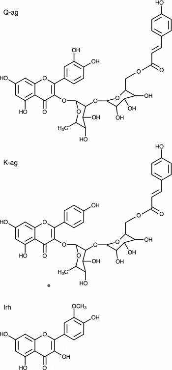
The major flavonol constituents of Ginkgo biloba are essentially flavonol-O-glycosides of quercetin, kaempferol and isorhamnetin forming mono-, di- and triglycosides with D-glucose and L-rhamnose (Hasler, Reference Hasler and van Beek2000; Bedir et al., Reference Bedir2002). Besides differences in their basic structure and the glycosylation pattern, flavonoids exhibit an enormous diversity due to glycosylation, methylation and acylation. In particular, acylation has been shown to significantly modify physico-chemical and biological properties of these substances. Acylation of flavonoids with malonic, coumaric, caffeic or ferulic acid is common, especially among anthocyanins, but is less frequent for flavonols (Viskupicova et al., Reference Viskupicova, Ondrejovic and Sturdik2009). The acylated flavonol glycosides in Ginkgo biloba consist of di- and triglycosides which are esterified with coumaric acid. They are substitued with a diglycoside unit (biloside) at C3. Acylated quercetin- and kaempferol-3-O-bilosides are specific constituents of Ginkgo leaves (Hasler, Reference Hasler1993). The two acylated flavonol glycosides, 3-O-(2’’-O-(6’’’-O-(p-hydroxy-trans-cinnamoyl)-β-D-glucosyl)-α-L-rhamnosyl)quercetin (Q-ag) and 3-O-(2’’-O-(6’’’-O-(p-hydroxy-trans-cinnamoyl)-β-D-glucosyl)-α-L-rhamnosyl)kaempferol (K-ag) (Figure 3), represent about 4.5% of EGb 761® content (Dr. F. Lang, Dr. Willmar Schwabe GmbH & Co. KG, personal communication).
The terpene trilactone constituents include ginkgolides A, B, C and J, which together account for 3.1%, and bilobalide, which comprises 2.9% of the total EGb 761® extract. The Ginkgo biloba extract and its numerous individual constituents have, among other activities, shown to protect and improve mitochondrial energy production, to ameliorate microcirculation, and to possess antioxidant, radical scavenging and neuroprotective properties (Wu and Zhu, 1999; DeFeudis and Drieu, Reference DeFeudis and Drieu2000; Ahlemeyer and Krieglstein, Reference Ahlemeyer and Krieglstein2003; Ahmad et al., Reference Ahmad2005; Abdel-Kader et al., Reference Abdel-Kader2007; Rojas et al., Reference Rojas, Serrano-García, Mares-Sámano, Medina-Campos, Pedraza-Chaverri and Ögren2008).
Experimental and clinical data suggest that EGb 761® exerts beneficial effects as a cognitive enhancer and in the therapy of age-related neurological disorders including Alzheimer's disease (AD) (for review, see DeFeudis and Drieu, Reference DeFeudis and Drieu2000; DeFeudis, Reference DeFeudis2003; Weinmann et al., Reference Weinmann, Roll, Schwarzbach, Vauth and Willich2010). In animal experiments, Ginkgo biloba extract was shown to improve spatial memory deficits in a transgenic mouse model of AD (Stackman et al., Reference Stackman, Eckenstein, Frei, Kulhanek, Nowlin and Quinn2003), as well as to improve acquisition of working memory in young (Satvat and Mallet, 2008) and aged rats (Wang et al., Reference Wang, Wang, Wu and Cai2006; Blecharz-Klin et al., Reference Blecharz-Klin, Piechal, Joniec, Pyrzanowska and Widy-Tyszkiewicz2009). However, the precise neurochemical correlates of these behavioral effects of Ginkgo biloba extract are not completely understood. A number of clinical studies have demonstrated that Ginkgo biloba, and particularly EGb 761®, ameliorate cognitive deficits associated with mild to moderate AD and vascular dementia (Wang et al., Reference Wang2010; Weinmann et al., Reference Weinmann, Roll, Schwarzbach, Vauth and Willich2010) and improve memory function in patients with very mild cognitive impairment (Grass-Kapanke et al., Reference Grass-Kapanke, Busmane, Lasmanis, Hoerr and Kaschel2011) as well as in middle-aged healthy volunteers (Kaschel, Reference Kaschel2011). Based on the evidence from randomized and controlled clinical trials, the German Institute for Quality and Efficacy in Health Care (IQWiG) concluded that EGb 761® is beneficial in AD (IQWiG, 2008). Two large-scale prospective cohort studies have suggested that long-term intake of EGb 761® might protect against development of dementia (Andrieu et al., Reference Andrieu2003; Dartigues et al., Reference Dartigues, Carcaillon, Helmer, Lechevallier, Lafuma and Khoshnood2007). A randomized placebo controlled pilot study highlighted that delay of conversion to dementia can only be expected with good compliance in long-term treatment (Dodge et al., Reference Dodge, Zitzelberger, Oken, Howieson and Kaye2008).The Ginkgo Evaluation of Memory (GEM) study was carried out to test whether Ginkgo biloba is effective in prevention of dementia (and especially AD) in elderly individuals with normal cognition or those with mild cognitive impairment (DeKosky et al., Reference DeKosky2008). The interpretation of this randomized, double-blind, placebo controlled clinical trial of 3069 participants aged 72–96 years is limited by the very poor treatment compliance of this tertiary care center trial (Jerant et al., Reference Jerant, Chapman, Duberstein, Robbins and Franks2011). A primary care based long-lasting clinical study on the use of EGb 761® for secondary prevention of AD involving participants without dementia aged 70 years or older who had spontaneously complained of memory problems (GuidAge) has been performed in Europe (Andrieu et al., Reference Andrieu2008). During the study 4.3% of the patients in the EGb 761® group developed AD compared to 5.2% in the placebo group. Because the dementia rate in this trial (4.8%) was much lower than expected for sample size planning (13.8%), this trend did not reach statistical significance. However, a clear difference was observed in a preplanned analysis of subjects treated for at least four years as only 1.6% of the EGb 761® treated participants converted to AD in comparison to 3.0% in the placebo group (Vellas et al., Reference Vellas2010).
Besides the proposed anti-dementia and pro-cognitive effects of Ginkgo biloba, a growing body of clinical evidence exists for the efficacy of EGb 761® in the treatment of neuropsychiatric symptoms of dementia (Bachinskaya et al., Reference Bachinskaya, Hoerr and Ihl2011; Ihl et al., Reference Ihl2011). Thus, EGb 761® at a daily dose of 240 mg effectively alleviated behavioral and neuropsychiatric symptoms including apathy/indifference, sleep/night-time behavior, irritability/lability, depression/dysphoria, and aberrant motor behavior in patients with mild to moderate dementia (Bachinskaya et al., Reference Bachinskaya, Hoerr and Ihl2011). A review and meta-analysis of the usage of Ginkgo extract as an adjunct therapy in chronic schizophrenia showed that its use as an add-on therapy to antipsychotic medication produced a significant improvement in total and negative symptoms of chronic schizophrenia (Singh et al., Reference Singh, Singh and Chan2010). In addition, EGb 761® was effective in reducing the symptoms of tardive dyskinesia in schizophrenia patients (Zhang et al., Reference Zhang, Tan, Zhang, Chan, Wu and Zhou2011).
The clinical studies demonstrating the efficacy of EGb 761® in alleviating neuropsychiatric symptoms in dementia, as well as the negative and cognitive symptoms in schizophrenia, suggest that the extract contains one or more active constituents, which may exert direct modulatory actions on central neurotransmitter systems. Of those, the most interesting candidates are the cholinergic and monoaminergic projections to the prefrontal cortex and hippocampus, the neuroanatomical structures strongly implicated in cognitive function and affective behavior. Indeed, cholinergic replacement therapy using the cholinesterase inhibitors (donepezil, rivastigmine, or galantamine) is currently the prevailing strategy for treatment of mild to moderate AD (O'Brien and Burns, Reference O'Brien and Burns2011). The new (second and third) generations of antipsychotic drugs aim to reduce the negative symptoms and ameliorate cognitive deficits in schizophrenia preferentially by increasing the cholinergic (Terry, Reference Terry2008) and dopaminergic transmission (Meltzer and Huang, Reference Meltzer and Huang2008; Kim et al., Reference Kim, Maneen and Stahl2009) in the frontal cortical structures. In view of these facts, it is hypothesized that the systemic administration of EGb 761® may modulate the levels of dopamine (DA) and acetylcholine (ACh) neurotransmitters in the prefrontal cortex. In order to investigate this hypothesis, the microdialysis technique, which allows monitoring of extracellular levels of neurotransmitters including DA, ACh, as well as noradrenaline (NA), serotonin (5-HT) and the metabolites 3,4-dihydroxyphenylacetic acid (DOPAC), homovanillic acid (HVA) and 5-hydroxyindoleacetic acid (5-HIAA) was applied in awake rats.
Effects of acute and chronic treatment with EGb 761® on monoamines and their metabolites in the rat brain
In an initial study, the effects of acute and subacute (14 days, once daily) treatment with EGb 761® on extracellular levels of the monoamines DA, NA and 5‑HT and their acidic metabolites DOPAC, HVA and 5-HIAA in the medial prefrontal cortex of awake rats were examined. A single oral administration of EGb 761® (100 mg/kg) had no effect on basal extracellular levels of monoamines and their metabolites (Yoshitake et al., Reference Yoshitake, Yoshitake and Kehr2010).
This finding is in disagreement with some earlier in vitro and ex vivo whole tissue studies, which have suggested that Gingko biloba extract reduces the activity of both MAO A and MAO B in the rat (White et al., Reference White, Scates and Cooper1996) and mouse brain (Wu and Zhu, 1999; Pardon et al., Reference Pardon, Joubert, Perez-Diaz, Christen, Launay and Cohen-Salmon2000). On the other hand, a number of microdialysis studies, including our earlier data (Yoshitake et al., Reference Yoshitake, Kehr, Yoshitake, Fujino, Nohta and Yamaguchi2004a), have demonstrated that the MAO inhibitors increase extracellular levels of all three monoamines and decrease the levels of their respective metabolites even after a single acute administration. These findings are also in agreement with the report by Shah and co-workers who found that an acute dose of Gingko biloba extract had no effect on brain tissue levels of monoamines and reduced the stress-induced increases in whole brain levels of catecholamines and 5-HT in the rat (Shah et al., Reference Shah, Sharma and Vohora2003).
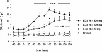
However, strikingly different effects were observed following subacute daily treatment with EGb 761® for 14 days. Here, the EGb 761® challenge at the doses of 100 and 300 mg/kg p.o. caused a marked elevation in extracellular DA levels within 40–180 minutes. The lower dose of EGb 761® caused a maximal increase of the DA levels to 185 ± 10% (mean ± SEM, p < 0.001) of the corresponding value of the control group at 160 minutes, whereas the dose of 300 mg/kg EGb 761® increased the DA levels to 234 ± 6% (p < 0.001) at 100 minutes after drug administration (Figure 1). The low dose (30 mg/kg) of EGb 761® had no effect on extracellular DA levels. In an earlier study (Yoshitake et al., Reference Yoshitake, Yoshitake and Kehr2010), it was shown that subacute treatment with EGb 761® had only a minor effect on the NA levels, while the levels of 5-HT were not affected. Interestingly, in spite of increased DA levels following the chronic treatment with EGb 761®, the concentrations of metabolites DOPAC and HVA were unchanged at any dose of EGb 761® (Yoshitake et al., Reference Yoshitake, Yoshitake and Kehr2010). In the rat striatum, the subacute treatment with EGb 761® at the highest dose caused only a minor increase in the DA levels to 125 ± 4% of the vehicle-treated group at 120 minutes after the EGb 761® administration. This finding is in agreement with behavioral recordings where no significant increase in locomotor activity was recorded. The EGb 761® doses of 100 and 300 mg/kg used in the study are considered to be relevant to the currently recommended clinical doses of 240 mg/day (Schwabe, Tebonin® konzent, Summary of Product Characteristics, European Medicines Agency, 2011).
Effects of subacute (14 days, once daily) treatment with EGb 761® (30, 100 and 300 mg/kg p.o.) on extracellular concentrations of dopamine in the mPFC of awake rat. The arrow indicates the time of EGb 761® administration on day 14, the levels are expressed as mean ± SEM, n = 5; (***) p < 0.001; (*) p < 0.05; two-way repeated measures ANOVA followed by Bonferroni multiple comparison test.

Effects of EGb 761® main constituents on extracellular levels of DA: a major role of the flavonol glycosides
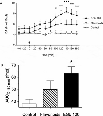
The following studies aimed to evaluate a potential role of the major constituents of EGb 761® on the observed effects of increased DA transmission in the rat mPFC. Three main constituent groups of the extract were tested: flavonol glycosides, ginkgolides, and bilobalide using the same dosing regimen as used for the whole extract. In separate groups of rats, each constituent was administered orally, once daily for 14 days at a dose corresponding to its respective content in 300 mg/kg of EGb 761®: flavonol glycosides 72 mg/kg, ginkgolides 8.7 mg/kg and bilobalide 9.3 mg/kg. Subacute treatment with flavonol glycosides caused a moderate increase in extracellular DA levels to a maximal value of 134 ± 14% at 120 min, as compared to the vehicle-treated group (Figure 2). Correspondingly, repeated administration of ginkgolides or bilobalide had only a minor or no effect on extracellular levels of DA (Yoshitake et al., Reference Yoshitake, Yoshitake and Kehr2010). The relative AUC(0–180 min) value calculated as the sum of the individual AUC(0–180 min) increments of all three constituents was 140% of the vehicle-treated group, which was similar to the value of 153% observed for the treatment with 300 mg/kg of EGb 761® (Yoshitake et al., Reference Yoshitake, Yoshitake and Kehr2010). Previously, it was proposed that EGb 761® may exert synergistic effects or a “polyvalent” action (DeFeudis and Drieu, Reference DeFeudis and Drieu2000), i.e. a potentiation of the effects caused by the multiple active constituents of the EGb 761®, a mechanism of action often proposed for herbal remedies. However, the present findings indicate that the effects of EGb 761® on the DA levels in the mPFC are more likely only additive and not synergistic, reflecting the sum of the individual contributions of the main groups of constituents.
(A) Effects of subacute (14 days, once daily) treatment with EGb 761® (300 mg/kg, p.o.) extract and its flavonoid fraction (72 mg/kg, p.o.) on the extracellular concentrations of dopamine in the medial prefrontal cortex of awake rats. The arrow indicates the time of drug administration on day 14, the levels are expressed as mean ± SEM, n = 5; (***) p < 0.001; (**) P < 0.01; (*) p < 0.05; two-way repeated measures ANOVA followed by Bonferroni multiple comparison test. (B) The inset shows the overall effect of EGb 761® and flavonoids expressed as area-under-the-curve (AUC(0–180 min)) values for DA concentrations measured during the 180-min period after the treatment.

Effects of EGb 761®-specific acylated flavonols on extracellular levels of DA and ACh in the rat mPFC
Ginkgo biloba leaves contain at least two acylated glycosides of the aglycones quercetin and kaempferol: 3-O-(2’’-O-(6’’’-O-(p-hydroxy-trans-cinnamoyl)-β-D-glucosyl)-α-L-rhamnosyl)querce-tin (Q-ag) and 3-O-(2’’-O-(6’’’-O-(p-hydroxy-trans-cinnamoyl)-β-D-glucosyl)-α-L-rhamnosyl)kaempferol (K-ag). The chemical structures of Q-ag and K-ag are shown in Figure 3. Both Q-ag and K-ag are specific flavonols of Ginkgo biloba, as already described in the introduction section. The standardized extract EGb 761® contain typically 2.4–2.6% Q-ag and 1.8–2.2% K-ag (Dr. F. Lang, Dr. Willmar Schwabe GmbH & Co. KG, personal communication).
Chemical structures of acylated glycosides of quercetin, 3-O-(2’’-O-(6’’’-O-(p-hydroxy-trans-cinnamoyl)-β-D-glucosyl)-α-L-rhamnosyl)quercetin (Q-ag) and kaempferol, 3-O-(2’’-O-(6’’’-O-(p-hydroxy-trans-cinnamoyl)-β-D-glucosyl)-α-L-rhamnosyl)kaempferol (K-ag), as well as isorhamnetin (Irh).

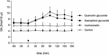
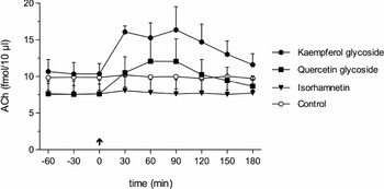
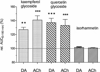
In the final experiments, the effects of chronic treatment with Q-ag, K-ag, and isorhamnetin (Irh) on extracellular levels of DA and ACh were evaluated in the mPFC of freely moving rats. Isorhamnetin, the O-methylated quercetin (Figure 3), was used as a reference substance. Q-ag, K-ag, and Irh were each administered at a dose of 10 mg/kg/day for 14 days and the microdialysis experiments were performed following Q-ag, K-ag or Irh application on the last day of treatment. At this point, the levels of DA increased within the first 30–60 minutes following treatment with Q-ag and K-ag, but not with Irh, as shown in Figure 4. The maximal increases to 181 ± 16% and 160 ± 14% (mean ± SEM, p < 0.001) for Q-ag-treated and K-ag-treated rats, respectively, were observed at 90 minutes after the drug administration (Figure 4). The levels remained markedly increased even at the end (180 min) of the sampling period. Similar effects of Q-ag and K-ag treatment were observed for the extracellular ACh levels (Figure 5). Thus, Q-ag increased the ACh levels to a maximal value of 121 ± 25% of controls at 90 minutes and K-ag caused a rapid increase in ACh values to 158 ± 5% at 30 minutes after its administration. The overall effects of Q-ag, K-ag, and Irh expressed as relative AUC(0–180 min) values calculated from the respective time courses (Figures 4 and 5) are depicted in Figure 6. As can be seen, Q-ag and K-ag increased the DA and ACh levels to about the same levels, whereas Irh had no effect on any of the neurotransmitters. Similarly, as in the case of the whole extract, the chronic treatment with the flavonols Q-ag, K-ag, and Irh did not affect the extracellular levels of the DA metabolites DOPAC and HVA.
Effects of treatment with 10 mg/kg/day of quercetin glycoside (Q-ag), kaempferol glycoside (K-ag) and isorhamnetin (Irh) for 14 days on the extracellular concentrations of DA in the mPFC of awake rats. The arrow indicates the time of drug administration on day 14, the levels are expressed as mean ± SEM, n = 5.

Effects of treatment with 10 mg/kg/day of quercetin glycoside (Q-ag), kaempferol glycoside (K-ag) and isorhamnetin (Irh) for 14 days on the extracellular concentrations of ACh in the mPFC of awake rats. The arrow indicates the time of drug administration on day 14, the levels are expressed as mean ± SEM, n = 5.

The overall effects of each 10 mg/kg/day of quercetin glycoside (Q-ag), kaempferol glycoside (K-ag) and isorhamnetin (Irh) for 14 days on the extracellular levels of DA and ACh in the mPFC of awake rats. The relative AUC(0–180 min) values are expressed as percentage of the AUC(0–180 min) value of the control group; mean ± SEM, n = 5; (***) p < 0.001; (**) p < 0.01; one-way ANOVA followed by Dunnett's multiple comparison test.

Summary
A major finding of the study is that the subacute treatment with EGb 761® or its main constituent group the flavonols, as well as the individual acylated flavonol glycosides Q-ag and K-ag, increase the DA and ACh transmission in the mPFC of awake rats. Ginkgolides and bilobalide had only a minor or no effect on the extracellular levels of the monoaminergic neurotransmitters. These results suggest that the flavonol constituents and, in particular, the Ginkgo-specific acylated quercetin and kaempferol glycosides account for the direct modulatory effects of EGb 761® on the central neurotransmitters DA and possibly ACh in brain areas, which are neuroanatomically relevant to learning and memory processing.
Chronic treatment with EGb 761® had no effect on the activities of MAO-A or MAO-B in homogenates of mouse brains (Fehske et al., Reference Fehske, Leuner and Müller2009), which is also supported by the findings of the present microdialysis study showing unchanged extracellular levels of DOPAC and HVA in rat brains. One possible explanation of the effects exerted by the subacute treatment with EGb 761® and flavonol glycosides could involve desensitization or downregulation of receptors modulating the DA, NA, and ACh release in prefrontal cortical structures. Only limited data are available on the binding affinity of EGb 761® and its main constituents to central neurotransmitter receptors. Thus, the Ginkgo biloba extract reduced [3H]ketanserin binding to 5-HT2A receptors in the frontal cortex of MAO-A knock-out mice (Shin et al., Reference Shin, Chen, Ridd and Seif2000). Chronic treatment with EGb 761® increased binding (Bmax values) to muscarinic acetylcholine receptors (Taylor, Reference Taylor1986) and α2‑adrenoceptors (Huguet and Tarade, Reference Huguet and Tarrade1992) in hippocampal membranes and 5-HT1A receptors (Huguet et al., Reference Huguet, Drieu and Piriou1994) in cortical membranes of aged but not of young rats. Administration of EGb 761® to adult rats increased NA turnover in the cerebral cortex only after subacute treatment (Brunello et al., Reference Brunello, Racagni, Clostre, Drieu and Braquet1985). In addition, it was shown that chronic but not acute treatment with sertraline caused an increase in the NA levels in the frontal cortex but not in the hippocampus of rats, most likely as a consequence of desensitization of cortical α2‑adrenoceptors (Thomas et al., Reference Thomas, Nutt and Holman1998). Blockade of α2-adrenoceptors with the α2‑antagonist idazoxan was shown preferentially to increase extracellular DA levels in the PFC (Hertel et al., Reference Hertel, Nomikos and Svensson1999), to potentiate the effects of venlafaxine on DA and NA levels (Weikop et al., Reference Weikop, Kehr and Scheel-Krüger2004), and to raise cortical ACh levels (Tellez et al., Reference Tellez, Colpaert and Marien1999). Modulation of other receptors such as 5-HT or muscarinic acetylcholine receptors by chronic treatment with EGb 761® or its flavonol constituents may also account for the observed increases in extracellular DA and ACh levels in the rat mPFC.
Cognitive decline is associated with natural processes of brain aging, but is pathologically accelerated in neurodegenerative diseases including AD or vascular type dementia and psychiatric disorders, such as schizophrenia or depression. Progressive impairment of learning and memory functions in AD is primarily attributed to the atrophy and loss of the cholinergic neurons in the basal forebrain (Bartus et al., Reference Bartus, Dean, Beer and Lippa1982; Coyle et al., Reference Coyle, Price and DeLong1983; Terry and Buccafusco, Reference Terry and Buccafusco2003) and cholinergic replacement therapy is currently the prevailing strategy for treatment of mild to moderate AD (O'Brien and Burns, Reference O'Brien and Burns2011). In addition, a body of evidence exists for the role of prefrontal cortical DA and DA receptors in modulating neuronal networks, which are essential for working memory (Castner and Goldman-Rakic, Reference Castner and Goldman-Rakic2004; Goldman-Rakic, Reference Goldman-Rakic1995; Goldman-Rakic et al., Reference Goldman-Rakic, Muly and Williams2000; Paspalas and Goldman-Rakic, Reference Paspalas and Goldman-Rakic2005).
Taken together, these reports support the findings of the current study on a putative mechanism of action of EGb 761®. The present data demonstrate for the first time that repeated administration of the Ginkgo biloba extract EGb 761®, and in particular, its two specific acylated flavonol glycosides, results in increased release of DA and ACh in the rat prefrontal cortex. Increased dopaminergic and cholinergic transmission in the frontocortical brain areas may be one of the underlying mechanisms behind the clinically observed effects of EGb 761® on improved cognitive function in aged and healthy people, as well as in the treatment of neuropsychiatric symptoms of dementia.
Conflict of interest
S. Yoshitake and J. Kehr are employed by Pronexus Analytical AB, and E. Koch and M. Nöldner are employees of the company Dr. Willmar Schwabe GmbH & Co. KG. The study was supported by Dr. Willmar-Schwabe GmbH & Co. KG, Karlsruhe, Germany.
Description of authors' roles
E. Koch, M. Nöldner, and J. Kehr designed the study and participated in the discussion of the results; S. Ijiri and S. Yoshitake performed the microdialysis experiments and, together with T. Yoshitake, the analysis of microdialysis samples; T. Yoshitake and J. Kehr carried out the data processing and statistical analysis; J. Kehr wrote the paper.