INTRODUCTION
The genus Oncholaimus Dujardin, 1845 is a large genus with approximately 100 species described so far (Gerlach & Riemann, Reference Gerlach and Riemann1974; Huang & Zhang, Reference Huang and Zhang2006). Eleven species have been recorded in the New Zealand region by Allgen (Reference Allgen1928, Reference Allgen1932) and Ditlevsen (Reference Ditlevsen1921, Reference Ditlevsen1930). Three of these species (O. cylindricaudatus Allgen, Reference Allgen1932, O. tenuicaudatus Allgen, Reference Allgen1932 and O. conicaudatus Allgen, Reference Allgen1928) cannot be considered valid because their descriptions were based on only one juvenile specimen. Three other species (O. microdon Ditlevsen, Reference Ditlevsen1930, O. bollonsi Ditlevsen, Reference Ditlevsen1930 and O. incurvatus Ditlevsen, Reference Ditlevsen1930) were found to be indistinguishable from other Oncholaimus species already recorded in the region (Allgen, Reference Allgen and Odhner1959). This leaves five valid species: O. aegypticus Steiner, 1921, O. brachycercus De Man, 1889, O. chiltoni Ditlevsen, Reference Ditlevsen1930, O. dujardini De Man, 1878 and O. viridis Bastian, 1865. One new species was found during a study of meiofaunal trophic dynamics in Papanui Inlet, southern New Zealand.
In a review of marine nematode feeding ecology, Moens & Vincx (Reference Moens and Vincx1997) described oncholaimid nematodes as facultative predators. It appears that oncholaimids, though capable of feeding on other nematodes, obtain the majority of their nutrition from other sources such as dissolved organic matter and detritus (Moens et al., Reference Moens, Verbeeck and Vincx1999). Stable isotope data from the field, however, suggested little difference between the trophic level of Enoploides longispiculosus (a supposedly strict predator) and Adoncholaimus fuscus (Moens et al., Reference Moens, Bouillon and Gallucci2005).
In recent years stable isotopes have helped improve our knowledge of nematode trophic dynamics (Carman & Fry, Reference Carman and Fry2002; Riera & Hubas, Reference Riera and Hubas2003; Moens et al., Reference Moens, Bouillon and Gallucci2005). Fatty acids have also been used to study the diet of invertebrates in marine systems (Meziane & Tsuchiya, Reference Meziane and Tsuchiya2000; Kharlamenko et al., Reference Kharlamenko, Kiyashko, Imbs and Vyshkvartzev2001). Although some information is available on the fatty acid composition of cultured nematodes (Abu Hatab & Gaugler, Reference Abu Hatab and Gaugler1999; Schlechtriem et al., Reference Schlechtriem, Tocher, Dick and Becker2004; Brock et al., Reference Brock, Browse and Watts2007), there are no data available on the fatty acid composition of marine nematodes. Oncholaimid nematodes lend themselves well to fatty acid analysis due to their large size and often high densities (e.g. Meyers et al., Reference Meyers, Hopper and Cefalu1970). This study combines stable isotope and fatty acid analysis to investigate the diet of Oncholaimus moanae sp. nov.
MATERIALS AND METHODS
Papanui Inlet is an unpolluted sheltered bay with an area of 3.5 km2. Tides are semidiurnal with a mean tidal range of 1.15 m (Albrecht & Vennell, Reference Albrecht and Vennell2007). Sampling was carried out in January 2007 in a 20 × 10 m plot (45º50′53.1″S 170º42′40.9″E) situated near the edge of a channel which drains a nearby Zostera muelleri meadow at low tide. Sediments consisted of well sorted fine sand with very low silt/clay and organic matter content (<1% and <0.5%, respectively).
Nematodes were obtained by scraping the top two centimetres of the sediment and extracted by decantation over a 125 µm sieve. Specimens for taxonomic study were fixed in 5% formalin and stained with Rose Bengal. Specimens for stable isotope and fatty acid analysis were kept frozen (−80°C) until analysis (see below). Descriptions were made from glycerin mounts (Riemann, Reference Riemann, Higgins and Thiel1988) using interference microscopy and drawings made with the aid of a camera lucida. All measurements are in µm and all curved structures were measured along the arc. Slides with type specimens of the new species are deposited in the National Nematode Collection of New Zealand (NNCNZ), Landcare Research New Zealand Ltd (Auckland) and in the Australian Helminthological Collection (AHC) of the South Australian Museum (Adelaide). Additional specimens are held in the reference collection of the Portobello Marine Laboratory. Abbreviations are as follows: a, body length/maximum body diameter; a.b.d., anal body diameter; b, body length/oesophagus length; c, body length/tail length; c.b.d, corresponding body diameter; V, vulva distance from anterior end of body; V%, V/total body length.
Samples for stable isotope and fatty acid analysis were obtained by picking Oncholaimus moanae sp. nov. individuals with a fine needle. Samples for both isotopic and fatty acid analysis weighed between 400 and 600 µgDW, and consisted of several hundred individuals each. Nematodes were transferred to a watch glass containing distilled water twice to remove impurities. Samples were centrifuged and excess water was removed. Samples for stable isotope analysis (N = 3) were dried at 60°C for 24 hours, transferred into aluminium cups, and kept in a dehydrator until analysis. It was decided not to freeze-dry the samples for fatty acid analysis because this method often led to loss of material during handling. Instead, samples for fatty acid analysis (N = 3) were kept frozen (−80°C) until analysis.
Isotopic analyses were done at Iso-trace NZ (Dunedin, New Zealand) on a Europa 20-20 Update stable isotope mass spectrometer (Europa scientific, Crewe, UK) interfaced to a Carlo Erba analyser (NA1500; Carlo Erba, Milan, Italy) in continuous flow mode (precision: 0.2‰). Analysis was calibrated to EDTA laboratory standard reference (Elemental Microanalysis, Cheshire, UK) and standardized against international standards (IAEACH-6 for carbon, IAEAN1 and IAEAN2 for nitrogen). Results are expressed relative to standards (Vienna PDB for carbon and atmospheric N for nitrogen) in standard delta notation according to the formula: δ13C or δ15N = [(RSAMPLE/RSTANDARD)−1] × 103‰, where R is the ratio of 13C/12C or 15N/14N.
Lipid extractions from nematode samples were carried out using a modified Bligh and Dyer method (Bligh & Dyer, Reference Bligh and Dyer1959). Lipids were extracted using chloroform/methanol (2:1 v/v). The lipid fraction was treated with sulphuric acid (AnalaR, BDH Chemicals, Poole, UK) in methanol solution and underwent acid-catalysed transesterification at 80°C for 12 hours. Fatty acid esters were extracted in hexane/water. The hexane-containing layer was removed, dried using N2 gas and reconstituted in 100 µl hexane. Fatty acid composition was determined by gas chromatography on a 5890 GC equipped with an HP7673 autosampler (Hewlett-Packard, Palo Alto, California, USA). Fatty acid methyl esters were separated on a DB225 capillary column, 30 m × 0.53 mm ID, 0.25 µm film (Agilent Technologies, Wilmington, Delaware, USA). The column oven temperature was ramped from 180–200°C over 50 minutes. Fatty acid peaks were identified by retention time matching with a composite standard made from commercially available methyl esters (NUCheck Prep, Elysian, Minnesota, USA, and Sigma, St Louis, Missouri, USA) and additional specific standards (Sigma-Aldrich: PUFA No. 1 and Supelco 37 component FAME mix). Peak areas were calculated and reported as percentage of total fatty acids.
Fatty acids were expressed as the ratio of the number of carbon atoms to the number of double bonds. Double bond position (n) is numbered from the methyl end of the fatty acid.
SYSTEMATICS
Oncholaimus moanae sp. nov. (A) Male head; (B) female head; (C) male anterior region; (D) demanian system; (E) male tail and reproductive structures; (F) female tail. Abbreviations: d.u., ductus uterinus; os, osmosium; d.e., ductus entericus; uv, uvette.

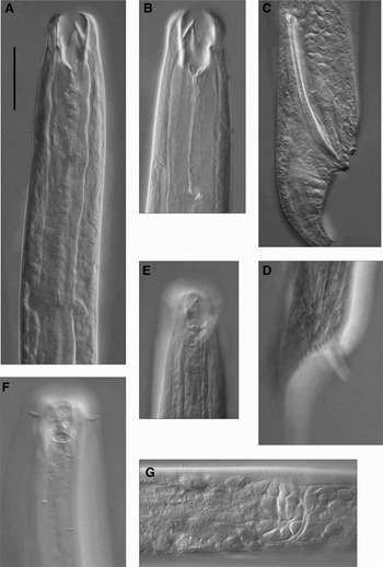
Oncholaimus moanae sp. nov. (A) Lateral view of male anterior region, showing buccal cavity and nerve ring; (B) lateral view of female mouth region, showing teeth and excretory pore; (C) lateral view of male reproductive structures, showing spicule and papillae; (D) lateral view of male circumcloacal setae; (E) lateral view of male amphid; (F) lateral view of female amphid, cephalic setae, and somatic setae; (G) lateral view of uvette, showing portion of ductus entericus and ductus uterinus. Scale bar = 30 µm.

TYPE MATERIAL
Holotype: adult male, formalin-fixed, mounted on slide in glycerin. Collected in January 2007 by D. Leduc from Papanui Inlet, Otago Peninsula, southern New Zealand (45º50′53.1″S 170º42′40.9″E). Upper two centimetres of intertidal sediments, sediment mean grain size = 163 µm, silt/clay content <1%, organic matter <0.5%, water content = 22%, deposited in the National Nematode Collection of New Zealand (NNCNZ 255).
Paratypes: one male and two females, same date and site (NNCNZ 2520-2). Two males and two females, same date and site, deposited in the Australian Helminthological Collection (AHC 29473-6)
ETYMOLOGY
The species is named after the author's daughter. Moana is also the Maori word for ocean.

DESCRIPTION
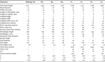
Males: body length 2043–2750 µm; maximum diameter 40–41 µm, with a smooth cuticle. Six rounded lips, each bearing a minute rounded labial papilla. Ten cephalic setae in one circle, six short (6 µm) and four slightly longer (7 µm). Head slightly constricted at level of amphids; head diameter 25–26 µm. Buccal cavity 30 µm deep with three teeth, left subventral tooth larger than right subventral and dorsal tooth. Amphids pocket-like with elliptical openings, 9–10 µm wide (0.3–0.4 c.b.d.), and 7 µm in height. Oesophagus muscular and cylindrical, 1/7–1/8 times body length. Excretory pore opening at about 0.2 times oesophagus length from anterior end, nerve ring at 0.45 times oesophagus length. A few sparse, short somatic setae situated anterior to the nerve ring. Tail conico-cylindrical (though cylindrical portion is very short), ventrally bent, 2.5–2.9 a.b.d., with two small terminal setae and several scattered caudal setae. One small caudal papilla located at about 1/3 of tail length with a pair of short setae. Tail tip slightly swollen and rounded.
Spicules equal, 73–78 µm (2.0 a.b.d.), slightly ventrally curved, with pointed tips and slightly cephalated proximal ends. One circle of circumcloacal setae, 11–12 pairs. One pre- and post-cloacal papilla. Pre-anal papilla bears four pairs of short, stout spines. Two outstretched testes leading to vesicula seminalis in mid-region of the body. Vas deferens set off from ductus ejaculatorius by a sphincter about 450 µm anterior of cloaca.
Females: very similar to males, but larger. Body length 2798–3288 µm, maximum body diameter 53–58 µm. Amphid somewhat smaller than in males, 8–9 µm wide and 5 µm in height. Tail not bent as in males, with longer cylindrical portion, no caudal setae. Single anterior ovary, reflexed. Up to 11 mature eggs (100–133 µm long) observed in gravid females. Vulva at 75% of body length. Demanian system connects to exterior through two lateral openings (seen as small round elevated regions on cuticle) at level of uvette. Unidentified amorphous material is often seen projecting out of the openings. Ductus entericus 141–178 µm long, ductus uterinus 402–434 µm.
DIAGNOSIS AND DISCUSSION
Oncholaimus moanae sp. nov. is characterized by the presence of long (>70 µm) spicules and a demanian system with two lateral openings at level of uvette. Oncholaimus moanae sp. nov. is most similar to O. appendiculatus Cobb 1930 in body length and shape, and male reproductive structures. Oncholaimus moanae sp. nov. differs from the latter species by the presence of spines on the precloacal papilla, the presence of a post-cloacal papilla, and the relative position of the nerve ring and of the excretory pore openings. Oncholaimus moanae sp. nov. differs from other members of the genus by the combination of the following traits: long (>70 µm) spicules, spine-bearing pre-cloacal papilla and demanian system with two lateral openings at level of uvette.
ISOTOPIC AND FATTY ACID COMPOSITION
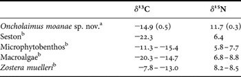
The mean δ13C and δ15N signatures of Oncholaimus moanae sp. nov. were −14.9 and 11.7‰, respectively (Table 2). A comparison with the δ13C signature of potential food sources indicates that microphytobenthos (which consisted mostly of dinoflagellates and diatoms at the time of sampling, personal observation) and macroalgae were the most likely food sources for O. moanae sp. nov. No macroalgae, however, were observed at the study location at the time of sampling. Seston can be ruled out as a major component of nematode diet due to its low δ13C signature (Table 2). Microphytobenthos, therefore, is the most likely carbon source for O. moanae sp. nov., which agrees with previous stable isotope studies on nematodes carried out in unvegetated intertidal sediments (Riera et al., Reference Riera, Richard, Gremare and Blanchard1996; Moens et al., Reference Moens, Luyten, Middelburg, Herman and Vincx2002, Reference Moens, Bouillon and Gallucci2005). The δ15N signature of O. moanae sp. nov. was elevated by 2.9–5.9‰ relative to all primary producers, and by 4.0–5.9‰ relative to microphytobenthos alone. This represents approximately one to two trophic levels if a 3.4‰ enrichment per trophic level is assumed (Vander Zanden & Rasmussen, Reference Vander Zanden and Rasmussen2001; Post, Reference Post2002), or up to almost three trophic levels if a 2.1‰ enrichment is assumed (mean value for animals analysed whole, see McCutchan et al., Reference McCutchan, Lewis, Kendall and McGrath2003). This level of enrichment is similar to that found between Adoncholaimus fuscus and particulate organic matter and microphytobenthos (Moens et al., Reference Moens, Bouillon and Gallucci2005).
Mean (SD) δ13C and δ15N signatures of Oncholaimus moanae sp. nov. and range of mean δ13C and δ15N signatures of potential food sources. Sources: athis study, bLeduc et al. (Reference Leduc, Probert, Frew and Hurd2006).

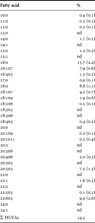
The fatty acid composition of O. moanae sp. nov. was dominated by (in order of importance) 16:0, 22:6n3, 18:0, 16:1n7, 20:5n3, and 18:1n7 (Table 3). All other fatty acids represented ≤2% of total fatty acid composition. The predominance of 22:6n3, 16:1n7 and 20:5n3 suggests that microalgae (i.e. dinoflagellates and diatoms) represent an important proportion of the diet of O. moanae sp. nov. (Ackman et al., Reference Ackman, Tocher and McLachlan1968; Sargent et al., Reference Sargent, Parkes, Mueller-Harvey, Henderson and Sleigh1987). The presence of 18:1n7, as well as smaller amounts of 15:0 and 17:0, indicate that bacteria also contribute (Volkman et al., Reference Volkman, Johns, Gillan, Perry and Bavor1980; Findlay et al., Reference Findlay, Trexler, Guckert and White1990). Macrophyte biomarkers such as 18:2n6 and 18:3n3 (Kharlamenko et al., Reference Kharlamenko, Kiyashko, Imbs and Vyshkvartzev2001) were very low. The fatty acid 18:1n9, which is often used as a biomarker of metazoan carnivory (Nyssen et al., Reference Nyssen, Brey, Dauby and Graeve2005, and references therein), was only found in small (<2%) amounts.
Fatty acid composition of Oncholaimus moanae sp. nov. (mean (SD)). nd, not detected; Σ HUFAs, sum of highly unsaturated fatty acids.

The apparent contradiction between the fatty acid data (which suggest a diet consisting of microalgae and bacteria) and the δ15N data (suggesting that O. moanae sp. nov. is not a primary consumer) could be explained in different ways. First, it is possible that 18:1n9 is not always a reliable biomarker of metazoan carnivory, and that O. moanae sp. nov. is, in fact, a predator. Phylogeny, for example, has been suggested as an important factor affecting the fatty acid composition of some animals, independent of diet (Nyssen et al., Reference Nyssen, Brey, Dauby and Graeve2005). This possibility is not considered likely since O. moanae sp. nov. made up 75% of nematode biomass at the study site (D. Leduc, unpublished data); nematode prey, therefore, would not be sufficient to sustain a strictly predatory feeding habit. Another possible explanation is that the trophic enrichment in δ15N signatures for nematodes is higher than 3.4‰. Trophic shifts in consumers δ15N values can vary greatly, with some values over 5‰ (McCutchan et al., Reference McCutchan, Lewis, Kendall and McGrath2003). In a study by Ruess et al. (Reference Ruess, Haggblom, Langel and Scheu2004), however, fungal feeding nematodes showed depleted or similar δ15N values compared to their diet. Another explanation could be the ingestion of ciliates and/or flagellates by O. moanae sp. nov. Heterotrophic protists have been shown to biosynthesize highly unsaturated fatty acids (HUFAs, defined as fatty acids having carbon chain length of ≥C20 and with ≥3 double bonds) such as 20:5n3 and 22:6n3 (Boechat et al., Reference Boechat, Schuran and Adrian2005; Veloza et al., Reference Veloza, Chu and Tang2006). Feeding on protists would represent an additional trophic step and could lead to elevated δ15N values. High δ15N signatures, for example, have been reported for Ascolaimus elongatus, a nematode with an unarmed buccal cavity similar to the ciliate feeder Anoplostoma viviparum (Moens & Vincx, Reference Moens and Vincx1997; Moens et al., Reference Moens, Bouillon and Gallucci2005). In addition, experimental evidence has shown that ciliates are important food items for the nematode Enoploides longispiculosus (Hamels et al., Reference Hamels, Moens, Mutylaert and Vyverman2001). Predation on ciliates has also been documented for other nematode species (Bouwman et al., Reference Bouwman, Romeyn, Kremer and Vanes1984; Moens & Vincx, Reference Moens and Vincx1997). Predation on heterotrophic protists (and ciliates in particular) is considered the most likely explanation for the isotopic and fatty acid composition of O. moanae sp. nov. at Papanui Inlet.
The high level of HUFAs observed in O. moanae sp. nov. has implications beyond the interpretation of food web relationships. HUFAs cannot be biosynthesized by most metazoans (but see below), and are essential for the normal functioning of cell membranes and membrane-bound enzyme systems (Bishop, Reference Bishop and Rechigl1976; Wantanabe et al., Reference Wantanabe, Arakawa, Kitajima, Fukusho and Fujita1978; Phillips, Reference Phillips1984). HUFA content is, therefore, an important indicator of food quality for consumers (e.g. Langdon & Waldock, Reference Langdon and Waldock1981; Read, Reference Read1981; Wantanabe et al., Reference Wantanabe, Kitajima and Fujita1983; Kanazawa, Reference Kanazawa, Cowsley, Mackie and Bell1985). It has previously been hypothesized that free-living marine nematodes should be poor in HUFAs as a result of their supposed bacterial diet (Coull, Reference Coull1999). Predators were suggested to favour preys with a high nutritional value such as harpacticoid copepods (rich in HUFAs) over nematodes (low in HUFAs). This would explain, in turn, why nematodes are seldom seen in fish gut contents (Nelson & Coull, Reference Nelson and Coull1989). It is obvious from the data presented here, however, that marine nematodes can be rich in HUFAs. Marine nematodes can therefore be a food source of high nutritional value, at least in terms of fatty acid composition. The low occurrence of nematodes in fish gut contents could be due to the rapid digestion of nematodes relative to copepods and the predominantly epibenthic lifestyle of many harpacticoid copepod species (Coull, Reference Coull1999; Nelson & Coull, Reference Nelson and Coull1989), instead of prey choice based on HUFA content (Coull, Reference Coull1999). Large nematodes living near or at the sediment surface may be particularly susceptible to predation by hyperbenthic predators. The large nematode Mesacanthion diplechma, for example, was the most common nematode species found in the stomachs of sand gobies despite their low abundance in the sediments (Hamerlynck & Vanreusel, Reference Hamerlynck and Vanreusel1993). Large nematodes such as O. moanae sp. nov. may, therefore, represent an important trophic link between the microbial community (e.g. ciliates and bacteria) and higher trophic levels.
Nematodes, unlike most metazoans, are able to biosynthesize HUFAs from acetate (Rothstein & Gotz, Reference Rothstein and Gotz1968; Bolla, Reference Bolla and Zuckermann1980). Whether marine species also have this ability remains unknown. Nematodes could represent a potentially important source of HUFAs in benthic food webs due to their high abundance and widespread distribution.
Very few data are available on the fatty acid composition of marine and freshwater meiofaunal organisms, although studies have been made on cultured freshwater nematodes such as Panagrellus redivivus (e.g. Lower et al., Reference Lower, Willet and Hansen1970; Schlechtriem et al., Reference Schlechtriem, Tocher, Dick and Becker2004), as well as on harpacticoid copepods in cultures (Norsker & Stottrup, Reference Norsker and Stottrup1994; Nanton & Castell, Reference Nanton and Castell1999; Williams & Biesiot, Reference Williams and Biesiot2004; Caramujo et al., Reference Caramujo, Boschker and Admirall2008) and from the field (Coull, Reference Coull1999). This lack of information is probably due to the difficulties associated with obtaining sufficient material for analysis. Several hundred nematodes (each weighing about 1–1.5 µgDW) were required in order to obtain sufficient biomass for reliable fatty acid analysis in this study. The use of fatty acid analysis in future studies of meiofaunal trophic dynamics is, therefore, likely to be limited to relatively large species until improvements are made to existing methods.
ACKNOWLEDGEMENTS
I am greatly indebted to Ashley Duncan (Department of Human Nutrition, University of Otago) for his expertise with fatty acid analysis, and to P. Keith Probert (Department of Marine Science, Univeristy of Otago) for his support throughout this study. I would also like to thank two anonymous referees who provided useful comments. This study was funded by a University of Otago Postgraduate Scholarship.