




No CrossRef data available.
Article contents
Hepatozoon griseisciuri in grey squirrels (Sciurus carolinensis): changes of blood leucocyte numbers resulting from infection
Published online by Cambridge University Press: 09 October 2003
Abstract
Numbers of leucocytes in squirrels with gametocytes of Hepatozoon in their blood (infected) were compared with animals without gametocytes (uninfected). Typical values for leucocytes/mm3 blood in uninfected squirrels were: leucocytes 5·7×103, granulocytes 3·4×103, lymphocytes 2·0×103 and monocytes 0·3×103 cells. Infection caused an increase in monocytes, lymphocytes and granulocytes, and there was a significant positive association between parasitaemia level and numbers of both total leucocytes and monocytes. Infected animals had more uninfected monocytes/mm3 blood than did uninfected animals. The proportions of monocytes were more variable over time in infected animals, but no shift between infected and uninfected status was detected. Transfer of serum from infected squirrels to mice resulted in elevated counts of total blood leucocytes, monocytes and granulocytes, but not of lymphocytes, as compared with controls. Serum from squirrels with high parasitaemias had a more marked effect than serum from squirrels with low parasitaemias. Results indicate an infection-related monocytosis, possibly controlled by cytokines, that increases the number of cells available for invasion by gametocytes, thus enhancing the chances of parasite transmission.
Information
- Type
- Research Article
- Information
- Copyright
- 2003 Cambridge University Press
INTRODUCTION
Gametocytes of Hepatozoon (probably H. griseisciuri) are found in the blood monocytes of grey squirrels (Sciurus carolinensis) (Hendricks, 1972; Britt & Molyneaux, 1979). Asexual development prior to the gametocytes occurs in various fixed tissues (Hendricks, 1975; Davidson & Calpin, 1976; Desser, 1990) and transmission probably involves a haemophagic arthropod host (Watkins & Nowell, 1991). We have reported high prevalence rates of Hepatozoon in squirrels trapped near Reading (UK) and repeated sampling showed that individuals had a chronic parasitaemia lasting many months, often with more than 50% of the monocytes being parasitized (Watkins & Nowell, 1991).
Except for Hepatozoon canis of dogs (e.g. Gondim et al. 1998), information concerning the blood picture of vertebrates infected with Hepatozoon is sparse. A previous study of Hepatozoon in S. carolinensis, recorded a differential leucocyte count in a single infected animal (Hendricks, 1972). Hepatozoon infection of squirrels may lead to destruction of monocytes and hence the numbers of monocytes and macrophages will be reduced and their functions may be impaired. The aim of the present paper is to describe the effects of Hepatozoon on levels of leucocytes, especially monocytes in squirrels. We also describe experiments involving transfer of squirrel serum to mice to check if serum factors are involved in such changes.
MATERIALS AND METHODS
Blood picture in squirrels
The methods for trapping and husbandry of squirrels, and for basic haematology are described in our previous paper (Watkins & Nowell, 1991).
Leucocyte counts were made with a haemocytometer and from blood smears. For total and differential leucocyte counts and for counts of cells parasitized by Hepatozoon (cells/mm3 blood), cardiac blood (0·2 ml) was drawn from a terminally anaesthetized animal (carbon dioxide), mixed with an aqueous solution of formalin (2%) and methylene blue (0·2%) (3·8 ml) and cells were counted in an Improved Neubauer haemocytometer (Dacie & Lewis, 1984). Thin blood smears obtained from the caudal vein were air dried, fixed in methanol and stained with Giemsa's stain before examining at 1000× magnification. If gametocytes were not seen in 2 blood smears taken from an animal, that animal was recorded as uninfected. Leucocyte counts were made from a central strip along the length of a blood smear. Differential leucocyte counts were made from a sample of 300 leucocytes per smear, which preliminary experiments had shown to provide reproducible results. Levels of monocyte (MNP) parasitization by Hepatozoon were estimated from 100 MNPs/smear.
Experimental transfer of squirrel serum to mice
Blood was collected by cardiac puncture from squirrels under terminal anaesthesia using carbon dioxide. The blood was allowed to clot at room temperature for 10 min before being centrifuged for 10 min in a refrigerated centrifuge (MSE Chilspin) to obtain the serum. Serum was stored at −70 °C until used. Serum from hamsters (Mesocricetus auratus) was similarly obtained. Male T.O. strain mice (Tucks, Rayleigh, Essex), 60–69 days of age were used. The mice were maintained at 21 °C, on a 12 h light/12 h dark cycle and allowed food (Diet 86A) and water ad libitum. Injected mice were given 0·2 ml of freshly thawed serum i.p. Blood was taken either from the tail vein of unanaesthetized mice or from the heart of terminally anaesthetized mice in the same way as for squirrels.
In a preliminary experiment, 5 groups of mice (3 animals/group) were subjected to one of the following treatments: (A) no serum injection; (B) injected with hamster serum (to check effect of foreign serum on blood picture); (C) injected with squirrel serum (negative parasitaemia); (D) injected with Hepatozoon-infected squirrel serum (low parasitaemia: <30% parasitized MNPs); (E) injected with Hepatozoon-infected squirrel serum (high parasitaemia: >50% parasitized MNPs). Blood smears for differential leucocyte counts were taken from these mice on days 2, 3, 4, 5, 6, 7, 14 and 21.
In the main experiment, 5 groups of randomized mice (6 or 7 mice/group) were subjected to the following treatments: R, no serum injection; S, no serum injection; T, injected with hamster serum; U, injected with pooled Hepatozoon-infected squirrel serum (low parasitaemia <30% parasitized MNPs); V, injected with pooled Hepatozoon-infected squirrel serum (high parasitaemia >50% parasitized MNPs). There was no group equivalent to group C of the preliminary experiment since there was a lack of uninfected squirrels at the time. Injections were all given on day 0 and mice were all killed on day 3 except for group R which were killed on day 0. Blood leucocyte counts were made using a haemocytometer.
Results were analysed usually by 2×2×2 factorial analysis of variance and Student's t-test. Percentage data were arcsin square root transformed.
RESULTS
Animals are categorized as ‘infected’ or ‘uninfected’ depending on whether or not they showed gametocytes in the blood monocytes.
Blood picture in squirrels
In both types of blood preparation, numbers of 3 types of leucocytes were estimated: lymphocytes, mononuclear phagocytes or monocytes (MNPs), and granulocytes (usually polymorphonuclear neutrophils (PMNs), but occasional basophils and/or eosinophils were present). Gametocytes of Hepatozoon could easily be discerned and these only occurred in MNPs.
In the first field study, total and differential leucocyte counts were made for 75 animals immediately after first capture: 35 uninfected and 40 infected (Table 1). Typical levels of leucocytes (cells/mm3 blood) in uninfected squirrels were: total leucocytes 5·7×103, granulocytes 3·4×103, lymphocytes 2·0×103 and MNPs 0·3×103. Factorial analysis of variance showed that the infected/non-infected status of the squirrels, but not age nor sex, had a significant effect on leucocyte counts. Infection caused a significant increase of MNPs (P<0·001), of lymphocytes (P<0·05), of granulocytes (P<0·001) and of total leucocytes (P<0·001). There were no significant differences in mean parasitaemias (parasitized MNPs/mm3 blood) related to the age or sex of the infected host (P>0·05). Comparison of the numbers of non-parasitized MNPs showed that infected hosts had a significantly higher level than uninfected animals (P<0·01) (Table 2). Data from the 40 infected squirrels treated by analysis of variance on the regressions show positive significant associations of the parasitaemia level with the total leucocyte count (P<0·01) and with the MNP count (P<0·001), but not with the lymphocyte or the granulocyte counts (both P>0·05) (Fig. 1).

Fig. 1. Relationship of Hepatozoon parasitaemia (parasites/mm3 of blood) to total leucocyte counts (cells/mm3 of blood). The proportions of mononuclear phagocyte (=monocyte) (MNPs), lymphocytes and granulocytes are shown. These proportions are derived from regression lines plotted of data from blood counts of 40 infected squirrels at first capture. Correlation coefficients and equations for original regression lines for individual cell types: total leucocytes, 0·64, y=6·94+2·57x; MNPs, 0·90, y=0·67+1·09x; lymphocytes, 0·40, y=2·51+0·61x; granulocytes, 0·49, y=4·06+0·87x.
Table 1. Total and differential leucocyte counts for the blood of Hepatozoon-infected and -uninfected squirrels at first capture (N=75) (MNPs, mononuclear phagocytes (=monocytes). Numbers of parasitized MNPs are also indicated.)

Table 2. A comparison of the number of non-parasitized mononuclear phagocytes (MNPs) in the blood of Hepatozoon-infected and -uninfected individuals (Data are derived from 75 first-captured animals (as Table 1).)

Comparable results were obtained from a second field study, when differential leucocyte levels were estimated from stained smears of tail blood from live animals. Analysis of variance of results from 218 first-capture squirrels showed no significant difference (P>0·05) between age and sex categories for any of the differential leucocyte proportions. There was a significant increase in the proportions of MNPs in infected squirrels over uninfected squirrels; proportions of lymphocytes were lower in the infected squirrels (apparently in contrast to the previous study – see Discussion section); but granulocytes were not significantly different (Table 3). As shown in this table, there was considerable variation between animals. From blood smears, there was no significant difference of proportions of cell types in relation to host age or sex (P>0·05). However, overall, males had a significantly higher percentage of parasitized MNPs than did females (N, mean±S.D.: 100, 48±28; 54, 38±29 respectively; P<0·05).
Table 3. Differential leucocyte percentages in blood smears from Hepatozoon-infected and -uninfected squirrels at first capture (N=218) (MNPs, mononuclear phagocytes (=monocytes).)

Blood smears taken from 8 squirrels (2 uninfected, 6 infected) trapped at monthly intervals over at least 6 months of a 21-month period failed to show any change either way between infected and uninfected status in individual animals. Differential blood leucocyte counts from smears made at every capture indicated a higher and much more variable percentage of MNPs in the infected as compared with the uninfected animals (Fig. 2). However, there was no obvious relationship between the leucocyte numbers and the parasitaemia levels over the sampling period. Weekly blood smears from laboratory-maintained animals showed no obvious pattern in the parasitaemia over the 16 weeks of sampling (Fig. 3). Again, the percentages of MNPs were higher and were more variable in the infected animals.

Fig. 2. Examples of monthly changes in differential leucocyte percentages of repeatedly trapped individual adult squirrels. (A) Female squirrel not showing an Hepatozoon parasitaemia. Ranges: granulocytes 57–81%; lymphocytes 14–37%; MNPs 3–7%. (B) Female squirrel showing an Hepatozoon parasitaemia. Ranges: granulocytes 40–76%; lymphocytes 16–46%; MNPs 5–28%. (C) The monthly variations of the parasitaemia in the same squirrel as in (B). MNP, mononuclear phagocyte (=monocyte).

Fig. 3. Examples of weekly changes in differential leucocyte percentages of individual adult squirrels maintained in captivity. (A) Male squirrel not showing an Hepatozoon parasitaemia. Ranges: granulocytes 57–64%; lymphocytes 30–39%; MNPs 4–6%. (B) Male squirrel showing an Hepatozoon parasitaemia. Ranges: granulocytes 47–66%; lymphocytes 24–46%; MNPs 6–18%. (C) The weekly variations of the parasitaemia in the same squirrel as in (B). MNP, mononuclear phagocyte (=monocyte).
Transfer of squirrel serum to mice
The preliminary experiment indicated that the proportion of MNPs in the total leucocytes on blood smears increased from about 1% on day 0 to a peak of 4–5% on day 3 in mice injected with infected squirrel serum (groups D and E). This proportion was significantly higher (P<0·001) than those of control mice (groups A, B and C), all remaining at about 1%. Proportions of granulocytes were also significantly higher in groups D and E than in groups A, B and C between days 2 and 6 (P<0·01). By day 14, proportions of granulocytes and MNPs in mice of groups D and E had returned to similar levels to the controls. On the basis of these results, the main sampling time of the second experiment was set at 3 days after injection.
Leucocyte counts for the groups of mice in the main experiment are shown in Table 4. Analyses of variance indicated significant differences between the 5 groups in counts of the total leucocytes, the MNPs and the granulocytes (all P<0·001), but not for the lymphocytes (P>0·05). Student's t-tests showed that counts of total leucocytes, MNPs and granulocytes were significantly higher (all at P<0·05) in group U mice as compared with groups R, S or T. Mice of group V had significantly higher counts of these cell classes than mice of group U (P<0·001).
Table 4. Transfer of serum from Hepatozoon-infected squirrels or from controls to mice: total and differential leucocyte counts (counts×10−3/mm3 blood) obtained for the 5 treatment groups (See text for experimental details. MNPs, mononuclear phagocytes (=monocytes). Mean±S.D. stated.)

DISCUSSION
The sampling methods were of necessity a compromise. Ideally all cell counts should have been made with a haemocytometer, but the repeated removal of suitable volumes of tail blood (0·2 ml minimum) would have been stressful to a wild animal and may have induced localized inflammation which could have altered the cell counts (Dacie & Lewis, 1984). Also it was clearly not feasible or humane to remove samples of cardiac blood from live animals. Blood smears required a smaller blood sample (about 0·02 ml), the acquisition of which required less manipulation of the animal and when blood sampling was done once only, the effects induced by skin puncture and inflammation were avoided. In regularly recaptured animals any inflammation of the tail tip had apparently subsided in the time between samples.
Haemocytometer counts show that infections result in increased numbers of the 3 types of leucocytes, but in blood smears this shows up as increased proportions of MNPs and decreased proportions of the other cell types. The overall picture is of a leucocytosis induced by the infection, with the major component being the monocytosis. An interesting feature is the positive association between the parasitaemia level and the levels of both total MNPs and unparasitized MNPs. The high and fluctuating levels of MNPs in the regularly sampled infected squirrels may be a sign of the conflict between the squirrel's immune system and the parasite continuing to survive and multiply.
Monocytosis occurs in a number of infections (Territo & Cline, 1976) including H. canis of dogs (Gondim et al. 1998) and is probably part of the animal's defence reactions. However, in contrast to H. canis which invades neutrophils (Baneth et al. 1998), monocytosis induced by Hepatozoon of squirrels may well aid the parasite by providing more cells for the gametocytes to inhabit, thus enhancing the chances of transmission to the arthropod host. In fact the increased levels of unparasitized MNPs in infected squirrels in the first field study suggested an overcompensation for parasitization of MNPs. The monocytosis would normally be expected to result from enhanced monocytopoiesis, although other mechanisms might play a role. These mechanisms could include MNP cell division whilst in the blood circulation, or MNPs may be influenced to have an extended life-span in the blood. Mitosis of blood MNPs in infected squirrels has not been seen so there is no parallel to the situation of Theileria inducing cell division of parasitized leucocytes (Morrison, MacHugh & Lalor, 1996; Campbell & Spooner, 1999). The life-span of blood MNPs is apparently not recorded for squirrels, but for rats the half-life is about 61 h reducing by more than half during some infections e.g. Salmonella enteritidis (see Volkman & Collins, 1974). However, conceivably the life-span of Hepatozoon-parasitized MNPs may be extended, for instance as in Leishmania infections where there is production of an anti-apoptotic factor (Moore & Matlashewski, 1994).
The experiment involving transfer of serum from squirrels to mice supports the hypothesis that one or more factors in the serum are elaborated during Hepatozoon infections of squirrels and that the factor(s) can enhance levels of murine leucocytes, especially MNPs and gametocytes. Since serum from squirrels with high parasitaemia stimulated higher counts of murine leucocytes than did serum from squirrels with low parasitaemia, there are indications of a direct association between the levels of Hepatozoon in the squirrel blood, the levels of the serum factor produced and the enhancement of leucocyte numbers. The nature of the serum factor(s) involved is not certain. The obvious candidates are cytokines, a number of which are secreted by MNPs and can influence production of both MNPs and neutrophils. These cytokines include macrophage and granulocyte-macrophage colony stimulating factors (M-CSF and GM-CSF), which at least in some cases are known to be cross-reactive between species (Callard & Gearing, 1994). They may be secreted by parasitized MNPs or conceivably by cells at sites of damage where the Hepatozoon gametocytes are released from schizonts in the host's tissues. GM-CSF also stimulates production of neutrophils and the increased level of granulocytes in both the infected squirrels and the mice given serum from infected squirrels must indicate that this cytokine is a likely candidate to be one of the mediators involved. As an alternative explanation, the parasite may play a more direct role by secreting molecules which either stimulate cytokine production by the host or even act as mimics of cytokines (Cox, 2001; Kaye, 1999).
Changes of the infection status of squirrels were not seen and therefore it was not possible to follow changes of leucocyte numbers as immunity developed. Also the presence of some squirrels without a parasitaemia but with schizonts in the lungs (Watkins & Nowell, 1991) suggests that immunity may be complicated by being stage specific. We have shown that numbers of gametocytes correlate with alterations of the blood picture, but it is possible that other stages of the parasite's life-cycle may also have an effect.
References
REFERENCES
BANETH, G.,
SHKAP, V.,
SAMISH, M.,
PIPANO, E. &
SAVITSKY, I.
(1998).
Antibody response to Hepatozoon canis in experimentally infected dogs.
Veterinary Parasitology 74, 299–305.CrossRefGoogle Scholar
BRITT, D. &
MOLYNEAUX, D. H.
(1979).
Parasites of grey squirrels in Cheshire, England.
Journal of Parasitology 65, 408.CrossRefGoogle Scholar
CALLARD, R. E. &
GEARING, A. J. H.
(1994).
The Cytokine Factsbook.
Academic Press/Harcourt Brace,
London.
CAMPBELL, J. D. M. &
SPOONER, R. L.
(1999).
Macrophages behaving badly: infected cells and subversion of immune responses to Theileria annulata.
Parasitology Today 15, 10–16.CrossRefGoogle Scholar
COX, F. E. G.
(2001).
Concomitant infections, parasites and immune responses.
Parasitology 122, S23–S38.CrossRefGoogle Scholar
DACIE, J. V. &
LEWIS, S. M.
(1984).
Practical Haematology,
6th Edn. Churchill Livingstone, Edinburgh.
DAVIDSON, W. R. &
CALPIN, J. P.
(1976).
Hepatozoon griseisciuri infection in gray squirrels of the southeastern United States.
Journal of Wildlife Diseases 12, 72–76.CrossRefGoogle Scholar
DESSER, S. S.
(1990).
Tissue cysts of Hepatozoon griseisciuri Clark 1958 in the grey squirrel Sciurus carolinensis: the significance of these cysts in species of Hepatozoon.
Journal of Parasitology 76, 257–259.CrossRefGoogle Scholar
GONDIM, L. F. P.,
KOHAYAGAWA, A.,
ALENCAR, N. X.,
BIONDO, A. W.,
TAKAHIRA, R. K. &
FRANCO, S. R. V.
(1998).
Canine Hepatozoonosis in Brazil: description of eight naturally occurring cases.
Veterinary Parasitology 74, 319–323.CrossRefGoogle Scholar
HENDRICKS, L. D.
(1972).
Schizogonic development of Hepatozoon griseisciuri of the Eastern grey squirrel. Ph.D. thesis, University of Maryland, USA.
HENDRICKS, L. D.
(1975).
Schizogonic development of Hepatozoon griseisciuri Clark 1958 (Sporozoa: Haemogregarinidae), of the grey squirrel (Sciurus carolinensis Gmelin 1788).
Journal of Parasitology 61, 458–461.CrossRefGoogle Scholar
KAYE, P.
(1999).
Parasite derived immunoregulatory molecules.
Parasite Immunology 21, 595–596.CrossRefGoogle Scholar
MOORE, K. J. &
MATLASHEWSKI, G.
(1994).
Intracellular infection by Leishmania donovani inhibits macrophage apoptosis.
Journal of Immunology 152, 2930–2937.Google Scholar
MORRISON, W. I.,
MacHUGH, N. D. &
LALOR, P. A.
(1996).
Pathogenicity of Theileria parva is influenced by the host cell type infected by the parasite.
Infection and Immunity 64, 557–562.Google Scholar
TERRITO, M. &
CLINE, M. J.
(1976).
Macrophages and their disorders in Man.
In Immunobiology of the Macrophage
(ed. Nelson, D. S.),
pp. 593–618.
Academic Press, New York.CrossRef
VOLKMAN, A. &
COLLINS, E. M.
(1974).
The cytokinetics of monocytosis in acute salmonella infection in rats.
Journal of Experimental Medicine 139, 264–277.CrossRefGoogle Scholar
WATKINS, B. M. &
NOWELL, F.
(1991).
Hepatozoon in grey squirrels (Sciurus carolinensis) trapped near Reading, Berkshire.
Journal of Zoology 224, 101–112.CrossRefGoogle Scholar

Fig. 1. Relationship of Hepatozoon parasitaemia (parasites/mm3 of blood) to total leucocyte counts (cells/mm3 of blood). The proportions of mononuclear phagocyte (=monocyte) (MNPs), lymphocytes and granulocytes are shown. These proportions are derived from regression lines plotted of data from blood counts of 40 infected squirrels at first capture. Correlation coefficients and equations for original regression lines for individual cell types: total leucocytes, 0·64, y=6·94+2·57x; MNPs, 0·90, y=0·67+1·09x; lymphocytes, 0·40, y=2·51+0·61x; granulocytes, 0·49, y=4·06+0·87x.

Table 1. Total and differential leucocyte counts for the blood of Hepatozoon-infected and -uninfected squirrels at first capture (N=75)
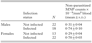
Table 2. A comparison of the number of non-parasitized mononuclear phagocytes (MNPs) in the blood of Hepatozoon-infected and -uninfected individuals

Table 3. Differential leucocyte percentages in blood smears from Hepatozoon-infected and -uninfected squirrels at first capture (N=218)
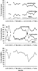
Fig. 2. Examples of monthly changes in differential leucocyte percentages of repeatedly trapped individual adult squirrels. (A) Female squirrel not showing an Hepatozoon parasitaemia. Ranges: granulocytes 57–81%; lymphocytes 14–37%; MNPs 3–7%. (B) Female squirrel showing an Hepatozoon parasitaemia. Ranges: granulocytes 40–76%; lymphocytes 16–46%; MNPs 5–28%. (C) The monthly variations of the parasitaemia in the same squirrel as in (B). MNP, mononuclear phagocyte (=monocyte).
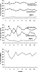
Fig. 3. Examples of weekly changes in differential leucocyte percentages of individual adult squirrels maintained in captivity. (A) Male squirrel not showing an Hepatozoon parasitaemia. Ranges: granulocytes 57–64%; lymphocytes 30–39%; MNPs 4–6%. (B) Male squirrel showing an Hepatozoon parasitaemia. Ranges: granulocytes 47–66%; lymphocytes 24–46%; MNPs 6–18%. (C) The weekly variations of the parasitaemia in the same squirrel as in (B). MNP, mononuclear phagocyte (=monocyte).

Table 4. Transfer of serum from Hepatozoon-infected squirrels or from controls to mice: total and differential leucocyte counts (counts×10−3/mm3 blood) obtained for the 5 treatment groups
 You have
Access
- 4
- Cited by