INTRODUCTION
Human alveolar echinococcosis (AE) is caused by the infection with the metacestode (larval stage) of E. multilocularis (see Gottstein and Reichen, Reference Gottstein, Reichen, Cook and Zumla2002; Gottstein, Reference Gottstein, Armstrong and Cohen2004; Ito and Craig, Reference Ito and Craig2003). Serological tests based on the enzyme-linked immunosorbent assay (ELISA) are useful for the early diagnosis of AE (i.e. before the disease becomes clinically manifest), at a time point when radical surgical resection of the parasitized tissue is more likely (Gottstein et al. Reference Gottstein, Saucy, Deplazes, Reichen, Demierre, Zürcher, Busato and Pugin2001). Several years ago, the early immunodiagnosis of AE was improved via the isolation and application in ELISA of an affinity-purified, native metacestode Em2 antigen (Gottstein et al. Reference Gottstein, Eckert and Fey1983) and the recombinant antigen II/3–10 (Müller et al. Reference Müller, Gottstein, Vogel, Flury and Seebeck1989; Felleisen and Gottstein, Reference Felleisen and Gottstein1993) representing a highly antigenic subfragment of E. multilocularis antigen II/3 (Vogel et al. Reference Vogel, Gottstein, Müller and Seebeck1988). The combined application of these antigens allowed discrimination between AE and cystic echinococcosis (CE, caused by the metacestode of E. granulosus) as well as cysticercosis (caused by the metacestode of Taenia solium) and other diseases caused by various parasitic nematodes and trematodes. Other research groups have also produced recombinant E. multilocularis antigens (EM10, Frosch et al. Reference Frosch, Frosch, Pfister, Schaad and Bitter-Suermann1991; Em4, Hemmings and McManus, Reference Hemmings and McManus1991; rEm18, Xiao et al. Reference Xiao, Mamuti, Yamasaki, Sako, Nakao, Nakaya, Gottstein, Schantz, Lightowlers, Craig and Ito2003) which exhibited similar diagnostic operating characteristics and all proved to represent the same ERM-gene family (Brehm et al. Reference Brehm, Jensen, Frosch and Frosch1999). Despite the excellent diagnostic qualities of, for example, the ELISA utilizing the antigens Em2 and II/3–10, either a lack of reactivity or some cross-reactivity was observed in a small number of confirmed AE cases, thus hampering interpretation in view of these peculiar cases (Gottstein et al. Reference Gottstein, Jacquier, Bresson-Hadni and Eckert1993). Weak antibody reactivity is a relatively common feature in AE cases, particularly when related to certain clinical scenarios, such as a recently acquired infection or an abortive course of infection (i.e. degradation or death of the parasite in tissues). Another issue is that cross-reactive serum antibodies, frequently occurring in cancer patients (e.g. Pfister et al. Reference Pfister, Gottstein, Cerny and Cerny1999; Poretti et al. Reference Poretti, Felleisen, Grimm, Pfister, Teuscher, Zürcher, Reichen and Gottstein1999) may complicate the interpretation of results.
Immunoblot analysis is an important approach that suitably complements the ELISA-based immunodiagnosis of CE (Poretti et al. Reference Poretti, Felleisen, Grimm, Pfister, Teuscher, Zürcher, Reichen and Gottstein1999; Liance et al. Reference Liance, Janin, Bresson-Hadni, Vuitton, Houin and Piarroux2000; Doiz et al. Reference Doiz, Benito, Sbihi, Osuna, Clavel and Gomez-Lus2001; Furuya et al. Reference Furuya, Kawanaka, Yamano, Sato and Honma2004; Akisu et al. Reference Akisu, Bayram Delibas, Yuncu, Aksoy, Ozkoc, Bicmen, Sevinc and Yaldiz2005). An immunoblot for CE, previously developed in our laboratory based on the use of E. granulosus hydatid cyst fluid (EgHF) antigen, relied on the detection of a genus-specific 8 kDa subunit of antigen B and 2 other immunodiagnostic antigens of 29 and 34 kDa (Poretti et al. Reference Poretti, Felleisen, Grimm, Pfister, Teuscher, Zürcher, Reichen and Gottstein1999). In this study, antigen B and, if detectable, the 29 kDa and 34 kDa antigens were shown to be genus-specific and allowed the diagnosis of AE and CE, with a sensitivity of 91% and a specificity of 97% (antigen B) or 94% (29 and 34 kDa antigens), respectively. Some years ago, Ito et al. (Reference Ito, Ma, Schantz, Gottstein, Liu, Chai, Abdel-Hafez, Altintas, Joshi, Lightowlers and Pawlowski1999) established 2 immunoblots for the diagnosis of AE, which were based on the use of fractions containing either semi-purified 8 kDa E. granulosus antigen B or an 18 kDa antigen (Em18), enriched by isoelectric focusing from an E. multilocularis protoscolex extract. A large-scale serological evaluation of this ‘dual immunoblot system’ revealed the genus-specific characteristics of antigen B in that 92% of the CE sera and 79% of the AE sera reacted with this antigen. Conversely, the Em18 antigen was shown to detect AE with a diagnostic sensitivity of 97% and a moderate specificity of 74%, and had potential to differentiate AE from CE. Liance et al. (Reference Liance, Janin, Bresson-Hadni, Vuitton, Houin and Piarroux2000) evaluated a commercial immunoblot (Echinococcus Western Blot IgG™, LDBIO Diagnostics, Lyon, France) that employs a whole larval antigen from E. multilocularis. In this test system, the serological reactivity with a 7 kDa band and/or a diffuse 26 to 28 kDa band was indicative of the presence of Echinococcus-specific antibodies. The diagnostic sensitivities of the immunoblot were determined to be 96·7% for AE and 98% for CE, respectively. Cross-reactivity was shown using sera from patients with neurocysticercosis (7 of 20) or Schistosoma mansoni infection (3 of 18 sera). In a more recent study, Korkmaz et al. (Reference Korkmaz, Inceboz, Celebi, Babaoglu and Uner2004) tested 2 E. multilocularis metacestode antigens, Em70 and Em90, and achieved a sensitivity of 100% and a specificity of 99·1% for the diagnosis of AE. Most of these studies did or could not include (due to unavailability) ‘diagnostically critical’ sera, such as those derived from patients with abortive AE or with very low levels of anti-E. multilocularis serum antibodies due to degenerative or dead metacestodes. Such sera frequently remain negative when tested against native or recombinant E. multilocularis antigens. Therefore, any improvement of the sensitivity and specificity of an antigen or a test for the serodiagnosis of AE is considered a significant advance. In the present study, we developed an improved immunoblot assay for the specific diagnosis of AE, irrespective of the form of disease. This assay utilizes an in vitro-produced E. multilocularis vesicle fluid (EmVF) antigen, as an alternative to native antigens usually obtained from alveolar cysts from experimentally infected rodents. Together with the present Em2- and II/3–10-ELISA (Gottstein et al. Reference Gottstein, Jacquier, Bresson-Hadni and Eckert1993), this immunoblot method allows the accurate diagnosis of both clinical and subclinical cases of AE.
MATERIALS AND METHODS
Preparation of E. multilocularis vesicle fluid (EmFV) antigen
The EmVF antigen was obtained by aseptic aspiration of fluid from fertile vesicular cysts, axenically cultivated in vitro as described previously (Hemphill and Gottstein, Reference Hemphill and Gottstein1995). EmVF was complemented with 1 mmol of the proteinase inhibitor phenyl-methyl-sulfonyl fluoride (PMSF), centrifuged at 14 000 g for 10 min and the supernatant subsequently stored frozen at −80°C until use. In order to validate individual batches of fluid, the protein/carbohydrate ratio was required to range between 4:1 and 5:1, the protein concentration between 0·7 and 0·9 mg/ml (EmVF with higher protein concentrations were discarded). Also, the immunoblot characteristics of the EmVF had to be consistent with those described herein.
Human sera
Well-defined sera from 62 Central European AE patients (54 of them with active hepatic lesions, and 8 of them had fully calcified lesions and thus represented inactive or ‘abortive’ cases) were used for assessing the primary diagnostic sensitivity of the tests for AE. Inactivity of metacestodes was proven either by post-diagnostic surgical removal of parasite material, with a subsequent viability assessment as described by Rausch et al. (Reference Rausch, Wilson, Schantz and McMahon1987) or by extensive clinical and subsequent computer tomography or X-ray imaging of the patients (see Gottstein et al. Reference Gottstein, Saucy, Deplazes, Reichen, Demierre, Zürcher, Busato and Pugin2001). Therefore, all sera were sampled prior to any therapeutic or surgical intervention. Each diagnosis of AE had been confirmed by clinical, histological (if feasible) and serological means.
Sera used for assessing test specificities were obtained from patients with clinically, parasitologically and/or histologically proven infections, involving the following parasites (disease and number of sera obtained from patients are in parentheses): E. granulosus (cystic echinococcosis, 22 sera), Fasciola hepatica (fascioliasis, 7 sera), Schistosoma spp. (schistosomiasis, 7 sera), Taenia solium (cysticercosis, 10 sera), Ascaris lumbricoides (ascariasis, 7 sera), Strongyloides stercoralis (strongyloidiasis, 6 sera), Toxocara canis (toxocariasis; visceral larva migrans, 7 sera), Trichinella spiralis (trichinellosis, 7 sera); Onchocerca volvulus (filariasis, 6 sera), and Entamoeba histolytica (amoebiasis, 7 sera). All of these sera had been pre-selected based on their ‘positivity’ using homologous parasite antigens in tests carried out routinely in our diagnostic laboratory in the University of Berne. Sera from 18 cancer patients were also selected from a previous study (Pfister et al. Reference Pfister, Gottstein, Cerny and Cerny1999) based on their serological reactivity in Echinococcus tests, as described by Poretti et al. (Reference Poretti, Felleisen, Grimm, Pfister, Teuscher, Zürcher, Reichen and Gottstein1999).
Enzyme-linked immunosorbent assays (ELISAs)
These assays were carried out essentially as described by Gottstein et al. (Reference Gottstein, Jacquier, Bresson-Hadni and Eckert1993), by testing sera at a 1:100 dilution and using the following antigens (at optimized coating concentrations): EmVF-antigen (2·8 μg protein per ml); EgHF antigen (5 μg protein per ml); Em2 antigen (2·8 μg carbohydrates per ml); II/3–10 antigen (1 μg protein per ml). All Echinococcus-antigen-ELISAs employed in the present study were first calibrated to determine the threshold for the discrimination between positive and negative results. The actual cut-off value was thus determined by testing blood donor sera, and the threshold was set at a mean of the respective OD405 nm values plus 3 standard deviations (cf. Gottstein et al. Reference Gottstein, Saucy, Deplazes, Reichen, Demierre, Zürcher, Busato and Pugin2001). When plotted, the respective OD405 nm values essentially represented a normal distribution. The blood donor sera were from healthy Swiss persons and were selected based on an even male-female ratio and an age range of between 20 and 65 years. By using the present selection criteria, the negative control groups thus matched the corresponding parameters of the 62 sera from the Central European AE-patients. Inter-test and intra-test variations were assessed by determining the coefficients of variation for reference negative and positive sera, all having been tested in triplicate on each test plate; variation of ⩽15% was recorded, which is considered acceptable for immunological assays (Pfister et al. Reference Pfister, Gottstein, Cerny and Cerny1999). Since this latter finding was essentially the same as described previously (Gottstein et al. Reference Gottstein, Jacquier, Bresson-Hadni and Eckert1993), no modification was required for the present study.
Immunoblotting
Immunoblotting was performed as described previously for E. granulosus hydatid fluid (EgHF) antigen (Poretti et al. Reference Poretti, Felleisen, Grimm, Pfister, Teuscher, Zürcher, Reichen and Gottstein1999), with the exception that EmVF (7 μg per cm slot) was used instead of EgHF. Sera were used at a dilution of 1:100.
Affinity purification of specific antibodies against the 20–22 and 8 kDa antigens
The affinity purification of specific antibodies against the 2 immunodominant bands in the EmVF antigen was performed essentially as described by Müller et al. (Reference Müller, Hemphill, Imboden, Duvallet, Dwinger and Seebeck1992). Briefly, the region corresponding to the 20–22 and 8 kDa antigens was excised from the nitrocellulose, following sodium dodecyl-sulfate polyacrylamide gel electrophresis (SDS-PAGE) and blotting of the EmVF antigen. After washing and blocking with phosphate-buffered saline (PBS, pH 7·2) containing 0·3% Tween 20 (Sigma) for 1 h at room temperature (22–24°C), a strongly positive serum from an AE patient, applied at a dilution of 1:20 in PBS containing 0·3% Tween 20, was incubated overnight at 4°C. After washing the strips in PBS containing 0·3% Tween 20 (3 times for 10 min each), the bound antibodies were eluted in 900 μl of low-pH buffer (50 mm Tris, 50 mm glycine, 0·05% Tween 20, pH 2·6) for 5 min on ice, with occasional vortexing. Subsequently, the strips were removed, and 100 μl of 1 m Tris base was immediately added to neutralize the pH. The eluted antibody fractions were centrifuged (10 000 g for 20 min) to remove any small nitrocellulose particles, and bovine serum albumin (BSA) was added to a final concentration of 0·1%. The affinity-purified antibodies were stored at −20°C and used for immunoblotting at a dilution of 1:10.
Determination of protein and carbohydrate concentrations
Soluble protein concentrations were determined using a commercial assay (Bradford Kit, Bio-Rad, Switzerland), employing bovine plasma gamma-globulin as a standard. Carbohydrate concentrations were estimated using the orcinol-sulfuric acid assay, employing Dextran T-2000 as a standard (Miguez et al. Reference Miguez, Baz and Nieto1996).
RESULTS
Comparative serological analyses of various Echinococcus antigen-ELISAs
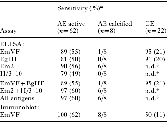
The sensitivities for the ELISAs using different antigens for the serological diagnosis of AE or CE are given in Table 1. The testing of ‘crude’ Echinococcus metacestode antigens using well-defined sera from untreated AE patients revealed that the EmVF-ELISA had a better diagnostic sensitivity than the EgHF-ELISA (89% vs 81%); most of the sera yielded positive results in the 2 ELISAs and also exhibited similar values. Furthermore, all sera positive in the EgHF-ELISA were also positive in the EmVF-ELISA. However, the accumulation of results achieved from both assays did not improve the diagnostic sensitivity. The individual diagnostic sensitivities of the Em2-ELISA and the II/3–10-ELISA were 90% and 79%, respectively. Interestingly, the Em2-ELISA scored positive for 6 of 8 sera from inactive AE cases with necrotic lesions (see Fig. 1). The cumulative diagnostic sensitivity from the Em2- and II/3–10-ELISAs (as achieved in the Em2plus-ELISA reported earlier, see Gottstein et al. Reference Gottstein, Jacquier, Bresson-Hadni and Eckert1993) was high, and allowed detection of 97% of AE cases included in the present study (Table 1). Hence, no further improvement in diagnostic sensitivity was achieved when the results from all 4 ELISAs assessed were accumulated.
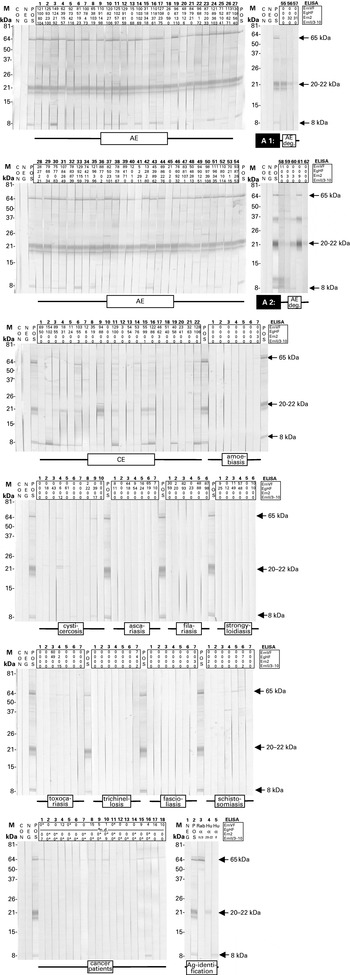
EmVF-immunoblot with sera from 62 patients with active alveolar echinococcosis (AE) (54 sera); degenerate alveolar echinococcosis (non-viable metacestode) (AE, deg; 8 sera in total: 3 tested in analysis 1 [A1, 2nd blot strip panel] and 5 in analysis 2 [A2, 4th blot strip panel]); cystic echinococcosis (CE, 22 sera); amoebiasis (7 sera); cysticercosis (10 sera); ascariasis (7 sera); filariasis (6 sera); strongyloidiasis (7 sera); toxocariasis (7 sera); trichinellosis (7 sera), fascioliasis (7 sera); and schistosomiasis (7 sera), and sera from cancer patients (18 sera). Molecular weight markers (M) (left) are given in kilodaltons (kDa) and positions of the major immunoreactive bands (right) representing 65 kDa, 20–22 kDa, and 8 kDa antigens are indicated by arrows. As controls, blot strips incubated exclusively with anti-human immunoglobulin-peroxidase conjugate (CON), or with negative (NEG) or postive (POS) serum were included. EmVF-, EgHF, Em2-, and II/3–10-ELISA values of the individual sera are listed on top of each strip. For sera from cancer patients, borderline-negative ELISA results are indicated with asteriscs. Identification of the major antigens (Ag-identification) and assessment of serological cross-reactivities among the corresponding bands on the blot was performed by demonstrating the immunoreactivity with a polyclonal rabbit anti-II/3–10 antibody (Rab α II/3) and antibodies affinity-purified on bands representing the 22–20 kDa (Hu α 20–22), and 8 kDa (Hu α 8) antigens. a Not done (n.d.), as pre-selected based on EgHF-positivity according to Pfister et al. (Reference Pfister, Gottstein, Cerny and Cerny1999).

Diagnostic sensitivities of crude, purified, and recombinant Echinococcus antigens for immunodiagnosis of alveolar (AE) and cystic (CE) echinococcosis by ELISA and immunoblot in patients with confirmed AE or CE
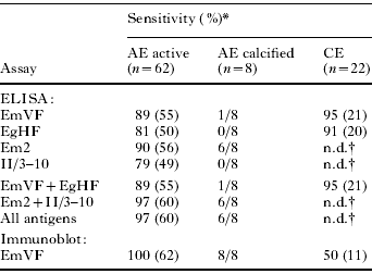
* The number of positive samples is given in parenthesis; individual test results, see Fig. 1.
† Not done (n.d.), diagnostically irrelevant.
Upon diagnosis, patients with CE demonstrated EmVF-ELISA reactivity, in that 21 of 22 sera scored positive values. Conversely, 20 sera from this panel were positive in the EgHF-ELISA (see Table 1). Accordingly, the EmVF-ELISA exhibited a better diagnostic sensitivity than the EgHF-ELISA (95% vs. 91%). Furthermore, most of sera positive in the 2 ELISAs showed similar values, and the accumulation of the results from both tests did not improve the diagnostic sensitivity (Table 1). As expected, the antigens Em2 and II/3–10 previously proven to be suitable for the specific serodiagnosis of AE did not react with the majority of sera from CE patients (see Fig. 1).
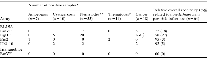
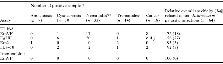
Diagnostic specificities linked to the different ELISAs and to potentially cross-reactive sera from patients with other parasitoses are listed in Table 2. Sera from patients with Taenia solium cysticercosis (n=10) showed low to moderate cross-reactivity in the EmVF-, Em2- and II/3–10-ELISAs (⩽2 cross-reactive sera) but high cross-reactivity in the EgHF-ELISA (6 cross-reactive sera). Also, some cross-reactivity was observed for sera from patients with nematode (n=33; 17, 20, 0 and 2 cross-reactive sera in the EmVF-, EgHF-, Em2-, and II/3–10-ELISAs, respectively) and trematode infections (n=14; 0, 1, 2 and 1 cross-reactive serum/sera in the EmVF-, EgHF-, Em2- and II/3–10-ELISAs, respectively). Sera from patients with hepatic amoebiasis were also assessed for possible cross-reactivity, because diagnostic images (by computer tomography or X-ray) of liver lesions due to this disease can be confused with those caused by AE. In this case, cross-reactivity was restricted to 1 serum sample, which was weakly positive in the Em2-ELISA. In summary, the overall specificities of the different ELISAs related to non-Echinococcus parasitic infections were determined to be 72% (EmVF-ELISA), 58% (EgHF-ELISA), 95% (Em2-ELISA) and 92% (II/3–10-ELISA).
Diagnostic specificities of crude, purified, and recombinant Echinococcus antigens in view of potential cross-reactions with serum antibodies from patients with various parasite infections or cancer malignancies
* Individual test results, see Fig. 1.
** Diseases (n): ascariasis (7), filariasis (6), strongyloidiasis (6), toxocariasis (7), trichinellosis (7).
† Diseases (n): fascioliasis (7), schistosomiasis (7).
§ The number of positive samples is given in parenthesis.
∫ Not done (n.d.), as preselected upon EgHF-positivity according to Pfister et al. (Reference Pfister, Gottstein, Cerny and Cerny1999).
Since the EgHF-ELISA used previously for the screening of CE was associated with relatively frequent false-positive reactions in cancer patients (Pfister et al. Reference Pfister, Gottstein, Cerny and Cerny1999; Poretti et al. Reference Poretti, Felleisen, Grimm, Pfister, Teuscher, Zürcher, Reichen and Gottstein1999), 18 such cross-reactive sera were specifically selected and tested in the different E. multilocularis-ELISAs. In the EmVF-ELISA, 8 of these sera scored positive and another 3 exhibited marginal reactivity (ELISA-value: ⩽−2 antibody units, AU). Conversely, in the Em2-ELSA, all samples from the panel of cross-reactive sera were test negative, but 9 of them showed marginal reactivity (⩽−2 AU). Within this panel, 2 sera were test positive in the II/3–10-ELISA, whereas 14 other sera gave low values (⩽−2 AU).
Serological evaluation of the EmVF-immunoblot
EmVF-immunoblot analysis using sera from AE patients revealed major immunoreactive bands of ∼65, 20–22 (triplet), and 8 (doublet) kDa (Fig. 1). While the molecules representing the 20–22 triplet and the 8 kDa doublet require biochemical characterization (see Discussion section), the 65 kDa band represents a full-length antigen II/3 (Felleisen and Gottstein, Reference Felleisen and Gottstein1993), supported by demonstrating the reactivity of this band employing a hyperimmune serum raised against antigen recII/3–10, a recombinant subfragment of E. multilocularis antigen II/3 (see Vogel et al. Reference Vogel, Gottstein, Müller and Seebeck1988; Müller et al. Reference Müller, Gottstein, Vogel, Flury and Seebeck1989). A preliminary evaluation of the blotting method in our diagnostic laboratory (data not shown) had indicated that neither the 65 kDa antigen II/3 nor the 8 kDa antigen achieved optimum diagnostic sensitivity or specificity. Therefore, all immunoblot strips obtained in the present study were scored with the criterion of positivity being a detectable signal for the 20–22 kDa triplet.
Immunoblot data and the corresponding ELISA-results of individual sera are shown in Fig. 1. The performance of immunoblotting with regard to sensitivity and specificity is summarized in Tables 1 and 2, respectively. When 62 sera from AE patients were tested in terms of their reactivity with the 20–22 kDa band triplet, the immunoblot revealed a diagnostic sensitivity of 100%. In particular, the immunoblot also detected all 8 inactive AE cases with degenerate liver lesions (Fig. 1). Here, respective testing consisted of 2 separate analyses (see Fig. 1; analysis 1 [panel of strips indicated as A1] and 2 [panel of strips indicated as A2]) that were performed with 2 different batches of EmVF antigen. As displayed in Fig. 1, in the second immunoblot analysis (A2), a few diagnostically irrelevant bands (e.g. a 10 kDa and 35 kDa bands) were more pronounced as compared with the first analysis (A1). Furthermore, the 20–22 kDa band triplet was not adequately resolved in the blot strips from analysis A2. However, despite this inconsistency, sera from all 8 inactive cases exhibited an appropriate profile for the 20–22 kDa band triplet.
The relatively high rate of cross-reactivity (50%) determined for sera from patients with CE (Table 1) demonstrated that the immunoblot was not suitable for a serological differentiation between AE and CE. However, the specificity of the immunoblot, considering sera from patients with parasitic infections other than Echinococcus, was 100% (Table 2). In this case, the scoring of strips incubated with sera from patients with T. solium cysticercosis was somewhat affected by the occasional detection of 2 faint bands above the diagnostically relevant 20–22 kDa triplet. However, strictly considering the diagnostic criteria (i.e. the position and nature of the immuno-reactivity of bands), such sera were scored as negative.
None of the 18 sera from cancer patients tested had non-specific reactivity with the diagnostic 20–22 kDa triplet. However, some of them reacted weakly with the 65 kDa band (9 positive sera) and the 8 kDa doublet (1 positive serum).
A possible antigenic relationship between the 65 kDa antigen II/3, the 20–22 kDa and the 8 kDa antigens was tested by incubating EmVF-blot strips with polyclonal antibodies specific to E. multilocularis antigen II/3- and affinity-purified polyclonal antibodies directed against the 20–22 or 8 kDa antigen. In this immunoblot (see Fig. 1, last panel), each of the 3 monospecific antibody fractions reacted individually with the corresponding antigen but did not exhibit detectable cross-reactivity with any of the other antigens on the blot. This result indicated that the 65, 20–22 and 8 kDa antigens are distinct and do not represent different breakdown products and/or conformational variants or isoforms of the same molecule.
DISCUSSION
Substantial efforts have been undertaken to maximize, or at least optimize, the perfomance of serological tests for the diagnosis of echinococcosis, and many studies have yielded novel test systems or antigens to enhance both diagnostic sensitivity and specificity (Gottstein et al. Reference Gottstein, Jacquier, Bresson-Hadni and Eckert1993; Poretti et al. Reference Poretti, Felleisen, Grimm, Pfister, Teuscher, Zürcher, Reichen and Gottstein1999; Liance et al. Reference Liance, Janin, Bresson-Hadni, Vuitton, Houin and Piarroux2000; Sako et al. Reference Sako, Nakao, Nakaya, Yamasaki, Gottstein, Lightowers, Schantz and Ito2002; Ito and Craig, Reference Ito and Craig2003; Xiao et al. Reference Xiao, Mamuti, Yamasaki, Sako, Nakao, Nakaya, Gottstein, Schantz, Lightowlers, Craig and Ito2003; Kormaz et al. Reference Korkmaz, Inceboz, Celebi, Babaoglu and Uner2004; Mamuti et al. Reference Mamuti, Yamasaki, Sako, Nakao, Xiao, Nakaya, Sato, Vuitton, Piarroux, Lightowlers, Craig and Ito2004). New epidemiological and clinical findings have shown unusual presentations of infections that also exhibit unusual serological profiles upon diagnosis. For example, hepatic lesions containing degrading or dead E. multilocularis cysts may stimulate weak anti-Em2-antibody responses, whereas anti-II/3–10 (and thus also anti-EM10, Em4 and/or Em18) responses are not detectable in such patients (Gottstein et al. Reference Gottstein, Bettens, Parkinson and Wilson1996; Zingg et al. Reference Zingg, Renner-Schneiter, Pauli-Magnus, Renner, van Overbeck, Schläpfer, Weber, Weber, Opravil, Gottstein and Speck2004; Matsumoto et al. Reference Matsumoto, Müller, Hemphill, Oku, Kamiya and Gottstein2006). Also, the technique of immunoblotting has gained increased attention for the diagnosis of echinococcosis (Ito et al. Reference Ito, Ma, Schantz, Gottstein, Liu, Chai, Abdel-Hafez, Altintas, Joshi, Lightowlers and Pawlowski1999; Poretti et al. Reference Poretti, Felleisen, Grimm, Pfister, Teuscher, Zürcher, Reichen and Gottstein1999; Kormaz et al. Reference Korkmaz, Inceboz, Celebi, Babaoglu and Uner2004). Crude or semi-purified antigens produced from the metacestode stage obtained from experimentally infected rodents have been used for these immunoblots. Two problems can be associated with this approach. One relates to the presence of host proteins in such extracts, which may relate to non-specific antibody reactivity to host epitopes or which may sterically cover and thus hide parasite epitopes otherwise accessible to antibodies.
In the present study, a significant improvement was achieved in that a highly sensitive (in relation to AE) and specific (regarding diseases other than AE and CE) immunoblot using axenically in vitro-produced vesicle fluid from E. multilocularis metacestodes as an antigen was developed. Although the corresponding E. granulosus hydatid fluid antigen can be readily obtained from fertile hydatid cysts, it has been shown to contain substantial amounts of host proteins that can lead to false-positive (anti-host-protein) reactions (Gottstein et al. Reference Gottstein, Eckert, Michael and Thompson1987). The in vitro culture of E. multilocularis metacestodes has been established as a valuable and commonly used alternative to animal experimentation, as reflected by numerous publications in the field (e.g. reviewed by Hemphill et al. Reference Hemphill, Stettler, Walker, Siles-Lucas, Fink and Gottstein2002; Brehm et al. Reference Brehm, Spiliotis, Zavala-Gongora, Konrad and Frosch2006). Preliminary experience with the EmVF antigen produced using the same procedure has already been gained previously by ELISA (Romig et al. Reference Romig, Kratzer, Kimmig, Frosch, Gaus, Flegel, Gottstein, Lucius, Beckh and Kern1999) and within the context of cell-immunological investigations of cytokines in both AE patients (Sturm et al. Reference Sturm, Menzel, Gottstein and Kern1995) and mice experimentally infected with E. multilocularis metacestode vesicles (Dai et al. Reference Dai, Hemphill, Waldvogel, Ingold, Deplazes, Mossmann and Gottstein2001).
The present culture system serves as a continuous source for an almost unlimited supply of EmVF antigen from a well-defined line of E. multilocularis, which is devoid of potentially interfering host components. This offers an excellent basis for the standardization of serological tests, as we have shown that antigenic variation can be expressed among different isolates of E. multilocularis from naturally infected rodents (Gottstein, Reference Gottstein1991). Compared with the other Echinococcus immunoblots established thus far (Ito et al. Reference Ito, Ma, Schantz, Gottstein, Liu, Chai, Abdel-Hafez, Altintas, Joshi, Lightowlers and Pawlowski1999; Poretti et al. Reference Poretti, Felleisen, Grimm, Pfister, Teuscher, Zürcher, Reichen and Gottstein1999; Liance et al. Reference Liance, Janin, Bresson-Hadni, Vuitton, Houin and Piarroux2000; Kormaz et al. Reference Korkmaz, Inceboz, Celebi, Babaoglu and Uner2004), the EmVF-blot exhibits a relatively simple immunoreactive banding profile, dominated by the diagnostic 20–22 kDa triplet. Although not yet proven experimentally, the similarity in antigenic abundance among the bands within the triplet suggests that the 20–22 kDa antigen represents a protein with differential conformations, post-translational modification (such as glycosylation) and/or degradation forms rather than a set of different proteins of similar sizes. Consistent with differential glycosylation is the finding that individual bands within this pattern are smeary.
In the present study, we tested the hypothesis that the protein(s) represented in the 20–22 kDa triple band correspond to the E. multilocularis protoscolex antigen Em18, identified previously (Sako et al. Reference Sako, Nakao, Nakaya, Yamasaki, Gottstein, Lightowers, Schantz and Ito2002) as an 18 kDa major proteolytic degradation product of the 65 kDa antigen II/3. However, specific antibodies purified to antigen II/3 bound to intact antigen II/3 (appearing on the EmVF-blot as a 65 kDa band) did not react with the 20–22 kDa triplet, refuting this hypothesis. Hence, the 20–22 kDa triplet does not appear to contain any antigen II/3 components. Interestingly, the 18 kDa band did not represent components of the EmVF-antigen. This may either be due to 1 or more biological characteristics of E. multilocularis, in that this band is detected in crude extracts of protoscoleces and/or when PMSF is added as a protease inhibitor to prevent proteolytic degradation (following the isolation of the fluid). Also, by demonstrating a lack of antibody cross-reactivity between the 20–22 kDa and the 8 kDa antigen (a subunit of antigen B) in the same immunoblot analysis, any immunological relationship between these 2 abundant antigens was not supported.
The present and previous investigations (Müller et al. Reference Müller, Gottstein, Vogel, Flury and Seebeck1989; Gottstein et al. Reference Gottstein, Jacquier, Bresson-Hadni and Eckert1993) have shown that recombinant antigen II/3–10 is an excellent diagnostic reagent, allowing a sensitive and specific diagnosis of AE by ELISA, confirmed by other researchers (Frosch et al. Reference Frosch, Frosch, Pfister, Schaad and Bitter-Suermann1991; Hemmings and McManus, Reference Hemmings and McManus1991; Xiao et al. Reference Xiao, Mamuti, Yamasaki, Sako, Nakao, Nakaya, Gottstein, Schantz, Lightowlers, Craig and Ito2003). Using immunoreactivity to the native 65 kDa antigen II/3 as a diagnostic criterion, we showed herein that the majority of AE and some CE sera tested positive, but also sera from patients with some other disorders did not completely lack reactivity. Such apparent cross-reactivity was also observed with the 8 kDa band known as the 8 kDa subunit of E. multilocularis antigen B (cf. Mamuti et al. Reference Mamuti, Yamasaki, Sako, Nakao, Xiao, Nakaya, Sato, Vuitton, Piarroux, Lightowlers, Craig and Ito2004). Since cross-reactivity complicates the scoring of the EmVF-immunoblot and the interpretation of the results, only the abundant 20–22 kDa bands were considered as diagnostic markers. This approach allowed the diagnosis of 100% of AE cases and also the serological detection of all cases with an inactive E. multilocularis infection. The immunoblot analysis did not consistently discriminate between AE and CE but exhibited a maximal specificity compared with other parasitoses. This finding was supported by results obtained by testing sera from patients suffering from various cancers. In a previous evaluation focused on the diagnostic specificity of the EgHF-ELISA (Poretti et al. Reference Poretti, Felleisen, Grimm, Pfister, Teuscher, Zürcher, Reichen and Gottstein1999), a substantial number (6·3%) of these sera tested false-positive, and this result was considered to complicate the serological diagnosis of echinococcosis. In the present study, only the EmVF- and II/3–10-ELISAs occasionally produced similar false-positive results, but this was not the case for the EmVF-immunoblot.
Taken together, the present study revealed that the EmVF-immunoblot ideally complements the Em2- and II/3–10-ELISAs for the serological diagnosis of AE. In combination with these 2 ELISAs, a reliable serodiagnosis of both active and inactive E. multilocularis infections in humans could be achieved. Prospectively, we plan to identify the nature of these 20–22 kDa proteins, to address their role in the host-parasite interplay and, if appropriate, to clone respective gene(s). We also plan to determine the value of the EmVF-immunoblot to monitor AE patients serologically after surgery and during long-term benzimidazole chemotherapy.
This work was supported by grants from the Swiss National Science Foundation (grant no. 31-11780/1), the Hans-Sigrist-Foundation of the University of Berne and the EU EchinoRisk-project QLK2-CT-2001-01995 (BBW no. 00·0586-1).