Introduction
International treatment guidelines recommend trauma-focused cognitive behavior therapy (TF-CBT) as the recommended treatment for post-traumatic stress disorder (PTSD) (Institute of Medicine, 2008; National Institute of Clinical Excellence, 2005). Despite the promise of TF-CBT, approximately one-third of patients still have PTSD after completing treatment (Bradley, Greene, Russ, Dutra, & Westen, Reference Bradley, Greene, Russ, Dutra and Westen2005). Accordingly, there is a need to determine the factors that prospectively identify patients who are not responsive to TF-CBT. One of the tenets of TF-CBT is the capacity to reappraise events and responses associated with the traumatic experience, such that they are experienced with less anxiety (Foa, Reference Foa2006). In healthy individuals, cognitive reappraisal of emotional information involves the activation of neural circuits within cognitive control networks that extend across the dorsomedial, dorsolateral, and ventrolateral prefrontal cortex, and posterior parietal lobe and modulation of the amygdala (Buhle et al., Reference Buhle, Silvers, Wager, Lopez, Onyemekwu, Kober and Ochsner2014; Ochsner, Silvers, & Buhle, Reference Ochsner, Silvers and Buhle2012). In contrast, attempted reappraisal of aversive emotional states in mood and anxiety clinical populations is associated with reduced recruitment of the dorsolateral prefrontal cortex (dlPFC) and ventrolateral prefrontal cortex coupled with enhanced activation of the amygdala (Zilverstand, Parvaz, & Goldstein, Reference Zilverstand, Parvaz and Goldstein2017). Consistent with this pattern, individuals with PTSD are deficient in the recruitment of the prefrontal cortical networks, particularly the dlPFC, and in the attenuation of the amygdala during reappraisal (New et al., Reference New, Fan, Murrough, Liu, Liebman, Guise and Charney2009; Rabinak et al., Reference Rabinak, MacNamara, Kennedy, Angstadt, Stein, Liberzon and Phan2014), which accords with the models that PTSD is characterized by deficits in emotion regulation (Etkin & Wager, Reference Etkin and Wager2007; Seligowski, Lee, Bardeen, & Orcutt, Reference Seligowski, Lee, Bardeen and Orcutt2015).
Previous neuroimaging studies have investigated the pre-treatment neural markers of TF-CBT response; poor response to TF-CBT has been associated with pre-treatment greater amygdala response during viewing fear faces (Bryant et al., Reference Bryant, Felmingham, Kemp, Das, Hughes, Peduto and Williams2008), enhanced anterior cingulate cortex activation in the anticipation of affective stimuli (Aupperle et al., Reference Aupperle, Allard, Simmons, Flagan, Thorp, Norman and Stein2013), and activation in the dorsal striatum and the orbitofrontal cortex during a Go-No Go inhibition task (Falconer, Allen, Felmingham, Williams, & Bryant, Reference Falconer, Allen, Felmingham, Williams and Bryant2013). Notably, only one study has investigated the capacity to engage reappraisal networks prior to TF-CBT as a predictor of TF-CBT response in PTSD patients (Fonzo et al., Reference Fonzo, Goodkind, Oathes, Zaiko, Harvey, Peng and Etkin2017); this study found that whereas better treatment response was predicted by greater dorsal prefrontal and less amygdala activation during emotional reactivity, activation during intentional regulation did not predict the treatment outcome. It has also been shown that pre-treatment activation of the bilateral insula and left dlPFC during reappraisal of aversive images predicts response to CBT in panic disorder patients (Reinecke, Thilo, Filippini, Croft, & Harmer, Reference Reinecke, Thilo, Filippini, Croft and Harmer2014). In summary, available evidence suggests that engagement of regulatory networks and downregulation of amygdala may be associated with a successful response to CBT (Goldin, Manber, Hakimi, Canli, & Gross, Reference Goldin, Manber, Hakimi, Canli and Gross2009; Klumpp, Fitzgerald, & Phan, Reference Klumpp, Fitzgerald and Phan2013; Paquette et al., Reference Paquette, Levesque, Mensour, Leroux, Beaudoin, Bourgouin and Beauregard2003; Straube, Glauer, Dilger, Mentzel, & Miltner, Reference Straube, Glauer, Dilger, Mentzel and Miltner2006).
The goal of the current study was to identify the neural markers of a successful response to TF-CBT in PTSD patients by having patients cognitively reappraise distressing stimuli during functional magnetic brain resonance imaging (fMRI) prior to the commencement of treatment. On the basis of evidence that reappraisal is associated with increased dlPFC and diminished amygdala activation during reappraisal in healthy individuals (Buhle et al., Reference Buhle, Silvers, Wager, Lopez, Onyemekwu, Kober and Ochsner2014), we predicted that heightened activation of the dlPFC and diminished recruitment of the amygdala during reappraisal prior to treatment would predict greater symptom reduction in PTSD patients treated with TF-CBT. Further, some evidence suggests better treatment outcome may be associated with reduced amygdala activation during emotional reactivity.
Materials and method
Participants
Participants were 46 treatment-seeking patients, 37 of whom were included in imaging analyses (with nine excluded for failing to complete the relevant task). The mean age of the 37 patients (17 females, 20 males) was 41.4 ± 11.1 years who developed PTSD following an assault, childhood abuse, motor vehicle accidents, or police duties which occurred on average 36.9 ± 31.9 months prior to treatment. PTSD was diagnosed by clinical psychologists using the Clinician Administered PTSD Scale (CAPS Blake et al., Reference Blake, Weathers, Nagy, Kaloupek, Gusman, Charney and Keane1995); a symptom is endorsed following the ‘2/1’ method, indicating at least ‘moderate’ distress and frequency of at least ‘once or twice a month’ (Blake et al., Reference Blake, Weathers, Nagy, Kaloupek, Gusman, Charney and Keane1995). Participants with a history of neurological disorder, moderate or severe traumatic brain injury, psychosis, bipolar disorder, or substance dependence were excluded. The protocol permitted prescribed medication if the dosage had remained stable for 2 months prior to the scan and was not altered during the course of the study; 10 (27.8%) participants were taking selective serotonin reuptake inhibitors. The study also included a comparison group of 24 healthy participants (12 females, 12 males) of mean age 35.0 ± 13.9 years who had never experienced a criterion A stressor and with no current Axis I disorders, as assessed by the Mini International Neuropsychiatric Interview (MINI version 5.5) (Sheehan et al., Reference Sheehan, Lecrubier, Harnett-Sheehan, Amorim, Janavs, Weiller and Dunbar1998). Depression and anxiety levels were also assessed by self-report on the Depression, Anxiety, and Stress Scale (DASS). Participant characteristics are described in Table 1.
Participant characteristics

Note. Standard deviations appear in parentheses.
PTSD, post-traumatic stress disorder; CAPS; Clinician Administered PTSD Scale,
** p < 0.001.
Procedure
The study was approved by the Western Sydney Area Health Service Human Research Ethics Committee and written informed consent was obtained from participants. Participants were initially assessed for PTSD (as defined by DSM-IV) by clinical psychologists using the CAPS and for comorbid Axis I disorders using the MINI was used to assess for a current major depressive episode, generalized anxiety disorder, social phobia, panic disorder, agoraphobia, obsessive-compulsive disorder, and a substance use disorder.
Reappraisal task
Participants were scanned using fMRI 2–3 weeks prior to the commencement of TF-CBT. During scanning, participants engaged in a reappraisal paradigm derived from prior neuroimaging studies of reappraisal (Ochsner, Bunge, Gross, & Gabrieli, Reference Ochsner, Bunge, Gross and Gabrieli2002). The task was presented in an fMRI block design comprising of two fMRI runs. Stimuli included 20 negative (mean valence ratings, 2.18; mean arousal ratings, 6.91) and 20 neutral (mean valence ratings, 5.05; mean arousal ratings, 3.2) images selected from the IAPS image collection (Lang, Bradley, & Cuthbert, Reference Lang, Bradley and Cuthbert2005) used across the two task runs. In each run, images were presented in three blocks (THINK, NEUTRAL, and WATCH) of 10 image trials with each trial 10 s long. Negative images were used for the THINK and WATCH trials, whereas neutral images were used for the NEUTRAL trials. Each negative image was used once in the THINK trial for one run and then for a WATCH trial in the second run. For each trial, the word WATCH or THINK appeared on the screen for 1.5 s, immediately followed by the image for 5 s and then by a rating screen for 3.5 s where participants had to rate each image based on how ‘negative you feel’ using a five-point scale (1 = not at all, 5 = extremely) via a button box in the scannerFootnote i. For the THINK trials, participants were instructed to down-regulate emotional responses by using the strategies of cognitive reappraisal that were modelled on previous reappraisal studies (Goldin, McRae, Ramel, & Gross, Reference Goldin, McRae, Ramel and Gross2008); for example, participants were instructed to assume the perspective of a movie director focusing on technical aspects of the scene. These were explained to participants prior to the scanning session, and examples are given to ensure that participants understood the procedure. On WATCH and NEUTRAL trials, participants were instructed to simply view the image. The order of the THINK and WATCH blocks was counterbalanced across the participants with the NEUTRAL block always presented in the middle. The block order was randomized across participants, and the same block order was used across the two task runs for each participant.
Within 2–3 weeks of scanning, participants commenced a course of 9 once-weekly individual 60–90 min sessions of TF-CBT that were delivered by experienced doctoral-level or masters-level clinical psychologists. Therapy involved an initial session of psychoeducation about psychological responses to trauma, then six sessions of 40 min imaginal exposure to the trauma memory, instructions regarding in vivo exposure to avoided situations, and cognitive restructuring of thoughts related to the traumatic event. An additional session reinforced cognitive restructuring exercises, and a final session focused on relapse prevention (Bryant et al., Reference Bryant, Mastrodomenico, Hopwood, Kenny, Cahill, Kandris and Taylor2013). This therapy procedure is consistent with gold standard TF-CBT protocols (McLean, Asnaani, & Foa, Reference McLean, Asnaani, Foa, Schnyder and Cloitre2015); although some treatment protocols allow up to 12 sessions of therapy, many require a minimum of nine sessions. Independent clinicians rated the fidelity of 130 sessions (18%), indicating full adherence to the treatment protocols and high level of quality on a seven-point scale (mean = 6.11, s.d. = 1.32). There were no treatment effects according to a different therapist or therapist qualifications. A post-treatment assessment using the CAPS was conducted by an independent clinical psychologist 1 week following completion of the course of treatment.
Imaging acquisition
Functional imaging was performed on a 3.0T GE Signa HDx scanner (GE Healthcare, Milwaukee, Wisconsin) using an echo-planar imaging protocol and an eight-channel head coil. A total of 120 functional T2*-weighted volumes were acquired in each task run, comprising 40 axial contiguous slices parallel to the intercommissural (AC-PC) line, with 3.5 mm thickness and TR = 2.5 s, TE = 27.5 ms, Flip angle = 90°; with FOV 24 × 24 cm2, matrix size 64 × 64. Three additional dummy scans were also acquired at the start of the sequence to allow magnetization to stabilize to steady state. A high-resolution T1-weighted anatomical structural image was also acquired in the sagittal plane using a 3D spoiled gradient echo (SPGR) sequence: TR = 8.3 ms; TE = 3.2 ms; flip angle = 11°; TI = 500 ms; NEX = 1; ASSET = 1.5; Frequency direction: S/I; matrix size = 256 × 256; 180 contiguous 1 mm slices covering the whole brain resulting in a 1 mm3 isotropic voxel resolution. This sequence was collected for use in the normalization of the fMRI data to standard space.
Data analyses
Pre-processing (realignment and unwarping, spatial normalization into standardized MNI space, smoothing using an 8 mm FWHM isotropic Gaussian kernel), connectivity, and statistical analysis of fMRI data were conducted using Statistical Parametric Mapping (SPM8, Wellcome Department of Neurology, London). Motion correction was done by realigning and unwarping fMRI images to the initial image of each task run. For normalization to stereotactic MNI space, the T1-weighted SPGR images were normalized to standard space using the FMRIB non-linear registration tool and the fMRI EPI data were coregistered to the T1 data using FMRIB linear registration tool. The normalization warps from these procedures were stored for use in functional to standard space transformations. To account for any physiological noise, a signal corresponding to CSF and white matter was estimated using a mask in the ventricles and white matter, and was removed from the motion-corrected fMRI time series. All fMRI data were smoothed using an 8 mm Gaussian kernel. In the first-level analysis, a hemodynamic response convolved boxcar function was used to model the Blood Oxygenation Level-Dependent (BOLD) response for each of the three conditions (THINK, NEUTRAL, and WATCH) in a general linear model framework. As done in previous reappraisal fMRI studies, contrast images were calculated for THINK v. WATCH trials to determine BOLD responses to cognitive reappraisal, and for WATCH v. NEUTRAL trials to determine BOLD responses to emotional reactivity. These contrast images were normalized to standard space using normalization warps estimated in the preprocessing steps outlined above.
To examine the change in PTSD severity independent of initial severity, residual change was calculated from a regression of pre-treatment total CAPS scores on post-treatment total CAPS scores (Siegle, Carter, & Thase, Reference Siegle, Carter and Thase2006), and were correlated with neural measures.
Firstly, we evaluated group-specific activations and group differences in neural activations for the two contrasts for the PTSD and healthy control groups as voxel-wise single- and two-sample t tests. Then voxel-wise regressions analyses of residual PTSD severity on fMRI signal for both contrasts were used to detect pre-treatment neural activity associated with treatment recovery. To test the specific hypotheses regarding neural activation associated with cognitive reappraisal, we employed coordinates specified in the meta-analyses of neuroimaging studies of cognitive reappraisal in healthy participants, which identify the dlPFC and dmPFC as the primary prefrontal cortical regions subserving cognitive reappraisal in healthy individuals (Buhle et al., Reference Buhle, Silvers, Wager, Lopez, Onyemekwu, Kober and Ochsner2014) but deficient during reappraisal in PTSD patients (Rabinak et al., Reference Rabinak, MacNamara, Kennedy, Angstadt, Stein, Liberzon and Phan2014).
Regions of interest (ROIs) for the dlPFC were defined using 10 mm radius spheres centered on these coordinates: left – (−36, 15, 57) and (−51, 12, 21) and right – (51, 15, 48) and (42, 30, 39), and for the dmPFC using a 10 mm radius sphere centered on these coordinates: (0, 18, 42). In addition, we included the amygdala, defined from the Automated Anatomical Labelling (AAL) atlas (Tzourio-Mazoyer et al., Reference Tzourio-Mazoyer, Landeau, Papathanassiou, Crivello, Etard, Delcroix and Joliot2002), because of its role in emotional response.
To test the hypotheses regarding neural activation associated with emotional reactivity, we employed the use of the negative affect network [comprising of the subgenual (sgACC) and pregenual anterior cingulate cortex (pgACC), bilateral amygdala, insula, and hippocampus], chosen for its relevance to neural circuitry of negative emotion processing (Williams, Reference Williams2016). ROIs for this network were defined using the AAL atlas (for bilateral amygdala, insula, and hippocampus), and meta-analyses for sgACC/pgACC (Kober et al., Reference Kober, Barrett, Joseph, Bliss-Moreau, Lindquist and Wager2008). These two ROIs were defined using spheres centered on these coordinates: sgACC (0, 24, −8) and pgACC (0, 42, 4).
Next, we conducted functional connectivity analyses (generalized psychophysiological interactions (gPPI)] (McLaren, Ries, Xu, & Johnson, Reference McLaren, Ries, Xu and Johnson2012) using significant clusters from the activation analyses to determine whether neural connectivity for those seeds was similarly associated with treatment response. The details regarding the gPPI model and analyses are outlined in the online Supplementary Methods section. We evaluated connectivity related to both cognitive reappraisal (THINK v. WATCH) and emotional reactivity (WATCH v. NEUTRAL) contrasts in second-level voxel-wise ROI analyses testing for correlations between connectivity and residual PTSD severity as done for activation previously. The same ROIs described above were respectively included for cognitive reappraisal and emotional reactivity analyses. All voxel-wise statistical analyses were conducted at the family-wise error-corrected voxel p level of 0.05 (small volume corrected p FWE < 0.05). For both activation and connectivity, we also performed exploratory whole-brain voxel-wise analyses (p FWE < 0.05) to test any effects beyond our hypothesized ROIs.
A series of additional analyses were also conducted: (1) to evaluate generalizability to further samples and to determine accuracy, sensitivity, and specificity of the measures in predicting symptom improvement and treatment response (responders were defined as those with 50% reduction in pre-treatment CAPS) using cross-validation analyses, with a general model and a best-predictive fit model; (2) to compare activation and connectivity neural profiles for treatment responders and non-responders relative to healthy controls; (3) to determine whether neural measures predicted treatment response above and beyond traditional clinical and demographic variables; and (4) to evaluate any confounds due to medication use on neural measures.
Results
Clinical outcomes
All 37 included participants met DSM-IV criteria for PTSD prior to treatment, and all participants completed 7–9 sessions of TF-CBT. The mean CAPS score was 67.8 ± 19.1 at pretreatment and 28.4 ± 20.0 following TF-CBT treatment. Based on our set criteria, 25 (67.6%) participants classified as treatment responders and 12 (32.4%) were classified as non-responders. Participant characteristics are outlined in Table 1.
Cognitive reappraisal and emotional reactivity activations
The neural activations for cognitive reappraisal (THINK v. WATCH contrast) in the hypothesized brain regions for the healthy control group and as a comparison between the PTSD and healthy control groups are listed in online Supplementary Tables S3 and S4. As expected, controls and PTSD participants were found to significantly activate the dmPFC and dlPFC bilaterally (p FWE < 0.05) during reappraisal relative to passive viewing. Also, PTSD participants demonstrated significantly lower activations in both these cortical regions relative to controls (p FWE < 0.05). Activations in the amygdala were not significant for either group or significantly different between the two groups, although both healthy controls and PTSD exhibited the expected direction trend of decrease in activation during cognitive reappraisal at an uncorrected level.
For emotional reactivity (WATCH v. NEUTRAL contrast), there was a significant decrease in the right insula activation only for the control group, with no significance between group differences in either of the hypothesized ROIs. As above, both healthy controls and PTSD exhibited the expected direction trend of increase in amygdala activation during emotional reactivity only at an uncorrected level.
The PTSD group had significantly lower connectivity than controls between the left amygdala and left hippocampus only for emotional reactivity (p FWE < 0.05; online Supplementary Table S6). None of the connectivity measures were significantly different between the groups for cognitive reappraisal (online Supplementary Tables S5).
Exploratory whole-brain analyses for these comparisons are presented in the online Supplementary Section (Tables S1 and S2).
Between symptom change and neural measures
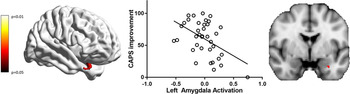
Significant correlations between PTSD symptom improvement (reduction in CAPS scores) and pretreatment neural measures were observed for both reappraisal and emotion reactivity contrasts. For cognitive reappraisal, a significant negative correlation was found between the reduction in PTSD severity and left amygdala activation (Table 2; Fig. 1). Left amygdala connectivity during reappraisal was not significantly related to symptom improvement.
Significant correlations between amygdala reactivity during cognitive reappraisal with a change in PTSD severity. Three-dimensional brain image shows amygdala region that is significantly correlated with a change in symptom improvement (at an uncorrected p value <0.05, for visualization purposes) for the cognitive reappraisal contrast (THINK v. WATCH). The scatter plot represents patient's symptom improvement with their brain activation at pre-treatment, using extracted β values (contrast estimates) for the significant cluster (p FWE < 0.05) displayed on the 2D coronal slice adjacent to the plot.

Summary of voxel-wise region of interest correlation analyses for cognitive reappraisal and emotional reactivity contrasts

Note. Regions of interest are reported significant at voxel-wise small volume corrected p FWE < 0.05.
PTSD, posttraumatic stress disorder; HC, healthy controls; L, left; R, right; sgACC, subgenual anterior cingulate cortex; pgACC, pregenual anterior cingulate cortex.
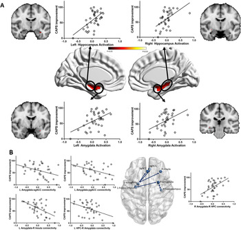
For emotional reactivity, significant positive correlations were found between reduction in PTSD severity and bilateral amygdala and hippocampal activations (Table 2; Fig. 2a). Connectivity of the left amygdala to the sgACC, pgACC, and right insula, as well as the left hippocampal connectivity to the right amygdala were all significantly negatively correlated with symptom improvement. In contrast, right amygdala connectivity to the right hippocampus was positively correlated with symptom improvement (Table 2; Fig. 2b).
Significant amygdala and hippocampus activation (a) and connectivity (b) during emotion reactivity correlated with improvement in PTSD symptoms. Three-dimensional brain images show amygdala and hippocampal regions (a) that are significantly correlated with a change in symptom improvement (at an uncorrected p value <0.05, for visualization purposes; 2D coronal brain images depict the p FWE < 0.05 significant cluster) for the emotional reactivity contrast (WATCH v. NEUTRAL). The brain map (b) depicts connectivity between the negative affect network that is significantly correlated with a change in symptom improvement. The scatter plots represent patient's symptom improvement with their brain activation (a) or connectivity (b) at pre-treatment, using extracted β values (contrast estimates) for the significant clusters (p FWE < 0.05).

To further evaluate if the correlations with treatment response were dissociable between the reappraisal and reactivity contrasts particularly for the amygdala which was significant for both these contrasts and also to evaluate if these effects were driven by a particular condition within each contrast, we evaluated amygdala associations for each condition separately relatively to the implicit baseline (i.e. THINK v. baseline, WATCH v. baseline, and NEUTRAL v. baseline) with symptom change. However, amygdala activation for neither of these contrasts was significantly associated with symptom improvement, which suggests that the significant correlations for the cognitive reappraisal and emotional reactivity contrasts are dissociated and are evident only for the difference between the respective conditions.
These effects were significant controlling for medications (online Supplementary Section). To index whether low baseline symptom scores affected significant results, these findings were re-analyzed controlling for baseline symptom severity; all originally observed effects remained significant. Similarly, to identify whether differences between the groups were driven by the difference in the DASS anxiety measures, the findings were re-analyzed controlling for these scores; all originally observed effects remained significant. To determine the power of our sample (n = 37) for detecting the observed effects, a post-hoc power analysis indicated that the achieved power was 45% for medium (Cohen's d = 0.3) and 90% for large (d = 0.5) effect sizes; suggesting there was limited power to detect small to medium effect sizes.
Comparison between treatment responders, non-responders, and healthy controls
To compare baseline patterns of responders and non-responders in relation to healthy controls, planned comparisons were conducted on the extracted β values from significant clusters of activation and connectivity for the cognitive reappraisal and emotional reactivity contrasts associated with a change in CAPS.
Of all the significant measures associated with symptom change in the main analyses, significant main effects of group (ANOVA) were present for eight of the 10 measures and as expected these were mainly driven by differences between responders and non-responders (Table 3). Post-hoc group differences relative to controls were observed only for the left amygdala–right insula connectivity for emotional reactivity contrast, with non-responders having greater pre-treatment connectivity than controls (and responders). In comparison, responders had similar connectivity levels as compared to controls.
Responders and non-responders v. controls in activation and connectivity for cognitive reappraisal and emotional reactivity

Note. Extracted betas for significant activation and connectivity results for each contrast are included. Brain measures were included in ANOVA comparing all groups, as well as planned comparisons between each group.
L, left; R, right; HPC, hippocampus; sgACC, subgenual anterior cingulate cortex; pgACC, pregenual anterior cingulate cortex.
Predictive models of treatment response
To illustrate how the neural markers we identified could be helpful in a treatment decision and to inform future studies, we evaluated the predictive value of neural measures compared to demographic and clinical measures, the generalizability of the models using cross-validation statistics, and the best predictive models to predict treatment outcome. These analyses are detailed in the online Supplementary Section.
In our cohort, the demographic or clinical measures model alone did not significantly predict treatment response. However, the addition of neural measures significantly improved predictive value (p < 0.001). Cross-validation linear regression analyses for a general model using mean fMRI signal from all our predefined ROIs (without any bias of our findings from the main analysis), replicated to some extent the correlation findings from the main analysis described above with moderate cross-validation predictive accuracies (online Supplementary Table S7). The best predictive model in classifying responders and non-responders, using the neural measures identified in our main analyses, identified left hippocampal activation and left amygdala–right insula connectivity for the emotional reactivity contrast to be the only two best predictive features (online Supplementary Table S9).
Discussion
This study aimed to identify the neural patterns during cognitive reappraisal of aversive material prior to treatment that can predict response to TF-CBT in a cohort of PTSD patients. The primary findings of this study were that better treatment response was (a) negatively associated with left amygdala activation during reappraisal at baseline, (b) positively associated with bilateral amygdala and hippocampal baseline activations during emotional reactivity, and (c) negatively associated with baseline connectivity of the left amygdala to the sgACC, pgACC, and right insula, and with connectivity between the left hippocampus and right amygdala.
The finding that reduced amygdala activation during reappraisal predicted better treatment response is consistent with evidence from meta-analytic studies that cognitive reappraisal involves reduced amygdala activation (Buhle et al., Reference Buhle, Silvers, Wager, Lopez, Onyemekwu, Kober and Ochsner2014). This also accords with evidence that cognitive reappraisal reduces anxiety (Gross, Reference Gross1998). This finding also fits with the proposition that TF-CBT aims to down-regulate anxiety, as part of the extinction process (Foa, Reference Foa2006). Clinical studies have shown that a poor response to TF-CBT is associated with very elevated anxiety prior to treatment (Blanchard et al., Reference Blanchard, Hickling, Malta, Jaccard, Devineni, Veazey and Galovski2003), which may limit the capacity of patients to regulate their distress during the therapy exercises. It is therefore possible that a positive response to TF-CBT is more likely when a patient is adept at down-regulating the amygdala during reappraisal. The prior investigation of neural processing during reappraisal predicting PTSD outcomes following TF-CBT found no evidence of amygdala activation during reappraisal predicting the outcome (Fonzo et al., Reference Fonzo, Goodkind, Oathes, Zaiko, Harvey, Peng and Etkin2017); it is difficult to identify the reasons for the different amygdala findings across these two studies because both used the same affective stimuli and comparable reappraisal instructions. It is possible that the results were influenced by each study embedding the reappraisal task among a range of diverse fMRI paradigms, which may have impacted amygdala response. In any case, the current finding is novel in that it provides the first evidence that being able to regulate the amygdala during reappraisal predicts treatment response. We note that an earlier study found that reduced amygdala activation was associated with better treatment response to TF-CBT in PTSD patients (Bryant et al., Reference Bryant, Felmingham, Kemp, Das, Hughes, Peduto and Williams2008); however, this study involved passive viewing of subliminal presentations of fearful faces and so involved markedly different task demands than the current reappraisal paradigm.
The observation that greater improvement following TF-CBT was associated with stronger activation of the amygdala during the WATCH trials of the reappraisal task suggests that treatment response is predicted by engagement with emotional processing. A central tenet of TF-CBT is that the individual can engage emotional experiences in order for the relevant affective responses to be processed and mastered (Parsons & Ressler, Reference Parsons and Ressler2013). In this sense, the current finding may reflect the difficulty of patients who do not readily engage the amygdala during emotional processing to utilize TF-CBT. This finding accords with the evidence of reduced activation of affective networks in PTSD patients with the dissociative subtype of PTSD (Lanius et al., Reference Lanius, Vermetten, Loewenstein, Brand, Schmahl, Bremner and Spiegel2010), who do not respond optimally to TF-CBT (Wolf, Lunny, & Schnurr, Reference Wolf, Lunny and Schnurr2016). This finding needs to be considered in the context of earlier studies have found that amygdala activation was not associated with treatment response following pharmacological (Nitschke et al., Reference Nitschke, Sarinopoulos, Oathes, Johnstone, Whalen, Davidson and Kalin2009; Whalen et al., Reference Whalen, Johnstone, Somerville, Nitschke, Polis, Alexander and Kalin2008) and psychotherapeutic (Ball, Stein, Ramsawh, Campbell-Sills, & Paulus, Reference Ball, Stein, Ramsawh, Campbell-Sills and Paulus2014) treatments. It is worth noting that none of these studies employed a reappraisal paradigm. The findings that better treatment response in the current study was associated with increased amygdala activation during emotional reactivity but decreased amygdala activation during reappraisal suggests that TF-CBT involves the dual abilities to engage emotions during a task that requires emotional processing and also to down-regulate emotions when required. Contrary to expectation, we did not observe that the predictive role of down-regulation of amygdala activation was accompanied by enhanced recruitment of regulatory networks. In contrast, activation of the bilateral insula and left dlPFC during reappraisal has been shown to predict response to CBT in panic disorder patients (Reinecke et al., Reference Reinecke, Thilo, Filippini, Croft and Harmer2014). Further, a study of patients with panic disorder and generalized anxiety disorder found that greater activation of the dlPFC during down-regulation of emotions predicted response to CBT (Ball et al., Reference Ball, Stein, Ramsawh, Campbell-Sills and Paulus2014). Interestingly, the only prior study of the role of reappraisal predicting PTSD psychotherapy outcome also found that regulatory networks did not predict treatment response (Fonzo et al., Reference Fonzo, Goodkind, Oathes, Zaiko, Harvey, Peng and Etkin2017), which was interpreted as being consistent with the proposition that successful mastery of fear during therapy involves engagement with emotions whilst not inhibiting emotional reactions with cognitive strategies (Craske et al., Reference Craske, Kircanski, Zelikowsky, Mystkowski, Chowdhury and Baker2008). The earlier study of reappraisal predicting psychotherapy response for PTSD did find that better treatment response was predicted by greater ventromedial prefrontal/ventral striatal activation during a task that involved implicit regulation of emotional conflict. This may suggest that individuals who respond well to therapy are those with a natural tendency to engage in the top-down regulation of emotional stimuli. This interpretation accords with our observation that better treatment response was associated with reduced amygdala activation during reappraisal, despite the absence of enhanced regulatory network activation during intentional reappraisal.
The finding that poor treatment response was associated with increased connectivity between the amygdala and regulatory networks (sgACC/pgACC/insula) during emotional reactivity trials can be considered in the context of the proposal that coupling of regulatory (frontal) networks and the amygdala are important for emotion regulation (Cisler et al., Reference Cisler, Steele, Lenow, Smitherman, Everett, Messias and Kilts2014; Goldin et al., Reference Goldin, Ziv, Jazaieri, Hahn, Heimberg and Gross2013). The direction of the relationship between these networks is mixed across studies. Whereas numerous studies have reported that down-regulation of aversive states is associated with negative connectivity between the amygdala and frontal cortical regions (Ochsner et al., Reference Ochsner, Bunge, Gross and Gabrieli2002; Urry et al., Reference Urry, van Reekum, Johnstone, Kalin, Thurow, Schaefer and Davidson2006), others report positive connectivity (Banks, Eddy, Angstadt, Nathan, & Phan, Reference Banks, Eddy, Angstadt, Nathan and Phan2007; Paschke et al., Reference Paschke, Dorfel, Steimke, Trempler, Magrabi, Ludwig and Walter2016). Use of reappraisal strategies is linked to the negative coupling of the amygdala with the frontal cortex (Pico-Perez et al., Reference Pico-Perez, Alonso, Contreras-Rodriguez, Martinez-Zalacain, Lopez-Sola, Jiminez-Murcia and Soriano-Mas2018). Moreover, successful emotion regulation during reappraisal is predicted by negative resting-state functional connectivity between the amygdala and the medial frontal cortex (Uchida et al., Reference Uchida, Biederman, Gabrieli, Micco, de Los Angeles, Brown and Whitfield-Gabrieli2015) and one previous smaller study reported negative connectivity between the amygdala and the insula during cognitive reappraisal predicted subsequent successful response to TF-CBT for PTSD (Cisler et al., Reference Cisler, Sigel, Steele, Smitherman, Vanderzee, Pemberton and Kilts2016). It is possible that decreased amygdala-regulatory circuit connectivity reflects successful inhibition of anxiety. Accordingly, patients who did not respond optimally to treatment may have a tendency to overregulate the amygdala because of their difficulty in top-down regulation. Put together with the finding that poorer treatment response was associated with decreased amygdala activation during reactivity, it is possible that optimal TF-CBT response occurs in patients who are able to engage emotion processing networks, and who have reduced connections between emotion and regulatory regions because of their capacity to also down-regulate their emotions.
It is worth noting that treatment non-responders were distinguished from responders in terms of activation of the left amygdala during reappraisal, and bilateral amygdala and hippocampus during emotion reactivity. These patterns underscore the aforementioned conclusion that PTSD patients who are able to benefit from TF-CBT are better able to down-regulate amygdala during reappraisal, and engage emotion processing networks during emotional reactivity tasks. The finding that treatment responders had reduced amygdala–insula connectivity relative to non-responders can be interpreted in terms of the latter needing to exert greater regulatory effort to reappraisal than responders. There is increasing evidence amygdala–insula pathways are implicated in fear conditioning and extinction (Shiba et al., Reference Shiba, Oikonomidis, Sawiak, Fryer, Hong, Cockcroft and Roberts2017), and elevated activation in both regions is noted in anxiety disorders (Greco & Liberzon, Reference Greco and Liberzon2016). The finding that responders had comparable connectivity between these two regions, but distinct from non-responders, points to the possibility that PTSD patients who are more likely to respond TF-CBT are those with the capacity to recruit networks, and connections between networks, that result in greater efficiency of emotion regulation.
A number of methodological limitations are noted. First, we emphasize that we did not include a wait-list comparison group, which would delineate the different predictors of TF-CBT and spontaneous remission; however, the latter explanation would not be expected considering that the mean time since trauma exposure was 36 months. Second, a proportion of participants were taking antidepressant medication prior to their course of TF-CBT, and it is possible that antidepressants may interact with TF-CBT; however, the medication dosage was stabilized prior to the commencement of therapy and the dosage was not altered throughout the study. Our supplementary analysis of patients who were medication-free did not find antidepressant use to significantly impact the relationship between pretreatment neural activity and symptom change. Third, the reappraisal paradigm used in this study targets only one mechanism involved in TF-CBT and may not reflect other core processes, including extinction learning (Reinhardt et al., Reference Reinhardt, Jansen, Kellerman, Schuppen, Kohn, Gerlach and Kircher2010). Fourth, despite the reasonable sample size, greater confidence in these findings would be achieved with a large sample size; despite the challenges of achieving large samples of treatment completers who have undergone fMRI testing, future studies predicting treatment outcomes should strive for significantly larger samples. Our sample size, combined with the number of factors that were entered into our predictive model, may have contributed to the modest prediction in the regression analyses after cross-validation. We deliberately tested this using all our predefined ROIs instead of the neural features identified in our main analysis to avoid double dipping. However, the best predictive neural model identified in our cohort is likely to improve this predictive accuracy and warrants testing in an independent cohort. Relatedly, we note that whereas we observed task effects for the PTSD sample for both reappraisal and reactivity tasks, these did not survive the more stringent p FWE < 0.05 threshold (where only right insula and bilateral hippocampal activation during emotion reactivity survived); this pattern suggests that these effects may have been observed with greater statistical power. Fifth, all patients underwent TF-CBT; future studies could usefully compare neural predictors of different forms of psychotherapy and pharmacotherapy to determine if there are overlapping or distinct neural networks predictive of outcome. Sixth, due to technical reasons, we did not obtain sufficiently reliable ratings of emotion in the scanner, which precluded assessing the extent to which the neural responses corresponded with emotional regulation. Finally, the capacity of pre-treatment neural signals during reappraisal and reactivity to predict longer-term follow-up of TF-CBT would provide much-needed information about the prediction of sustainability of treatment effects. In light of these limitations, together with the modest effects observed, the current findings need to be interpreted cautiously, and it is important that these findings are replicated with larger sample sizes and appropriate no-treatment control conditions to confirm the current findings.
In summary, this study indicates that a capacity to engage the amygdala during emotion processing but also inhibit the amygdala during reappraisal may be an important marker of the propensity to respond successfully to TF-CBT. Considering the important role of being able to down-regulate emotional responses during TF-CBT, this finding elucidates an important neural mechanism underpinning treatment response. In the context of one-third of PTSD patients not responding to TF-CBT, identifying this network advances our knowledge of biomarkers of treatment response for PTSD and sheds light on the possibility of enhancing treatment outcome by compensating for deficits in reappraisal-related networks. For example, initial evidence suggests that transcranial magnetic stimulation targeting the dlPFC can enhance the effects of TF-CBT (Kozel et al., Reference Kozel, Motes, Didehbani, DeLaRosa, Bass, Schraufnagel and Hart2018); using this approach may activate networks relevant to optimizing response to TF-CBT. The finding that self-reported clinical measures did not predict treatment outcome underscores the relevance of biological predictors of therapy response. Noting the logistic barriers and costs of brain imaging, there is also a need to develop other readily accessible biological markers of treatment response, such as psychophysiological measures.
Supplementary material
The supplementary material for this article can be found at https://doi.org/10.1017/S0033291720001129.
Financial support
This study was supported by the National Health and Medical Research Council Program Grant (1073041), NHMRC CCRE Grant (455431), and the Centenary of Anzac Centre, a Department of Veterans' Affairs funded initiative of Phoenix Australia. The study sponsor had no role in the design and conduct of the study; collection, management, analysis, and interpretation of the data; preparation, review, or approval of the manuscript; and decision to submit the manuscript for publication.
Conflict of interest
No authors are declaring a conflict of interest.