Introduction
The spirurid nematodes, in the adult stage, are obligatory parasites of vertebrates and require one or more intermediate arthropod hosts (Stunkard, Reference Stunkard1953). Insects serve as the intermediate host of species of the genus Physaloptera Rudolphi, 1819 (Gray & Anderson, Reference Gray and Anderson1982), while reptiles and amphibians serve as paratenic hosts (Widmer, Reference Widmer1970). Nematodes of the Physaloptera genus have been reported as parasites of amphibians, reptiles, birds and mammals, mainly in the stomach but sometimes also in the small intestine (Ortlepp, Reference Ortlepp1922, Reference Ortlepp1937).
Physaloptera bispiculata Vaz & Pereira, Reference Vaz and Pereira1935 (Spiruridae: Nematoda) is a parasite of Nectomys squamipes Brants, 1827 (Rodentia: Cricetidae), a water rat that only occurs in Brazil. Two studies report a high prevalence of physalopterid species in N. squamipes, probably causing similar lesions as those described for Turgida turgida (Nettles et al., Reference Nettles, Prestwood and Davidson1975; Alden, Reference Alden1995), since they were also found attached to the stomach wall. The P. bispiculata species was re-evaluated by Mafra & Lanfredi (Reference Mafra and Lanfredi1998) based on morphometric and morphological data such as lips, papillae, teeth, amphids, and number and arrangement of the caudal papillae.
Nematode reproduction is usually sexual. Males are usually smaller than females (often much smaller) and often have a characteristically bent tail to hold the female for copulation. During copulation, one or more chitinized spicules move out of the cloaca and are inserted into the female's genital pore. Amoeboid sperm then crawl along the spicule into the female worm. Nematode sperm cells are thought to be the only eukaryotic cells without globular protein. Following fecundation, the female produces eggs, which can be embryonated or unembryonated when passed by the female, in the latter case meaning the fertilized eggs cannot develop (Blaxter et al., Reference Blaxter, De Ley, Garey, Liu, Scheldeman, Vierstraete, Vanfleteren, Mackey, Dorris and Frisse1998).
Materials and methods
Naturally infected rodents were captured in the municipality of Rio Bonito in Rio de Janeiro state, Brazil. The abdominal and thoracic cavities were opened and the organs were placed separately in Petri dishes, kept in physiological saline and dissected under a stereomicroscope to obtain the helminths. Physaloptera bispiculata was collected from the stomach. Worms were washed twice in saline to remove tissue debris and fixed in 3 ml glacial acetic acid, 7 ml of 37% formaldehyde and 90 ml of 70% ethanol (AFA) at 60°C and maintained in the same solution. For light microscopy, the nematodes were clarified in phenol 80% (70% ethanol and phenolic acid) and mounted on temporary slides. Morphological analyses were made using differential interferential contrast (DIC) microscopy and the images were captured with an Olympus DP-12 digital camera coupled to an Olympus BX-51 light microscope. For scanning electron microscopy, the worms were washed in 0.1m cacodylate buffer, pH 7.2, postfixed in 1% OsO4 and 0.8% K3Fe(CN)6, dehydrated in graded ethanol (30–100%) for 2 h, critical-point dried in CO2, sputter-coated with gold and examined under a JEOL JSM 5310 scanning electron microscope at 15 kV. The procedure adopted in this work was approved by the animal research committee of the Instituto de Biofísica Carlos Chagas Filho, UFRJ. Voucher specimens were deposited at the Instituto Biológico de São Paulo (numbers 1583 A and B, one male and one female, respectively) and at the Natural History Museum, London (numbers 1997.2.3.5–7). The remaining specimens are in the Laboratório de Biologia de Helmintos Otto Wucherer collection.
Results and discussion
Nematodes were collected from the stomach, where they were usually firmly attached to the stomach wall. The morphology of the anterior end in both sexes shows two large lateral simple pseudolips, delimiting the cephalic end from the body and provided with papillae; a cuticle forming a cephalic collaret; and deirids located anterior to the nerve ring. The pattern of the male caudal region and the cloacal papillae arrangement, observed by light microscopy, allowed the specimens to be identified as Physaloptera bispiculata Vaz & Pereira, Reference Vaz and Pereira1935. Eleven N. squamipes were parasitized by P. bispiculata, with a total of 45 nematodes (40 adults and 5 juveniles; 33 females and 12 males).
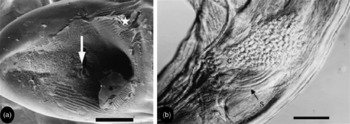
Scanning electron microscopy of the posterior end of the male P. bispiculata worms showed the morphological features described by Mafra & Lanfredi (Reference Mafra and Lanfredi1998). Some worms had an adhesive secretion, produced by the prostate glands, covering the ventral region, with some eggs adhering to the cloacal opening (fig. 1a).
Physaloptera bispiculata male. (a) Scanning electron microscopy showing the ventral side of the posterior end covered by cement (star) and the cloacal opening with eggs adhered to it (arrow) (scale bar 200 μm). (b) Light microscopy of cloacal region (C) full of eggs, and spicule (S) (scale bar 100 μm).

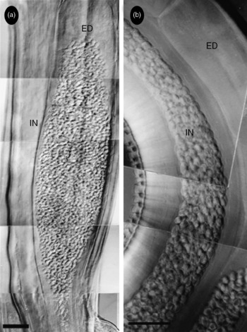
The male ejaculatory duct is dorsally situated to the digestive tract and located at the rear portion of the body (fig. 1b). This structure is elongated and piriform, with its thickest portion located away from the cloaca (fig. 1b). The intestine, ejaculatory duct and pouch containing a pair of spicules open at the cloaca (fig. 2a).
Physaloptera bispiculata. Light microscopy of the male posterior region. Ejaculatory duct (ED) and intestine (IN) with uncountable eggs. Scale bars: (a) 100 μm; (b) 250 μm.

Observations of male P. bispiculata worms by light microscopy revealed nine male worms containing embryonated eggs in the cloaca (fig. 1b), ejaculatory duct (fig. 2a and b) and rear portion of the intestine (fig. 2b). The number of eggs ranged from only a few to an uncountable number. This is an intriguing feature. Our suggestion is that the muscular contractions of the female associated with the back-and-forth movement of the spicules in the vagina and the body contraction of the male during copulation probably pump and/or suck the eggs from the female uterus and vagina into the reproductive tubule and sometimes into the intestine of the male. The male probably does not have the ability to expel the eggs, and for this reason a large quantity were found in these organs. On the other hand, this could be an important adaptation for the parasite, i.e. when male worms are expelled by the host they can carry a large number of eggs and, if eaten by intermediate hosts, this allows the larvae to infect these new hosts.
As far as we know this is the first report of physalopterid nematode eggs harboured in the cloacal region, ejaculatory duct or intestine.
Acknowledgements
Brazilian financial support: Conselho Nacional de Desenvolvimento Científico e Tecnológico (CNPq), Fundação Carlos Chagas Filho de Amparo a Pesquisa do Estado do Rio de Janeiro (FAPERJ) and Coordenação de Aperfeiçoamento de Pessoal de Nível Superior, CAPES-PROCAD.