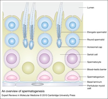
Animal models have been indispensable for the study of a number of biological processes, including fertility. Histological analysis in 1871 allowed Viktor von Ebner to describe the cycle of spermatogenesis in the rat (Refs Reference Russell and Griswold1, Reference Skinner and Griswold2). Seven years later, Enrico Sertoli provided detailed evidence of the origin of the cell types in the testis, correctly distinguishing the types of maturing germ cells – spermatogonia, spermatocytes and spermatids – and also identifying their support cell, which von Ebner later termed the ‘Sertoli cell’ (Refs Reference Russell and Griswold1, Reference Skinner and Griswold2, Reference Russell3) (see Fig. 1). For many years thereafter, understanding of the mechanisms of spermatogenic regulation was limited, and research was heavily focused on proteins that were affected by a few naturally occurring genetic mutations. However, knowledge of the molecular basis of infertility exponentially increased with the advent of mouse gene-knockout technology. Since up to 30% of male infertility cases are probably due to genetic causes, mouse models of infertility may be instrumental in directing treatment of these patients (Ref. Reference Matzuk and Lamb4). This review will focus on spermatogenesis, with an emphasis on how the local signalling environment regulates germ cells and how mouse models of infertility have enriched our knowledge of testis development. In many instances, these results directly relate to the genetic causes of human male infertility.
An overview of spermatogenesis. As germ cells mature, they progress from the basal compartment near the basal lamina (basement membrane) of the seminiferous tubule to the adluminal compartment in the centre of the tubule. Mitotic spermatogonia differentiate into preleptotene spermatocytes and enter meiosis. It is at this time of maturation that the germ cells transit the blood–testis barrier and enter the adluminal compartment. After a prolonged period in meiotic prophase, spermatocytes (4N) undergo meiotic divisions to form round spermatids (1N). Round spermatids undergo extensive morphological remodelling to differentiate into elongate spermatids, which are subsequently released into the lumen. As germ cells progress from mitotic spermatogonia to postmeiotic mature elongate spermatids, they depend on continuous signalling and support from Sertoli cells. Figure based on original drawing in Ref. Reference Russell3.

Spermatogonial regulation
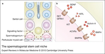
Spermatogonia are the progenitor population of male germ cells. Unlike the ovary, which has a fixed pool of oocytes for the lifetime of an organism, testicular germline stem cells continually proliferate. Similarly to other stem-cell types, the most important function of spermatogonial stem cells is to balance self-renewal with differentiation. If there is not adequate differentiation, sperm counts will suffer. However, if there is not adequate self-renewal, the pool of germ cells will progressively become exhausted. This balance is dependent on both extrinsic and intrinsic factors. The extrinsic factors are derived from the spermatogonial stem cell niche, an environment that is propagated by signalling factors from the vasculature (Ref. Reference Yoshida, Sukeno and Nabeshima5) and also those originating more locally in adjacent peritubular myoid cells, Leydig cells and, most importantly, Sertoli cells (Ref. Reference Oatley and Brinster6). A number of factors are important for the niche, including glial-cell-derived neurotrophic factor (GDNF), kit ligand (KITL), activin A and bone morphogenic protein 4 (BMP4), to name a few (Ref. Reference de Rooij7). In vitro and in vivo studies have shown the impact of these signalling factors on intrinsic spermatogonial stem cell factors and spermatogonial self-renewal versus differentiation (Fig. 2).
The spermatogonial stem cell niche. Sertoli cells, peritubular myoid cells and Leydig cells (not shown) all secrete signalling factors that form the spermatogonial stem cell environment, or niche. The basal lamina is a shared product of Sertoli and peritubular myoid cells that regulates integrins and harbours growth factors to facilitate regulation of the stem cell niche (a). Important signalling proteins of the stem cell niche include glial-cell-derived neurotrophic factor (GDNF), kit ligand (KITL), activin A and bone morphogenic protein 4 (BMP4). GDNF signals through RET and GDNF family receptor A1 (GFRA1) to promote self-renewal, and KITL, activin A and BMP4 signal through KIT receptor (KIT), activin receptors (ACVRs) and BMP receptors (BMPRs), respectively, to promote differentiation (b). Reviewed in Ref. Reference Oatley and Brinster6.

Glial-cell-derived neurotrophic factor
GDNF, which was first discovered as a prosurvival factor for midbrain dopaminergic neurons, is a distant member of the transforming growth family β (TGFβ) superfamily that is expressed and secreted by Sertoli cells (Refs Reference Meng8, Reference Yue9, Reference He10). A subset of spermatogonia express the major GDNF receptors, RET (ret proto-oncogene) and GFRA1 (GDNF family receptor α1) (Refs Reference Meng8, Reference He10, Reference Naughton11), and this led to investigation of the roles of GDNF signalling in spermatogonial stem cells in vitro. It was found that not only does GDNF enhance stem cell maintenance (Ref. Reference Nagano12), but it is also essential for the maintenance of stem cell activity in long-term cultures (Refs Reference Kubota, Avarbock and Brinster13, Reference Kanatsu-Shinohara14). These results led to interest in GDNF receptors as markers of ‘stemness’ in spermatogonial stem cell cultures. However, studies using GFRA1 antibodies in magnetic-activated cell-sorting procedures to enrich for GFRA1+ cells found that although a subset of spermatogonial stem cells did express this GDNF receptor, the overall stem cell pool was heterogeneous with respect to receptor expression (Refs Reference Buageaw15, Reference Ebata, Zhang and Nagano16). In contrast to those findings, siRNA-mediated knockdown of Gfra1 in mouse type A spermatogonial cultures led to suppression of proliferation and induction of markers of spermatogonial differentiation (Ref. Reference He10). In addition, phosphorylation of Tyr1062 of RET, a known binding site for many of its downstream signalling components, was reduced in the absence of GFRA1 (Ref. Reference He10), suggesting that RET also has a pivotal role in the prevention of differentiation and the maintenance of self-renewal of this cell type.
Transgenic mouse models were crucial in the discovery of the functional importance of GDNF to the spermatogonial stem cell niche and the translation of the in vitro findings to a live organism. Gdnf heterozygous mutant mice are fertile but contain a number of seminiferous tubules with progressive depletion of spermatogonial pools; conversely, transgenic mice that overexpress GDNF accumulate masses of undifferentiated spermatogonia postnatally (Ref. Reference Meng8). To circumvent the neonatal lethality of Gdnf-, Gfra1- and Ret-knockout mice, testes from these mice were transplanted into the back of castrated nude mice (Ref. Reference Naughton11). Consistent with signalling of GDNF through a receptor complex that includes RET and GFRA1, the null testes phenocopied each other and exhibited impaired spermatogonial stem cell proliferation, decreased markers of spermatogonia stem cells and increased markers of differentiation in the spermatogonial population (Ref. Reference Naughton11). Knock-in mice have also been used to replace Tyr1062 of RET with a phenylalanine codon, rendering it incapable of phosphorylation. The testes from these mice showed marked atrophy, and RET-expressing spermatogonia became almost undetectable by 3 weeks of age, revealing in vivo that Tyr1062 of RET is essential for the self-renewal of spermatogonial stem cells (Ref. Reference Jijiwa17).
Kit ligand
Spontaneous mutations in the mouse Steel (Sl) locus, which encodes the kit ligand (official gene symbol kitl, also known as stem cell factor) were the first clues in the identification of the importance of KITL in spermatogenesis (Refs Reference Mauduit, Hamamah and Benahmed18, Reference Bedell and Mahakali Zama19). KITL is a signalling factor synthesised by Sertoli cells in the postnatal testis (Ref. Reference Munsie20) that binds and activates the KIT receptor (official gene symbol Kit, also known as c-kit) located on differentiating spermatogonia and early spermatocytes (Ref. Reference Yoshinaga21). Similarly to KITL, the importance of KIT in spermatogenesis was well known owing to spontaneous mutations in the white spotting (W) locus that encodes it (Refs Reference Mauduit, Hamamah and Benahmed18, Reference Bedell and Mahakali Zama19). Early work using one such mutant (W v) led to the discovery that W v-heterozygous mice that were made cryptorchid to eliminate all differentiating germs cells and leave only undifferentiated spermatogonia, were never able to progress to differentiating spermatogonia after surgical reversal (Ref. Reference Koshimizu22). Injection of the ACK2 blocking antibody, which prevents KIT activation, led to reduced mitosis of differentiating spermatogonia, but had no effect on the mitosis of undifferentiated spermatogonia or the meiosis of spermatocytes (Ref. Reference Yoshinaga21). The effect on undifferentiated spermatogonia was further corroborated by later studies revealing that spermatogonial stem cells (SSCs) do not express Kit mRNA or KIT protein (Refs Reference Shinohara, Avarbock and Brinster23, Reference Prabhu24). When wild-type spermatogonial stem cells were transplanted into KITL mutant mice, undifferentiated spermatogonia were able to proliferate, but the cells were never able to differentiate into more mature spermatogonia (Ref. Reference Ohta25). However, these same cells when retransplanted to KIT mutant testes were able to differentiate and re-establish complete spermatogenesis (Ref. Reference Ohta25). Further proof of the importance of Sertoli cell expression of KITL and germ cell expression of KIT came when KITL mutant germ cells were transplanted into KIT mutant testes and were shown to successfully rescue the infertility in those mice (Ref. Reference Ogawa26).
Mouse models have also been crucial for discovering the relationship between intrinsic stem cell factors and KIT–KITL signalling. One such intrinsic protein is zinc finger and BTB domain containing 16 (ZBTB16 also known as Plzf or Zfp145), a DNA-sequence-specific transcriptional factor. ZBTB16 is expressed in undifferentiated spermatogonia, and mice with mutations in the Zbtb16 locus have progressive infertility owing to depletion of the spermatogonial stem cell pool (Refs Reference Costoya27, Reference Buaas28). ZBTB16 directly represses the transcription of Kit, and undifferentiated spermatogonia from Zbtb16-null mice have a relative enrichment of KIT-positive cells, leading to the hypothesis that ZBTB16-mediated repression of Kit is at least partially responsible for its function in maintaining the undifferentiated state in spermatogonia (Ref. Reference Filipponi29).
The previous studies emphasise the relationship between KIT–KITL signalling and spermatogonial differentiation. However, studies in Kit mutant mice indicate that this signalling is not necessary for the initial differentiation of juvenile undifferentiated spermatogonia into KIT-positive cells, but is necessary for the maintenance and proliferation of those cells (Ref. Reference Ohta, Tohda and Nishimune30). A recent study also discovered that KIT mutant testes contain spermatogonial stem cells that could be enriched in culture and reintroduced into recipient KIT mutant testes to successfully re-establish all stages of spermatogenesis (Ref. Reference Kubota31). This result casts doubt on the dogma that KIT–KITL signalling is an absolute requirement for spermatogonial differentiation. In another interesting study, it was shown that GDNF and fibroblast growth factor 2 (FGF2) can reprogram KIT-positive spermatogonia to dedifferentiate into spermatogonial stem cells, raising the possibility that ‘stemness’ can be acquired by differentiating progenitor cells in the testis (Ref. Reference Barroca32). Although the results are quite complicated, studies of the GDNF and KITL pathways support the hypothesis that there is a balance between self-renewal and differentiation of spermatogonial stem cells, and studies in humans support the hypothesis that alterations in this balance can lead to infertility and/or cancer. This topic will be discussed in greater length below.
Meiotic initiation
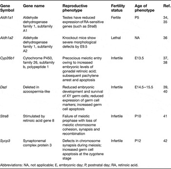
After undifferentiated spermatogonia commit to entering a differentiation pathway, these cells undergo a couple of mitotic divisions and then differentiate into primary spermatocytes, which subsequently undergo meiotic division. Similarly to spermatogonial maintenance, initiation of meiosis is dependent upon intrinsic as well as extrinsic signals. Sertoli cells have a crucial role in controlling the extrinsic signalling environment that influences meiotic initiation. Much of the work in this field has focused on the role of retinoic acid signalling and the regulation of the permissive meiotic environment (Ref. Reference Edson, Nagaraja and Matzuk33, Table 1).
Extrinsic retinoic acid signalling
Historically, it has been observed that vitamin A deficiency in rodents causes spermatogenic defects and complete loss of meiotic germ cells (Ref. Reference Griswold43). This was the first evidence that retinoids, the metabolic derivatives of vitamin A, are important for spermatogenesis. During embryogenesis, the mesonephros expresses a major enzyme of retinoic acid synthesis (ALDH1A2) and, consequently, is the main source of retinoic acid in the embryonic gonad (Ref. Reference Bowles37). Postnatally, Sertoli cell expression of ALDH1A1 might have a more prominent role in retinoic acid (RA) production because Aldh1a1-null testes have reduced expression of RA-sensitive genes at postnatal day 5 (Refs Reference Vernet35, Reference Bowles44). Although there appears to be abundant RA present throughout gonadal development, there are also prominent sex-specific differences in RA availability to the germ cells, which have helped to clarify the role of RA in meiotic initiation.
Meiosis begins embryonically in females and postnatally in males. A major retinoid-degrading enzyme – cytochrome P450, family 26, subfamily b, polypeptide 1 (CYP26B1) – is expressed sex-specifically in Sertoli cells of the male gonads (Ref. Reference Bowles37). Deletion of Cyp26b1 causes male germ cells to enter meiosis precociously at the same embryonic time point that female germ cells normally initiate meiosis (E13.5) (Refs Reference Bowles37, Reference MacLean38), and CYP26B1-null embryonic testes have elevated levels of RA (Ref. Reference MacLean38). Addition of a P450 antagonist to E12.5 gonadal cultures reduces the expression of spermatogonial stem cell markers and increases the expression of meiotic markers (Ref. Reference Bowles37). Meiosis is induced in gonadal cultures if they are treated with a synthetic retinoid that is resistant to CYP26B1-mediated metabolism (Ref. Reference MacLean38). Further cementing the importance of the role of RA in meiotic initiation is the observation that meiotic markers are suppressed in female gonadal cultures that have been treated with a synthetic antagonist of RA (Ref. Reference Bowles37).
Mouse models with knockout of meiotic genes

Abbreviations: NA, not applicable; E, embryonic day; P, postnatal day; RA, retinoic acid.
Intrinsic competency factors
Stimulated by retinoic acid gene 8 (Stra8) is an RA-responsive gene that is expressed in premeiotic germ cells in both the male and female gonads (Refs Reference Anderson41, Reference Zhou45, Reference Menke, Koubova and Page46). Treatment of cultured gonocytes, spermatogonia or whole neonatal testes with RA strongly induces Stra8 expression (Ref. Reference Zhou45). Stra8-knockout males show a block in spermatogenesis immediately preceding meiotic prophase and show no signs of meiotic recombination or chromosomal synapsis (Ref. Reference Anderson41). These results therefore beg the question – since RA is quite abundant in many tissue types in development, why are germ cells the only cell type to respond to RA by initiating meiosis? Meiotic competence must not only depend on an extrinsic signal but also an intrinsically permissive environment.
Deleted in azoospermia-like (Dazl) is a germ-cell-specific gene that is expressed in the embryonic male and female gonads (Refs Reference Seligman and Page47, Reference Cooke48). Dazl-knockout female gonads have defective meiotic chromosome condensation and synapsis and severely suppressed endogenous Stra8 mRNA levels (Ref. Reference Lin49). In contrast to fetal ovaries, wild-type fetal testes do not normally undergo meiotic initiation, but when cultured in the presence of RA, they respond by upregulating Stra8 mRNA (Ref. Reference Lin49). However, culture of Dazl-null male gonads with RA does not result in Stra8 mRNA upregulation (Ref. Reference Lin49). Interestingly, germ cells in Stra8-null testes express an important protein for meiotic synapsis, synaptonemal complex protein 3 (SYCP3), which is not loaded onto chromosomes as would normally occur during proper meiosis (Ref. Reference Anderson41). DAZL directly binds to the 3′-untranslated region of Sycp3 mRNA and enhances its translation (Ref. Reference Reynolds50), and Sycp3 is severely suppressed in Dazl-null germ cells (Ref. Reference Lin49). Taken together, these results suggest that DAZL enables meiotic competence of germ cells by promoting SYCP3 expression and that RA-induced STRA8 promotes meiotic progression by enabling the proper localisation of SYCP3.
Mouse mutants with defects in androgen receptor expression and/or function

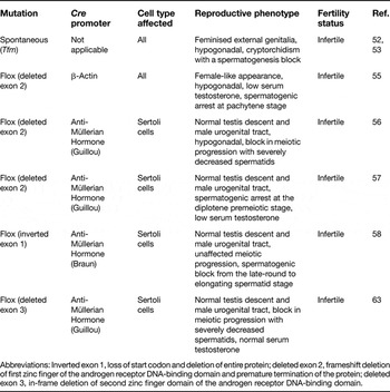
Abbreviations: Inverted exon 1, loss of start codon and deletion of entire protein; deleted exon 2, frameshift deletion of first zinc finger of the androgen receptor DNA-binding domain and premature termination of the protein; deleted exon 3, in-frame deletion of second zinc finger domain of the androgen receptor DNA-binding domain.
Spermatozoan maturation
As germ cells progress through the meiotic divisions of spermatogenesis, they migrate from the basal layer of the seminiferous tubule towards the adluminal compartment and are eventually released into the tubular lumen as mature spermatozoa. During this maturation process, Sertoli cells are responsible for a number of important functions including: (1) reforming the blood–testis barrier as the germ cells transit from the basal to adluminal compartments; (2) providing proper signalling and nutrients; (3) secreting tubular fluid to maintain the seminiferous tubular lumen; and (4) remodelling the constantly changing junctions between Sertoli cells and maturing germ cells (Refs Reference Skinner and Griswold2, Reference Russell3). Improper Sertoli cell functioning during spermatogenesis could cause a myriad of spermatozoan defects including mis-shaping of the sperm head, improper formation of the sperm tail leading to reduced fertility, or decreased sperm counts, to name just a few. Androgen signalling through the androgen receptor is a major signalling pathway that is crucial for proper Sertoli cell regulation and support of spermatogenesis (Ref. Reference Kerkhofs51, Table 2).
Androgen receptor
In 1970, Lyon and Hawkes isolated the testicular feminisation (Tfm) mouse mutant, an X-linked mutation that caused XY mice to develop external female genitalia, cryptorchidism and a spermatogenic block at the transition from spermatocytes to round spermatids (Ref. Reference Lyon and Hawkes52). Mapping of the Tfm mutation revealed a single base deletion in the 5′ end of the gene encoding the androgen receptor (Ar), which results in a frameshift mutation and premature termination of AR at amino acid 412 (Ref. Reference He, Kumar and Tindall53). Later studies showed that Tfm mutant mice had reduced Sertoli cell numbers at birth, but by puberty, Tfm mutant testes were no different from wild-type testes that were made cryptorchid surgically, as shown by their similar histology and gene expression levels (Ref. Reference Johnston54). Knockout mice that were engineered to have a null allele of Ar phenocopy the Tfm mutant mice, but it was unclear whether the spermatogenic defects in these mice were due to defects in androgen signalling or cryptorchidism (Refs Reference Kerkhofs51, Reference Yeh55).
Conditional-knockout mice have been developed using Cre-loxP technology to knock out the androgen receptor in specific cells or tissues. A number of different laboratories made Sertoli-cell-specific knockouts of Ar (Refs Reference De Gendt56, Reference Chang57, Reference Holdcraft and Braun58). None of these conditional-knockout mice had cryptorchidism, but they did vary in the severity of the spermatogenic block, with some experiencing a block between the spermatocyte and spermatid stages (Refs Reference De Gendt56, Reference Chang57) and another showing a block between the round spermatid and elongating spermatid stages (Ref. Reference Holdcraft and Braun58). Despite these differences, all of these Sertoli-cell-specific knockouts of Ar had defects in both the permissive and instructive environment driving the later stages of spermatogenesis. The permissive environment of late spermatogenesis refers to the adluminal compartment that is protected by the blood–testis barrier. The integrity of this barrier is essential to shield the developing germ cells from signalling from any source besides Sertoli cells or other germ cells, and also functions as an immunological barrier. mRNA transcript levels of a variety of genes involved in tubular restructuring and maintenance of the blood–testis barrier, including proteases and protease inhibitors, cell adhesion molecules and tight junction components, were affected in all of the Sertoli-cell-specific Ar-knockout mouse models (Refs Reference Eacker59, Reference Denolet60, Reference Wang61). One functional assay indeed showed that these mice experienced disruption of blood–testis barrier integrity (Ref. Reference Meng62). mRNAs encoding proteins involved in transport and metabolism were also misregulated (Refs Reference Eacker59, Reference Wang61). Interestingly, one of the upregulated genes was alcohol dehydrogenase 1 (Adh1), the rate-limiting enzyme of retinal-to-RA conversion, which raised the possibility of crosstalk between AR and RA pathways (Ref. Reference Eacker59).
Clinical implications
Research using mouse models has produced a wealth of knowledge about the signalling pathways and intrinsic factors that are essential to spermatogenesis. Applying this knowledge to human disease is vitally important, and some steps have been taken in that direction (reviewed in Ref. Reference Matzuk and Lamb4). There are a number of chromosomal abnormalities and gene mutations, including Klinefelter syndrome and cystic fibrosis transmembrane conductance regulator mutations, respectively, that are related to male infertility, but the following section will focus on the pathways that we have discussed in previous sections. We will also discuss the relationship of these molecular pathways in the pathogenesis of testicular cancer and the relationship between male infertility and cancer.
Genetics of male infertility
It is estimated that 15% of male infertility patients suffer from chromosomal alterations or single gene mutations (reviewed in Refs Reference Ferlin64, Reference Krausz and Giachini65). One common chromosomal aberration found in patients with severe infertility is microdeletion in the long arm of the Y chromosome, which is estimated to cause 10–15% of azoospermia (absence of sperm in the ejaculate) and 5–10% of severe oligozoospermia (low sperm counts) (Ref. Reference Ferlin64). This commonly deleted portion of the Y chromosome has been divided into three ‘azoospermia factor’ regions: AZFa, AZFb and AZFc. Deletions of AZFc are frequently found in patients with severe disease (3–7%), and this locus contains DAZ1–DAZ4, the Y chromosome paralogues of the autosomal gene DAZL (Ref. Reference Ferlin64). In one study that spanned 10 years of infertility treatment, 71.4% of patients with an AZFc deletion had sperm in their testes that could be retrieved, and two-thirds of those cases resulted in pregnancy via assisted reproductive techniques (Ref. Reference Stahl66). In a new twist, new AZFc haplotypes termed gr/gr or b2/b3, which both result in loss of DAZ3 and DAZ4, have been linked to infertility, but have also been found in normozoospermic males (Refs Reference Ferlin64, Reference Krausz and Giachini65). As suggested from knockout of Dazl in mice, which causes loss of germ cells and complete absence of gamete production (Ref. Reference Ruggiu39), DAZL in humans has been examined as a candidate for male infertility. One study identified four novel missense mutations in DAZL with one homozygous DAZL-null male patient presenting as infertile (Ref. Reference Tung67). This group hypothesised that DAZL mutations account for the variations in infertility found in patients with partial deletions in AZFc; however, they were not able to discern a link between autosomal DAZL function and severity of male infertility in the population they examined (Ref. Reference Chen68).
Idiopathic infertility that is not related to chromosomal alterations might be due to single gene mutations or polymorphisms. One of the most widely appreciated and best-characterised genes found to be mutated in infertile men is the androgen receptor, which is estimated to be associated with infertility in 2–3% of all cases of azoospermia or oligozoospermia (Ref. Reference Ferlin64). The AR gene can be mutated in a number of ways, resulting in mild-to-complete androgen insensitivity. The cases of complete androgen insensitivity phenocopy the Tfm mice, in which the 46XY patients present as phenotypic females with undeveloped gonads, whereas the mild forms present as phenotypic males with low sperm counts. In the phenotypic males with low sperm counts, there is a significant relationship between AR mutations and infertility; however, specific AR polymorphism has not yet been matched to disease severity (Refs Reference Gottlieb69, Reference Ferlin70). Polymorphisms that have been examined in the AR gene relate to two sites in exon 1 that exhibit variable stretches of CAG or GGC repeats (Ref. Reference Ferlin64). In a mouse model of Kennedy disease (a neurodegenerative disorder caused by CAG expansion in the AR gene), 113 glutamine codons were knocked into exon 1 of Ar (Ref. Reference Yu71). These mice experienced progressive infertility, not as a result of loss of AR function, but because of insoluble AR fractions created in Sertoli cell nuclei, which led to cytoskeletal abnormalities and decreased support of germ cells (Ref. Reference Yu71). When examining CAG polymorphisms in groups of infertile patients, different groups had different results; but overall, many studies found a relationship between CAG expansion and infertility (Ref. Reference Rajender, Singh and Thangaraj72). Unlike in the mouse, it is unclear whether this low level of CAG expansion affects AR transactivation.
Germ cell tumours and testicular dysgenesis syndrome
Type II testicular germ cell tumours (TGCTs), including seminomas and nonseminomas, arise from malignant primordial germ cells or gonocytes. They are the most prevalent type of male germ cell tumour and account for 1% of all cancers in male Caucasians and up to 60% of malignancies in those between 20 and 40 years of age (Ref. Reference Looijenga73). Several studies have linked TGCT occurrence to subfertility, cryptorchidism and genitourinary tract malformations (Ref. Reference Looijenga73). This knowledge has led to the testicular dysgenesis syndrome (TDS) hypothesis (Ref. Reference Sonne74). According to the TDS hypothesis, TGCTs form embryonically when a localised disturbance in niche environment impairs the differentiation of fetal gonocytes. These gonocytes do not mature and remain in the testis as carcinoma in situ. These cells proliferate during the peripubertal period and, with the help of abnormal signalling, acquire genetic changes that progress to TGCT in the adult. Environmental factors and genetic mutations and polymorphisms in the pathways discussed above are all hypothesised to be vital to the development of TGCTs as they all affect the gonocyte niche.
Hormonal disruption
A rat model of the early pathogenesis of TDS gave clues towards defining pathways that are affected in human disease (Ref. Reference Fisher75). Dibutyl phthalate administration to gravid mothers induced cryptorchidism, hypospadias, infertility and testis abnormalities in their male offspring, which were similar to those seen in human TDS (Ref. Reference Fisher75). Cryptorchid testes often show areas of focal dysgenesis containing partially formed testicular cords with mislocalised Sertoli cells, Leydig cells and gonocytes, which might have been caused by disrupted peritubular myoid cell function as a result of suppressed testosterone levels in utero (Ref. Reference Fisher75). Dysgenic and ‘Sertoli-cell-only’ tubules contained Sertoli cells expressing markers of immaturity (Ref. Reference Fisher75). Since this model of early pathogenesis is very similar to the human disease (Ref. Reference Sharpe76), gene expression analysis in this system could lead to the discovery of important pathways in human disease. One such study found that phthalate esters, including dibutyl phthalate, affect a number of systems, including genes necessary for proper steroid hormone synthesis (i.e. testosterone) and genes important for formation of junctions and signalling (including KIT) between germ cells and Sertoli cells (Ref. Reference Liu77). In humans, there has not been a definitive study linking phthalate ester exposure to TDS, but one group found that high exposure to prenatal phthalates corresponded to decreased anogenital distance, penile width and testicular descent in male infants, which are all signs of low prenatal testosterone (Ref. Reference Swan78).
Nonhormonal factors
One major finding from the study assessing gene expression changes in the rat model of TDS was the decreased expression of KIT receptor in the dysgenic testes (Ref. Reference Liu77). Conversely, Kitl-heterozygous mice have increased incidence of TGCTs (Ref. Reference Heaney79). Is it possible that KIT activating mutations are selected for in the Kitl-heterozygous mice and that the downregulation of the KIT receptor is the major reason why rat models of TDS using phthalate esters do not recapitulate the TGCTs seen in human disease? Activating mutations of KIT are often seen in TGCTs (Refs Reference Tian80, Reference Kemmer81) and have been associated with bilateral tumour development (Refs Reference Looijenga82, Reference McIntyre83). The use of KITL immunostaining has also been proposed as novel diagnostic marker for early malignant germ cells (Ref. Reference Stoop84). In a mouse model, mutation of the tyrosine codon of KIT that is necessary for phosphatidylinositol 3-kinase (PI3K) activation led to infertility (Ref. Reference Blume-Jensen85). This was in direct contrast to one human study that found a KIT mutation that was associated with TGCT pathogenesis and also constitutively activated PI3K in vitro (Ref. Reference Kemmer81). Another in vitro study indicated that KIT activation of PI3K upregulates cyclin D3 (CCND3) and promotes cell cycle progression, which would explain its dichotomous role in the aforementioned studies (Ref. Reference Feng, Ravindranath and Dym86).
One cohort study and one retrospective literature review found that even when excluding cryptorchidism as a confounding factor, there is still a significant positive association between subfertility and increased risk of testicular cancer (Refs Reference Jacobsen87, Reference Peng88). All the studies that have determined a genetic link for infertility and cancer raise the concern that better screening for genetic causes of infertility is vital so that these patients can be referred for genetic counselling and possibly preimplantation genetic screening if they are attempting to conceive using assisted reproductive technologies (ART).
Research in progress and outstanding questions
Although there has been a lot of research in both the clinical and basic science fields, more needs to be done to translate laboratory research into patient therapies. Testicular dysgenesis syndrome and embryonic stem (ES) cell technology are two fields that have both benefited from extensive basic science research.
Testicular cancer
We previously outlined a couple of areas that have seen major progress with regards to translational research, but we also would like to highlight some areas that need more attention. The first genetic study attempting to link AR mutations or trinucleotide repeat expansions to testicular cancer saw an increased incidence of null mutations of AR and certain trinucleotide repeat polymorphisms in TGCT patients (Ref. Reference Garolla89), but not much has been done in this area since then. Another important deletion in infertility, the gr/gr haplotype, has been associated with an increased risk of developing TGCTs (Ref. Reference Nathanson90). Studying the functions of AR and DAZ orthologues in humans will be vital to determine the causes of TDS.
Although we have convincing mouse models for the role of KIT and KITL in infertility and good human data for the role of this signalling system in TGCTs, very little is known about KIT or KITL mutations in human infertility. We were only able to find two studies that looked at the role of KIT in infertility (Refs Reference Feng, Ravindranath and Dym86, Reference Grimaldi91), with only one of these finding a positive association (Ref. Reference Feng, Ravindranath and Dym86). A mouse model in which GDNF was overexpressed demonstrated an early phenotype of infertility, but at later time points developed seminomatous tumours (Ref. Reference Meng92). Although this model does not accurately recapitulate human disease, it is unclear whether the GDNF pathway is important to human TDS, because we were not able to find any human studies examining this ligand or its receptors. Again, better understanding of the genetics of human male infertility with regards to the KITL and GDNF pathways will lead to better understanding of TDS. Research in this field is moving in the right direction, with the establishment of the International Testicular Cancer Linkage Consortium (Ref. Reference Mai93).
Embryonic stem cell technology
New studies using DAZ and its homologues are revolutionising the field of ES cell technology and highlighting some of the differences between mouse and human ES cells. Overexpression of DAZL in mouse ES cells causes cells to differentiate into phenotypic male and female germ cells without any extrinsic signalling (Ref. Reference Yu94). In a human ES cell study, however, researchers had to overexpress all DAZ homologues (DAZ1–DAZ4, DAZL and BOULE) and supplemented the differentiation medium with multiple BMP ligands (BMP4, BMP7 and BMP8b) to make the human cells meiotically competent (Ref. Reference Kee95). Interestingly, both culture systems seemed to result in the appearance of Sertoli-like cells, which raises the possibility that germ cells also influence the development of Sertoli cells, or alternatively, that the crosstalk between both cell types is necessary for their coordinated development. The surprisingly different requirements of mouse and human ES cells to achieve meiotic competence might just be a reflection of the innate differences between mouse and human ES cells, because mouse ES cells have many strong germ cell markers and may represent a germ-cell-like lineage, unlike human ES cells, which might be more like cells from the epiblast (Ref. Reference Tesar96). These studies also support the theories that DAZ homologues are the causative factor in infertility in patients with Y-chromosome microdeletions and that DAZ homologues are intrinsic meiotic-competence factors that control germ cell identity.
Future directions
Of the ~15% of all couples in the US that experience infertility, one- to two-thirds of these cases are due, at least in part, to male-factor infertility (Refs Reference Rebar and DeCherney97, Reference McLachlan98). Scientific breakthroughs in ART have revolutionised the field of reproductive endocrinology and allowed many of these couples to conceive biological offspring. However, there are still many uncertainties regarding the long-term consequences of ART, especially in light of the genetic issues that we have outlined throughout. Treatment for severe male-factor infertility is limited (Refs Reference Donoso, Tournaye and Devroey99, 100), and the need for more research in this area is great, especially for couples experiencing infertility due to male factor alone. Although the idea of gene therapy for the treatment of male infertility seems far-fetched (Ref. Reference Boekelheide and Sigman101) and genetic treatment of male factor infertility may or may not reduce the monetary cost of ART, it would mitigate the medical cost for the female patient who must endure hormonal injections ridden with side effects and might also improve the health of the fetus, because the long-term consequences of ART are unknown (Refs Reference Lamb102, Reference Neri, Takeuchi and Palermo103, Reference Alukal and Lipshultz104, Reference Alukal and Lamb105).
Acknowledgements
Research in the Matzuk laboratory on male reproduction and testicular cancer has been supported by National Institutes of Health (U01HD060496, R01HD057880, P01HD036289, and R01CA60651). R.L.N. is a student in the Medical Scientist Training Program at Baylor College of Medicine and has been supported in part by the Edward J. and Josephine G. Hudson Scholar Fund. We would also like to thank our peer reviewers for their help with improving this review.