Introduction
The large endolymphatic sac anomaly is a congenital abnormality which results in acquired hearing loss. It is one of the most frequent malformations of the inner ear recognisable on imaging studies.Reference Jackler and De La Cruz1–Reference Valvassori and Clemis3
The pathophysiological mechanism of the associated hearing loss is unconfirmed. It has been postulated that hyperosmolar fluid from the large endolymphatic sac may reflux into the cochlea and damage the hair cells.Reference Naganawa, Koshikawa, Iwayama, Fukatsu, Ishiguchi and Ishigaki4 Alternatively, cerebrospinal fluid pressure fluctuations may be transmitted to the inner ear by the patent endolymphatic sac, resulting in endolymphatic hydrops, perilymphatic fistula or rupture of the cochlear membranes.Reference Jackler and De La Cruz1, Reference Colvin, Beale and Harrop-Griffiths5 Alternatively, it has been proposed that cerebrospinal fluid pressure is transmitted into the labyrinth through an associated deficient modiolus, or that hearing loss is directly due to such concurrent inner ear anomalies.Reference Colvin, Beale and Harrop-Griffiths5–Reference Hirsch, Weissman, Curtin and Kamerer7
There have been attempts to correlate specific anatomical features (e.g. endolymphatic sac size, endolymphatic sac T2-weighted magnetic resonance imaging (MRI) signal, and associated labyrinthine anomalies) with patients' audiological findings; however, results have been inconsistent.Reference Naganawa, Koshikawa, Iwayama, Fukatsu, Ishiguchi and Ishigaki4, Reference Colvin, Beale and Harrop-Griffiths5, Reference Boston, Halsted, Meinzen-Derr, Bean, Vijayasakaren and Arjmand8, Reference Koesling, Rsainski and Amaya9
There have also been limited reports of the presence of internal compartments within large endolymphatic sacs, demonstrated on MRI scans.Reference Naganawa, Koshikawa, Iwayama, Fukatsu, Ishiguchi and Ishigaki4, Reference Naganawa, Ito and Iwayama10, Reference Romo, Casselman, Robson, Som and Curtin11 However, their significance and their impact on our understanding of the associated hearing loss have not been systematically explored.
Thus, the present study had four aims: (1) to document the prevalence of internal compartments in large endolymphatic sac anomalies, demonstrated by MRI; (2) to record the location, morphology and signal characteristics of these compartments; (3) to correlate the presence and MRI characteristics of these compartments, and their interfaces, with the presence of other large endolymphatic sac anomaly imaging features (i.e. size, associated labyrinthine anomalies and sac fluid signal) and with clinical and audiological data; and (4) to assess the direct correlation between these same large endolymphatic sac anomaly imaging features (i.e. size, associated labyrinthine anomalies and sac fluid signal) and clinical and audiological data.
Method
Patients with a diagnosis of large endolymphatic sac anomaly were retrospectively identified from a search of the radiology information management system and the cochlear implant programme database.
The study was reviewed by the local National Health Service research and ethics committee. Informed consent was not considered to be required for this retrospective study.
Magnetic resonance imaging with thin-section T2- and T2*-weighted sequences was available in digital format for 40 patients (imaging performed between 2002 and 2009). Two of these patients were excluded from the study due to adjacent pathology or incomplete volume coverage.
The remaining 38 patients (mean age, 16.9 years; age range, one to 65 years; standard deviation (SD), 15.2 years; 24 females and 14 males) were reviewed for the study. There were seven cases of unilateral large endolymphatic sac anomaly and 31 cases of bilateral large endolymphatic sac anomaly, defined in accordance with previously described criteria,Reference Boston, Halsted, Meinzen-Derr, Bean, Vijayasakaren and Arjmand8, Reference Vijayasekaren, Halsted, Boston, Meinzen-Derr, Bardo, Greinwald and Benton12 giving a total of 69 inner ears with imaging analysis. Comprehensive clinical data were obtained for 33 of the 38 patients, and audiometric data (performed within one year of the MRI study; mean interval, 3.9 months; SD, 3.2 months) was present for 62 ears (i.e. 31 of the 38 patients; audiology was performed within three months of the MRI study in 18 of the 38 cases).
Magnetic resonance imaging was performed on 1.5 Tesla systems. Due to the retrospective nature of the study, a variety of thin-section T2- and T2*-weighted sequences were utilised, i.e. driven equilibrium radiofrequency reset pulse (also known as DRIVE; n = 22); constructive interference in steady state (also known as CISS; n = 8); sampling perfection with application-optimised contrasts using different flip angle evolutions (also known as SPACE; n = 3); and turbo spin echo (also known as TSE; n = 5).
Radiological analysis
Digital MRI data were reviewed on a GE Centricity PACS workstation (GE Medical Systems, Milwaukee, Wisconsin, USA) by two independent neuroradiology observers. Only axial images were assessed.
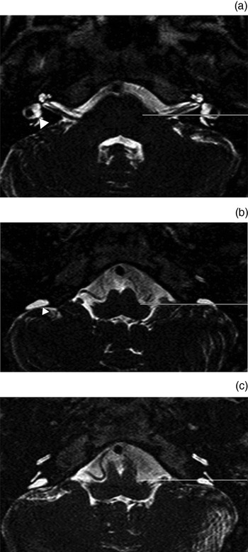
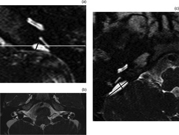
A series of endolymphatic sac size measurements were made, together with assessments of labyrinthine morphology and endolymphatic sac compartment interface anatomy (when present). For the purposes of measuring the endolymphatic sac size, the midpoint measurement required an initial delineation of the vestibular plane (a horizontal plane at the level of the dorsal common crus as it arises from the vestibule) (Figure 1a) and the opercular plane (a horizontal plane at the level of the superior opercular lip) (Figure 1b). The midpoint plane was defined as lying halfway between the vestibular and opercular planes (Figure 1c).Reference Boston, Halsted, Meinzen-Derr, Bean, Vijayasakaren and Arjmand8, Reference Vijayasekaren, Halsted, Boston, Meinzen-Derr, Bardo, Greinwald and Benton12 The midpoint measurement bisected the midpoint plane in the angle of the endolymphatic sac trajectory (Figure 2a). The operculum measurement was the maximum perpendicular endolymphatic sac width at the level of the operculum (Figure 2b). The extraosseous long measurement and extraosseous short measurement represented the maximum longitudinal and short axis dimensions perpendicular to the petrous ridge (Figure 2c).
Axial, T2-weighted, driven equilibrium radiofrequency reset pulse magnetic resonance imaging scans demonstrating the vestibular, opercular and midpoint planes in a patient with bilateral large endolymphatic sac anomaly but no septations. (a) Line corresponds to the vestibular plane, defined by the horizontal plane at the level of the dorsal common crus as it arises from the vestibule (indicated by arrowhead on contralateral side). (b) Line corresponds to the opercular plane, defined by the horizontal plane at the level of the superior opercular lip (indicated by arrowhead on contralateral side). (c) Line corresponds to the midpoint plane, defined as halfway anteroposteriorly between the vestibular and opercular planes.

Axial, T2-weighted, driven equilibrium radiofrequency reset pulse magnetic resonance imaging scans demonstrating the endolymphatic sac size measurements. (a) Cropped image showing the large endolymphatic sac anomaly. The midpoint measurement is demonstrated bisecting the midpoint plane in the angle of the endolymphatic sac trajectory, such that the measurement forms an equal angle with the lateral and medial walls of the endolymphatic sac. (b) Image demonstrating bilateral large endolymphatic sac anomaly with septations and small proximal compartments. The opercular measurements are shown on either side as the maximum endolymphatic sac widths at the level of the opercula. They extend perpendicular to the lateral wall of the endolymphatic sac. (c) Cropped image showing the extraosseous endolymphatic sac measurements, i.e. the extraosseous short measurement and the extraosseous long measurement.

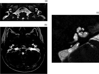
Labyrinthine morphology was recorded as regards the modiolus (i.e. normal, deficient or absent), the cochlear segmentation (i.e. normal or abnormal) and the vestibular-semicircular canal (i.e. normal, mild or severe dysplasia).13 Endolymphatic sac compartments were defined as visually apparent areas of differing signal within the endolymphatic sac, with a clear interface. The recorded MRI features of the compartments and the interfaces between the compartments were: angle of interface, orientation of interface, location of lower signal compartment, and the proportion of the endolymphatic sac filled by the lower signal compartment (Figure 3). Regions of interest were identified within the endolymphatic sacs (including separate compartments) and within the internal auditory meati (Figure 3).
Axial, T2-weighted, driven equilibrium radiofrequency reset pulse magnetic resonance imaging scans demonstrating imaging septation anatomy, labyrinthine anomalies and placement of regions of interest. (a) Image showing bilateral large endolymphatic sac anomalies. The right-sided, distal, lower signal compartment comprised 50–75 per cent of the volume (determined by examining adjacent images), whilst that on the left comprised 75–100 per cent. There is a right-sided, concave (with a 60–90 per cent angle) and a left-sided, straight (with 30–60 per cent angle) septation. (b) Image showing bilateral large endolymphatic sac anomalies with abnormal cochlear segmentation and vestibular dysplasia. Regions of interest (imposed ovals) are shown for the two left-sided endolymphatic sac compartments. The distal lower signal compartment occupies 75–100 per cent of the endolymphatic sac and there is a concave septation. (c) Cropped images of the enlarged right endolymphatic sac. Regions of interest (imposed ovals) are shown for the internal auditory meatus and the proximal and distal endolymphatic sac compartments. The region of interest corresponding to the distal compartment includes the region of the pars rugosa. Note the separate, high-signal ‘bubble’ within the distal compartment. Approximately 75–100 per cent of the endolymphatic sac is filled by the lower signal distal compartment, and the septation is concave.

Clinical and audiometric analysis
Clinical records were reviewed.
Pure tone audiograms, with or without conductive thresholds, were documented at 250, 500, 1000, 2500 and 4000 Hz, and the mean was calculated. A conductive or mixed component to the hearing loss was defined if the air–bone gap was more than 10 dB at one or more frequencies in the presence of a normal tympanogram; however, data were incomplete for 12/62 ears as the audiometry had been performed unmasked.
The course of the hearing loss was recorded as constant or progressive, from the clinical history. A diagnosis of progressive hearing loss was supported by pure tone audiometry showing a greater than 10 dB increase in hearing threshold over more than a three-month follow-up period.Reference Bagger-Sjoback, Jansson, Friberg and Rask-Anderson14 Episodes of sudden or fluctuating hearing loss and associated precipitating factors were assessed. We recorded family history, additional systemic syndromic associations, and, when available, the results of Pendred gene analysis and perchlorate discharge testing. We also recorded whether an ‘ooze’ or ‘gush’ of fluid was seen at cochlear implant surgery, upon entering the cochlea.
Statistical analysis
Interobserver reproducibility was assessed for MRI measurements of endolymphatic sac size and endolymphatic sac : internal auditory meatus signal ratio, using Pearson's correlation coefficient, difference/mean and absolute difference/mean. Combined mean values were used for subsequent analysis.
Audiological data (pure tone audiometry and patterns of hearing loss) were compared with endolymphatic sac size measurements and the lowest endolymphatic sac : internal auditory meatus signal ratio, using two-tailed Pearson correlation. Audiological data (pure tone audiometry and patterns of hearing loss), endolymphatic sac size measurements and the lowest endolymphatic sac: internal auditory meatus signal ratio were also compared with the presence of compartments and other labyrinthine abnormalities. This analysis was performed with the two-tailed Mann–Whitney U test or the Kruskal–Wallis test (depending on whether the labyrinthine abnormalities included two or more categorical values). The same comparisons were also performed for ears without other labyrinthine abnormalities.
Further correlations between categorical labyrinthine abnormalities were tested using the two-tailed Mann–Whitney U test or the Kruskal–Wallis test.
For endolymphatic sacs in which compartments were present, the orientation of the interface between compartments, the proportion of the endolymphatic sac filled with low-signal compartment, and the endolymphatic sac signal ratio measurements in proximal versus distal compartments were compared with the pure tone audiometry results and the pattern of hearing loss.
T-tests were used to compare the endolymphatic sac : internal auditory meatus signal ratio in the endolymphatic sacs without compartments with the endolymphatic sac : internal auditory meatus signal ratio in the proximal and distal compartments of sacs with compartments.
The presence of a fluid gusher at surgery was compared with the endolymphatic sac size measurements and the presence of modiolar deficiency, using the Mann–Whitney U test.
Results and analysis
Clinical and audiological data
In the 33 patients for whom full clinical data were available, there was progressive hearing loss in 46 per cent and sudden or fluctuating hearing loss in 30 per cent. In the 50 ears for which both comprehensive clinical and full audiometric data were available, mixed or conductive hearing loss was present in 76 per cent.
No patients had experienced episodes of vertigo.
Systemic associations were present in 27 per cent of patients (distal renal tubular acidosis in 9 per cent, Pendred's syndrome in 12 per cent and other associations in 6 per cent).
Hearing loss was categorised as normal in 2 per cent, mild (i.e. 30–49 dB) in 3 per cent, moderate (50–59 dB) in 3 per cent, severe (60–79 dB) in 16 per cent, near deafness (>80 dB) in 23 per cent, and deafness in 53 per cent.
Of the 15 patients who underwent cochlear implantation, six (40 per cent) had peri-operative oozing or gushing fluid.
Magnetic resonance imaging data
Large endolymphatic sac compartments were present in 39/69 ears (57 per cent) and were clearly demonstrated in 28/39 cases. When compartments were present, the lower signal compartment was always distal (i.e. posterolateral). The proportion of the endolymphatic sac occupied by the low-signal compartment was 0–25 per cent in 10 per cent of ears, 25–50 per cent in 21 per cent of ears, 50–75 per cent in 31 per cent of ears, and 75–100 per cent in 38 per cent of ears. Hence, the low-signal compartment was the dominant compartment in 69 per cent of ears. The compartmental interfaces were usually straight (32 per cent) or bowed away from the labyrinthine aspect (68 per cent).
The mean (SD) endolymphatic sac : internal auditory meatus signal ratio was 0.914 (0.09) for the proximal septated compartment, 0.489 (0.16) for the distal septated compartment, and 0.881 (0.12) for those endolymphatic sacs without compartments. The endolymphatic sac : internal auditory meatus signal ratio in the distal compartment of those sacs with compartments was significantly lower than the same ratio in endolymphatic sacs without compartments (p < 0.001).
The mean (SD) size measurements were: midpoint measurement, 1.99 (0.70) mm; opercular measurement, 2.63 (0.91) mm; extraosseous long measurement, 13.5 (6.4) mm; and extraosseous short measurement, 3.36 (3.1) mm.
The modiolus was deficient in 38 per cent of the large endolymphatic sac anomaly cases and absent in 4 per cent, the cochlear segmentation was abnormal in 51 per cent, and there was vestibular dysplasia in 41 per cent (mild in 35 per cent and severe in 6 per cent).
The interobserver reproducibility was excellent for all continuous data (i.e. endolymphatic sac size measurements and endolymphatic sac : internal auditory meatus signal ratio), with a Pearson's R coefficient of 0.9–0.99.
Correlation of clinical and audiometric data with magnetic resonance imaging data
The presence of compartments was significantly associated with larger extraosseous long measurements (p = 0.000).
Patients with sudden or fluctuating hearing loss had significantly larger extraosseous dimensions when only ears without labyrinthine anomalies were included (p = 0.034 for extraosseous long measurement and p = 0.043 for extraosseous short measurement). Sudden or fluctuating hearing loss was also associated with a lower endolymphatic sac : internal auditory meatus signal ratio in the distal compartment (p = 0.009).
There was a non-significant trend towards an association between concave bowed intercompartmental interfaces and progressive hearing loss (p = 0.071) when only those ears without labyrinthine anomalies were included.
Pure tone audiometry thresholds were higher in ears with larger opercular measurements (p = 0.022) and larger extraosseous measurements (p = 0.003 for the extraosseous long measurement and p = 0.004 for the extraosseous short measurement). These associations were also significant in ears without labyrinthine abnormalities.
The presence of a fluid gusher at cochlear implantation (in the 15 ears implanted) was significantly associated with the midpoint measurement (p = 0.05), but not with modiolar deficiency nor any other endolymphatic size measurement (p > 0.1 for all).
Discussion
The normal intraosseous endolymphatic sac contains only a few large folds and rugae; however, a multitubular appearance (termed the pars rugosa or multilobular portion) becomes more conspicuous within its extraosseous portion, distal to the confines of the bony vestibular aqueduct (Figure 3c).Reference Bagger-Sjoback, Jansson, Friberg and Rask-Anderson14 The contents of the lumen are heterogeneous and vary in their degree of staining with haematoxylin and eosin. The more distal areas, related to the pars rugosa, have been shown to be composed of mucopolysaccharide and hyaluronic acid, which are of unknown function but which may relate to inner ear fluid haemostasis.Reference Danckwardt-Lilliestrom, Laurent, Hellstrom, Friberg, Kinnefors and Rask-Andersen15
There is limited data on the histopathology of the large endolymphatic sac anomaly. An archived case of Pendred's syndrome with bilateral large endolymphatic sac anomaly demonstrated a prominent pars rugosa on one side but complete replacement of the pars rugosa on the other side.Reference Gill, Michaels, Phelps and Reardon16 Another case of an enlarged endolymphatic sac, in the setting of a Mondini defect, revealed replacement of the perisac connective tissue stroma.Reference Gussen17
Imaging studies have demonstrated that the entire endolymphatic sac may have a different MRI signal intensity or computed tomography density compared with cerebrospinal fluid and labyrinthine fluid.Reference Naganawa, Koshikawa, Iwayama, Fukatsu, Ishiguchi and Ishigaki4, Reference Okamoto, Ito, Furusawa, Sakai and Tokiguchi18 It has been postulated that this is a consequence of increased protein concentration. Normally, the endolymphatic sac is filled with endolymph which resembles intracellular fluid,Reference Mafee, Charlette, Kumar and Belmount2 with hyperosmolar protein concentrations of 1000–3000 mg/dl. In cases of large endolymphatic sac anomalies sampled at surgery, the endolymph protein concentration was reported as 335–660 mg/dl.Reference Jackler and De La Cruz1, Reference Couloigner, Grayeli, Sterkers and Ferrary19 It has been suggested that there may be abnormal, bidirectional fluid flow between the large endolymphatic sac and the cochleovestibular organ, leading to mixing and chronic contamination of the normally protein-poor cochleovestibular endolymph.Reference Couloigner, Grayeli, Sterkers and Ferrary19 This mixing may be responsible for the observed reduced protein concentration relative to the normal endolymphatic sac. Such a scenario implies that protein concentration may vary over time; in support of this theory, varying signals have been noted on serial MRI images.Reference Naganawa, Koshikawa, Fukatsu, Ishigaki and Nukashima20
Previous authors have also recognised that the distal (posterolateral) endolymphatic sac alone may have a lower signal on T2- and T2*-weighted MRI scans, compared with cerebrospinal fluid or labyrinthine fluid.Reference Naganawa, Koshikawa, Iwayama, Fukatsu, Ishiguchi and Ishigaki4 This feature has been described in other reports,Reference Koesling, Rsainski and Amaya9, Reference Romo, Casselman, Robson, Som and Curtin11 although its significance has not been explored. It has been proposed that this differing signal within the compartments represents the sub-epithelial connective tissue or multitubular tissue of the pars rugosa, rather than the hyperosmolar proteinaceous contents of the endolymphatic sac.Reference Romo, Casselman, Robson, Som and Curtin11, Reference Bagger-Sjoback, Jansson, Friberg and Rask-Anderson14 The morphology of the low-signal compartments with their well-defined interfaces, the known variation in endolymphatic sac signal over time,Reference Naganawa, Koshikawa, Fukatsu, Ishigaki and Nukashima20 and the impressive erosion of bone around the endolymphatic sacReference Gussen17 (consistent with hydraulic pressure) would be more indicative of a fluid-containing compartment than a solid tissue. The paucity of pars rugosa and connective tissue in the majority of previous pathological correlates, and the frequent extension of the low-signal compartment into the intraosseous endolymphatic sac away from the pars rugosa, would also argue against connective tissue or pars rugosa being responsible for this observation.
We postulate that, since the low-signal compartment equates to the position of the pars rugosa, it may correspond to mucopolysaccharide or hyaluronic acid secretion into the endolymph at this site, with separation from the proximal endolymphatic compartment by a rugal fold or septation. A dysfunctional enlarged endolymphatic sac may not be able to adequately remove such metabolites, particularly if they are compartmentalised. Such a concentration of metabolites may explain why lower signal was observed in the distal compartments of septated large endolymphatic sac anomalies, compared with large endolymphatic sac anomalies without compartments. It is appreciated that this theory remains speculative in the absence of any pathological correlation. Alternative explanations include haemorrhage or reduced signal due to fluid pulsatility within the distal compartment.
The presence of the septations provides another potential anatomical correlate with the audiovestibular phenotype, and the current study represents the largest series correlating large endolymphatic sac anomaly MRI features with audiological findings. We speculate that the bowing of the sac septation may indicate differing compartmental pressures or may suggest the direction of endolymph flow; furthermore, we hypothesise that both this and the proportion of the endolymphatic sac occupied by the low signal compartment (on T2- and T2*-weighted MRI) may correlate with the degree and the progression of hearing loss. However, apart from a trend towards progressive hearing loss in patients with a concave septation, these hypotheses are not supported by our results. Indeed, the presence of septations overall was not significantly associated with the degree of hearing loss or the pattern of hearing loss.
There is some evidence that intralabyrinthine reflux of low-signal fluid (seen on T2- and T2*-weighted MRI) may be implicated in hearing loss. A previous case report describes a patient scanned soon after hearing deterioration, in whom 3T MRI revealed low three-dimensional constructive interference in the steady-state signal within the endolymphatic space of the labyrinth.Reference Naganawa, Sone, Otake and Kakashima21 Our data concur with previous studies in which measures of signal intensity within the endolymphatic sac did not shown a correlation with the degree of hearing loss.Reference Colvin, Beale and Harrop-Griffiths5
We were particularly interested in the possibility that the presence and occasional rupture of a compartment, with reflux of accumulated debris and metabolites, could be associated with the well-described episodes of sudden or fluctuating hearing loss and vertigo. Sudden hearing loss may be triggered by coryzal illness, trauma, exercise and aeroplane travel,Reference Colvin, Beale and Harrop-Griffiths5 and associated variations in pressure could result in septation rupture and fluid leakage. Endolymphatic sac signal intensity in the distal compartment was indeed associated with sudden hearing loss. There were no documented episodes of vertigo in our patient group. Cases have been observed in which abnormal caloric responses have been related to low T2*-weighted MRI signal within the intraosseous endolymphatic sac.Reference Oh, Choi, Son, Lee, Chang and Kim22
Additional audiovestibular findings, such as a conductive component or mixed hearing loss,Reference Jackler and De La Cruz1, Reference Colvin, Beale and Harrop-Griffiths5, Reference Koesling, Rsainski and Amaya9, Reference Antonelli, Nall, Lemmerling, Mancuso and Kubilis23 may be described in the setting of large endolymphatic sac anomaly. The potential causes of a conductive component include increased cochlear fluid pressure, a ‘third window’ effect and stapes fixation. We found no relationship between the presence of septations or other MRI features and the presence of conductive or mixed hearing loss.
Perilymphatic gushers have previously been encountered during cochlear implant surgery in patients with a large endolymphatic sac anomaly.Reference Au and Gibson24 These were recorded in six of our 15 patients undergoing cochlear implantation. It has been previously suggested that this event may result from transmission of fluid either through the enlarged endolymphatic sac or through the cochlear aperture in the presence of a deficient modiolus. We demonstrated a significant relationship between the presence of a fluid gusher and the midpoint measurement (generally the narrowest part of the endolymphatic sac and hence a potential bottleneck), but not modiolar deficiency, hence favouring the former mechanism.
• Many large endolymphatic sac anomalies have compartments of differing magnetic resonance imaging (MRI) signal intensity
• Decreased distal compartment MRI signal intensity is associated with sudden or fluctuating hearing loss
• Larger extraossseous and opercular sac dimensions are associated with poorer audiometric results
• Larger intraosseous sacs are associated with fluid loss during cochlear implantation
• Such MRI features may aid prognosis and treatment planning
Although not our main focus, our data allowed us to analyse the relationship between endolymphatic sac size and audiological findings. We found that all four of our endolymphatic sac measurements, and in particular the extraosseous measurements, could be determined with excellent reproducibility from MRI images. Previous series have failed to demonstrate any association between the endolymphatic sac size (at the midpoint measurement, opercular measurementReference Colvin, Beale and Harrop-Griffiths5, Reference Koesling, Rsainski and Amaya9, Reference Naganawa, Ito and Iwayama10 or extraosseous measurementsReference Colvin, Beale and Harrop-Griffiths5, Reference Naganawa, Ito and Iwayama10) and the severity of hearing loss, although there has been a documented association with the progression of hearing loss.Reference Boston, Halsted, Meinzen-Derr, Bean, Vijayasakaren and Arjmand8 In our study, size measurements were related to pure tone audiometry threshold (significant in the case of the opercular measurement and the extraosseous measurements), and there was an additional relationship between extraosseous endolymphatic sac measurements and a history of sudden hearing loss, when patients without other labyrinthine abnormalities were studied alone.
Conclusion
This study documented the presence of compartments within large endolymphatic sac anomalies in 57 per cent of thin-section T2- and T2*-weighted MRI images; however, their presence did not correlate with patients' clinical or audiological data.
These compartmental septations were particularly frequent in the larger extraosseous endolymphatic sac anomalies, and were either straight or bowed towards the labyrinth. The distal compartment was usually larger, and was always of lower signal on T2- and T2*-weighted MRI scans.
The endolymphatic sac : internal auditory meatus signal ratio within the distal compartment was lower than that for endolymphatic sacs without compartments, and the lower signal was associated with a history of sudden or fluctuating hearing loss.
Finally, pure tone audiometry was lower in ears with larger opercular measurements and extraosseous measurements. The midpoint endolymphatic sac measurement, but not modiolar deficiency, was associated with a fluid gusher at the time of cochlear implantation.