Case presentation
A 60-year-old female with no significant past medical history presented to the emergency department with progressive onset cardiac sounding chest pain. She was not on any regular medications and was a non-smoker with no family history of ischaemic heart disease. She did not have a cough, fever, dyspnoea, or peripheral oedema, however her jugular venous pressure was elevated to 5 cm with quiet heart sounds on examination. Initial observations revealed a regular pulse of 90 beats per minute and a blood pressure of 90/50 mmHg. An initial electrocardiogram demonstrated diffuse ST depression with isolated ST elevation in aVR and V1. Her blood panel showed a mildly elevated troponin (0.2 ng/ml, 0–0.02 ng/ml), otherwise confirmed a normal kidney function, full blood count, and inflammatory markers were not raised with a negative D-dimer. An initial chest X-ray did not demonstrate any acute pathology and no cardiomegaly.
Diagnosis
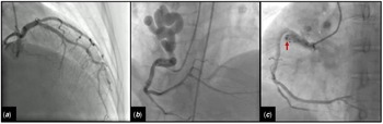
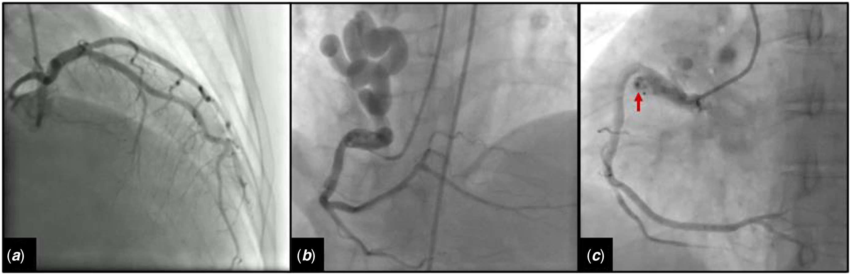
Given her chest pain, raised troponin and electrocardiogram changes, initial differential diagnosis was acute coronary syndrome, thus the patient was taken promptly to the catheter laboratory which ruled out coronary artery disease. However, confirmed a giant tortuous coronary fistula originated from the sinoatrial branch of the right coronary artery (Fig 1A+B, Supplemantary Video 1). To detect where the fistula is draning and for the treatment guidance, a CT coronary angiogram was performed and confirmed a giant tortuous coronary fistula connecting the sinoatrial branch of the right coronary artery and superior vena cava leading to myocardial steal and patients’ symptoms.
(a) Angiogram showing normal left coronary arteries. (b) Angiogram showing a giant tortuous coronary fistula originated from the sinoatrial branch of the right coronary artery (RCA). (c) Post-procedure angiography showed almost completely occluded fistula. Vascular plug is seen in the ostium of fistula (red arrow).
Initial management and prognosis
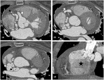
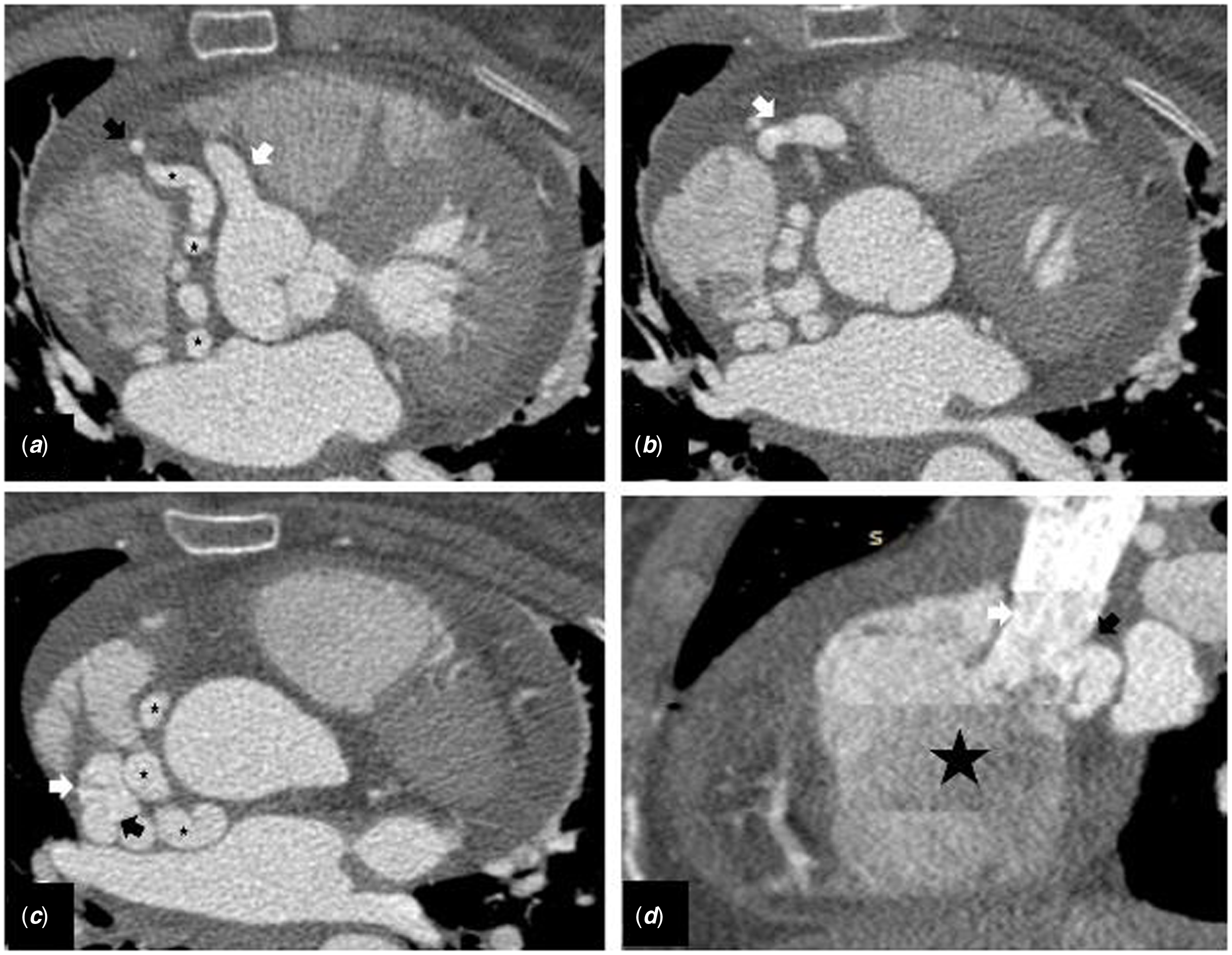
After initial fluid resuscitation and morphine to control pain, the patient was transferred to the coronary care unit. An urgent bedside echocardiogram demonstrated a global mild hypokinesia (ejection fraction 50%) with mild tricuspid regurgitation (pulmonary artery systolic pressure 40 mmHg) and a moderately dilated right atrium and ventricle with no pericardial effusion. Soon after echocardiography, she had two ventricular fibrillation events which were successfully treated with defibrillation and subsequently taken to the coronary catheter laboratory. Acute coronary syndrome protocol was initiated with administration of aspirin 300 mg and Clopidogrel 300 mg prior to catheterisation. Coronary catheterisation confirmed a giant coronary fistula originating from the sinoatrial branch of the right coronary artery and aneurysmal proximal ight coronary artery. Other coronaries were normal and right coronary artery was dominant. Post-angiogram, she was haemodynamically stable. Following this, the patient was taken for CT coronary angiogram (Fig 2) which confirmed a giant coronary fistula connecting the sinoatrial branch of the right coronary artery to the superior vena cava.
(a) The transverse slice at the level of aortic cusps shows dilated right coronary artery (white arrow), enlarged and tortuous vascular structures (black stars) heading towards atriocaval junction through aortoatrial and interatrial grooves which is thought to be the sinoatrial nodal branch of right coronary artery due to its course. RCA remains its normal diameter after the dilated sinoatrial branch (black arrow). (b) The division of sinoatrial branch from right coronary artery is seen (white arrow). (c) The fistulous communication between abnormally dilated sinoatrial nodal branch (black arrow) and superior vena cava (white arrow) at the level of slightly superior to atriocaval junction is accurately demonstrated. (d) The sagittal image also readily shows the fistulous connection of the sinoatrial branch (black arrow) with superior vena cava (white arrow). The right atrium is shown (black star).
Case progression and outcome
After reviewing both the angiogram and CT coronary angiogram, a multidisciplinary team discussion was held and a percutaneous management was opted for. An Amplatzer Occluder Device (Vascular plug) via a transcatheter approach was utilised in managing this patient successfully without any complications. Post-procedure angiography showed almost completely occluded fistula (Fig 1C, Supplementary 2). Right heart catheterisation was not performed before the fistula closure because it would not have changed our decision as this patient experienced critical ischaemia and ventricular fibrillation, due to definite closure being required. In this case, it was likely this patient had a congenital coronary artery fistulae as she had no recent intervention or past medical history and manifested myocardial steal and ischaemia. At 6 months follow-up, the patient felt well with no new symptoms with stable left ventricular heart function on repeat echocardiogram.
Discussion
Coronary artery fistulae are rare anomalies of the coronary tree, defined as abnormal communication between coronary arteries and vascular structures. According to the American Heart Association, the "Sakakibara" classification can be used to classify coronary artery fistulae. Reference Reddy, Davies, Holmes, Schaff, Singh and Alli1 Several studies have demonstrated the incidence of coronary artery fistulae ranging from 0.05% to 0.9% in the general population Reference Lim, Jung and Lee2 . Rare multiple coronary artery fistulae have also been reported in the literature. Reference Lim, Jung and Lee2 The most common site of origin of coronary artery fistulae is the right coronary artery and the right atrium, pulmonary artery, and right ventricle being draining end points. Reference Lim, Jung and Lee2 Whilst predominantly congenital (as in our case), coronary artery fistulae can also be acquired post-myocardial infarction or be iatrogenic post-endomyocardial biopsy or following percutaneous intervention. Reference Fernandes, Kadivar, Hallman, Reul, Ott and Cooley3 Depending on the size of the coronary artery fistulae patients can often be asymptomatic, thus be incidentally found during cardiac investigations. Large coronary artery fistulae can manifest with chest pain, shortness of breath, fluid overload, and a systolic crescendo–decrescendo murmur secondary to severe shunting. In 2018, the American Heart Association published guidelines on the investigation and management of coronary artery fistulae with an emphasis on multidisciplinary medical and surgical joint partnership. Reference Stout, Daniels and Aboulhosn4 However, in continental Europe, coronary artery fistulae are not specifically addressed in the international guidelines on adult CHD from the European Society of Cardiology. Reference Baumgartner, Bonhoeffer and De Groot5 Initial step of echocardiography is essential in evaluating the ventricular size, any regional wall abnormalities, shunting and left ventricular systolic function. Following this, coronary angiography is an ideal modality to look at coronary artery disease and fistulae, however CT coronary angiogram is considered to be the superior non-invasive modality as it allows enhanced visualisation of the coronaries with high-resolution and three-dimensional volume rendering. Reference Stout, Daniels and Aboulhosn4 Percutaneous closures of coronary artery fistulae involve occlusion coils, detachable balloons, and stents. Reference Stout, Daniels and Aboulhosn4 The question to opt for surgical or percutaneous management involves an multidisciplinary team approach to assess the anatomy and physiology of the coronary artery fistulae, the patient’s co-morbidities, and perioperative risk. For example, fistulae that are symptomatic, large, and complex are candidates for surgical management, whereas fistulae that is proximal, non-tortuous, and in older frail patient’s, percutaneous management can be opted for. Reference Stout, Daniels and Aboulhosn4 As our patient was relatively young with no comorbidities, both options for surgical and percutaneous management were given, of which after discussion with the patient percutaneous option was opted for.
Supplementary material
To view supplementary material for this article, please visit https://doi.org/10.1017/S1047951122000476
Financial support
This research received no specific grant from any funding agency, commercial, or not-for-profit sectors.
Conflicts of interest
None.