Introduction
Schistosomiasis, caused by trematode flatworms of the genus Schistosoma, is one of the most significant, neglected tropical diseases in the world. An estimated 779 million people are at risk for schistosomiasis, with 207 million infected in 76 countries and territories (Lammie et al., Reference Lammie, Fenwick and Utzinger2006; Steinmann et al., Reference Steinmann, Keiser, Bos, Tanner and Utzinger2006). Approximately 120 million people are symptomatic and 20 millions suffer from severe and debilitating disease (Chitsulo et al., 2000; Engels et al., Reference Engels, Chitsulo, Montresor and Savioli2002; Finkelstein et al., Reference Finkelstein, Schleinitz, Carabin and McGarvey2008). The most common anti-schistosomiasis drug used in endemic areas is praziquantel, which is effective against adult worms (World Health Organization, 1993). However, praziquantel does not prevent re-infection, is inactive against juvenile schistosomes and only possesses a limited effect on the already developed liver and spleen lesions (Chatterjee et al., Reference Chatterjee, Mbaye, De Man and Van Marck2002; Silva et al., Reference Silva, Menezes, De Oliveira and Andrade2003). A treatment strategy that relies on a single drug is not satisfactory due to the high probability of emergence of drug-resistant parasites (Ismail et al., Reference Ismail, Botros, Metwally, William, Farghally, Tao, Day and Bennett1999; Pica-Mattoccia & Cioli, Reference Pica-Mattoccia and Cioli2004). Furthermore, praziquantel was reported to induce haemorrhage in the lung tissue of the host (Flisser & McLaren, Reference Flisser and McLaren1989), as well as abdominal pain and diarrhoea (Kabatereine et al., Reference Kabatereine, Kemijumbi, Ouma, Sturrock, Butterworth, Madsen, Ornbierg, Dunne and Vennervald2003). In this context, the identification of new and effective schistosomicidal compounds is essential. The trend of using natural plant extracts as new, safe and effective drugs is promising (Magalhães et al., Reference Magalhães, Machado, Morais, Moreira, Soares, da Silva, Da Silva Filho and Rodrigues2009, Reference Magalhães, Kapadia, da Silva Tonuci, Caixeta, Parreira, Rodrigues and Da Silva Filho2010; Parreira et al., Reference Parreira, Magalhães, Morais, Caixeta, de Sousa, Bastos, Cunha, Silva, Nanayakkara, Rodrigues and da Silva Filho2010) and constitutes the basis for the development of lead chemicals for therapeutics.
Hesperidin (5,7,3′-trihydroxy-4′-methoxy-flavanone-7-rhamnoglucoside) is an abundant and inexpensive by-product of citrus cultivation and is the major flavonoid in the sweet orange and lemon (Barthe et al., Reference Barthe, Jourdan, McIntosh and Mansell1988). The effects of hesperidin in the prevention and treatment of several diseases have received considerable attention (Montanari et al., Reference Montanari, Chen and Widmer1998; Kamaraj et al., Reference Kamaraj, Anandakumar, Jagan, Ramakrishnan and Devaki2010). Hesperidin has been reported to possess antioxidant (Jovanovic et al., Reference Jovanovic, Steenken, Tosic, Marjonovic and Simic1994; Chen et al., Reference Chen, Gu, Ye, Lin, Sun, Deng, Zhang and Liu2010), anti-inflammatory and analgesic (Boisseau, Reference Boisseau2002; Olszanecki et al., Reference Olszanecki, Gebska, Kozlovski and Gryglewski2002), antifungal (Krolicki & Lamer-Zarawaska, Reference Krolicki and Lamer-Zarawaska1984), anticarcinogenic (Tian et al., Reference Tian, Miller, Ahmad, Tang and Patil2001; Winawer et al., Reference Winawer, Fletcher, Rex, Bond, Burt, Ferrucci, Ganiats, Levin, Woolf, Johnson, Kirk, Litin and Simmang2003; Kamaraj et al., Reference Kamaraj, Anandakumar, Jagan, Ramakrishnan and Devaki2010) and molluscicidal (Lahlou, Reference Lahlou2004) properties. Recently, Hamed & Hetta (Reference Hamed and Hetta2005) reported that treatment of S. mansoni-infected mice with ethanol extracts of the root of Citrus reticulata, which contains a high concentration of flavonoids, glycosides and polymethoxylated flavones, showed a reduction in egg count and worm burden. However, to date the effect of hesperidin on S. mansoni has not been studied and the aim of the present work was to investigate the potential schistosomicidal effect of hesperidin both in vitro and in vivo.
Materials and methods
In vitro bioassays
The life cycle of S. mansoni was maintained essentially as described by Standen (Reference Standen1949). Eight weeks post exposure to cercariae, mice were killed by cervical dislocation and a whole mouse was dipped in 10% ethanol to minimize contamination. Adult worm pairs were removed aseptically from the hepatic portal and mesenteric veins, washed in Roswell Park Memorial Institute (RPMI) 1640 medium (Invitrogen, Carlsbad, California, USA), and kept at pH 7.5 with HEPES 20 mm (the medium was supplemented with penicillin (100 IU/ml), streptomycin (100 μg/ml) and 10% heat-inactivated fetal calf serum). After washing, a pair of adult worms was transferred to each well of a 24-well Falcon plate containing 2 ml RPMI 1640 medium and incubated at 37°C in a humid atmosphere containing 5% CO2. At 24 h of incubation, hesperidin (Sigma Chemical, St. Louis, Missouri, USA) was dissolved in 1% dimethyl sulphoxide (DMSO) and used at concentrations of 50, 100 and 200 μg/ml. Control worms were assayed in RPMI 1640 medium with 1% DMSO (CM) as a negative control group and in 10 μg/ml praziquantel as a positive control group. The experiment was carried out in quadruplicate and repeated five times. Worm motor activity, tegumental alterations, mortality rate, egg output (oviposition) and egg development (based on the presence or absence of the miracidium) were monitored on a daily basis for 6 days, using an inverted microscope and a stereomicroscope (Nikon; Xiao et al., Reference Xiao, Keiser, Chollet, Utzinger, Dong, Endriss, Vennerstrom and Tanner2007; Magalhães et al., Reference Magalhães, Machado, Morais, Moreira, Soares, da Silva, Da Silva Filho and Rodrigues2009).
In vivo infection, treatment and worm recovery
Eight-week-old MF1 male mice were purchased from the King Fahd Centre for Medical Research, College of Medicine, King Abdulaziz University, Jeddah, Saudi Arabia. The mice were housed in stainless-steel cages under strict hygienic conditions at 25–28°C, with 8–12 h of light and free access to feed and drink. Schistosoma mansoni (John Bruce Egyptian strain) cercariae were collected by illumination of infected Biomphalaria alexandrina snails under a 60-watt lamp for 2 h, as previously described by Schubert (Reference Schubert1948).
Mice were infected by the tail immersion method according to Bruce & Radke (Reference Bruce and Radke1971) with some modifications. Briefly, mice were individually placed in a mouse-holding chamber with their tails outside the chambers. After cleaning with dechlorinated tap water, the tail was inserted into a vial containing 60 cercariae in 2 ml dechlorinated tap water. Mice were left in contact with the infective cercariae for 2 h, and then removed from the chamber and their tails were allowed to dry. The remaining cercariae were counted and mice with more than 5% of cercariae remaining were excluded from the experiment.
Hesperidin was dissolved in 1% DMSO and S. mansoni-infected mice were injected intraperitoneally (i.p.) with hesperidin at a total dose of 600 mg/kg body weight (bw; equal to 1/10 of LD50) divided equally into six injections, with two injections per week for three consecutive weeks, commencing on week 6 of the infection. The dose of hesperidin, namely 6 g/kg bw, was selected on the basis of a pilot study on LD50 of hesperidin in S. mansoni-infected mice: which was 6 g/kg bw. Praziquantel powder was dissolved in 2% Cremophor El, and the praziquantel control group was treated orally for two consecutive days with 500 mg/kg bw at week 6 following infection. At the end of week 9 following exposure, all mice were sacrificed and worm burdens were estimated by portal perfusion according to Duvall & DeWitt (Reference Duvall and DeWitt1967). Possible worm pairs were calculated by taking the number of males or females, whichever was the lowest. The percentage worm reduction was calculated as follows: % reduction = [(no. of adult worms from infected controls − no. of adult worms from treatd mice)/no. of adult worms from infected controls] × 100.
The number of S. mansoni eggs in the perfused liver and in the intestine of infected mice was estimated after alkali (5% KOH) digestion as described by Cheever (Reference Cheever1968). The mean eggs per gram (g) of liver, intestine and tissue (liver and intestine) and mean eggs/g tissue per possible worm pairs were calculated using the following formulae:
 $$Eggs/g\,tissue = \frac {eggs/g\,liver + eggs/g\,intestine}{2}Eggs/g\,tissue\,per\,possible\,worm\,pairs = \frac {eggs/g\,liver + eggs/g\,intestine}{2\times worm\,pairs} $$
Antibody response to S. mansoni antigens
Soluble worm antigen protein, soluble egg antigen and cercarial antigen preparation of S. mansoni were purchased from Schistosome Biological Supply Center, Theodore Bilharz Research Institute (TBRI, Imbaba, Giza, Egypt). Antigen preparations were suspended in 0.01 m phosphate-buffered saline (PBS), pH 7.2 and protein content was measured by Lowry's method (Lowry et al., Reference Lowry, Rosenbrough, Farr and Randall1951). The measurement of anti-soluble worm antigen protein, anti-soluble egg antigen or anti-cercarial antigen preparation antibodies was performed using indirect enzyme-linked immunosorbent assay (ELISA) according to Allam (Reference Allam2009) with some modifications. Briefly, Maxisorp 96-well microtitre plates (Nunc, Denmark) were coated with 5 μg/ml of either soluble worm antigen protein, soluble egg antigen or cercarial antigen preparation in 0.05 m carbonate-bicarbonate buffer, pH 9.6, for 12–16 h at 4°C, then blocked for 1 h at 37°C with 200 μl per well of 10% bovine serum albumin. Individual mouse serum was diluted 1:100 in 0.01 m PBS and 100 μl of each serum was added per well then incubated for 2 h at 37°C in a humidified atmosphere. Biotionylated rat anti-mouse IgM, IgG, IgA and IgE (eBioscience, Birmingham, Alabama, USA) were used at a concentration of 1 μg/ml in 0.01 m PBS with 0.01% Tween 20 (PBST) for the detection of IgM, IgG, IgA and IgE, respectively. After 1 h incubation at 37°C in a humidified atmosphere, unbound biotin was removed by five washings with 0.05% Tween 20 in PBS. The peroxidase-labelled streptavidin (eBioscience) diluted 1:4000 in PBST (100 μl/well) was dispensed and incubated for 1 h at room temperature. After five washings, 100 μl of ortho-phenylenediamine (Sigma) substrate solution was dispensed to each well and the plates were placed in the dark at room temperature for 30 min. The reaction was stopped by adding 100 μl/well of 2n sulphuric acid. The absorbance was measured at 492 nm using a microplate reader (BioTech, Winooski, Vermont, USA). Individual mouse sera were applied in triplicate, and the mean was calculated. Data were presented as mean optical density (OD).
The Ethics Review Committee in the College of Medicine and Medical Sciences at Taif University, KSA, reviewed and approved the study.
Data analysis
Statistical tests were performed with the SPSS (version 16) software (SPSS Inc., Chicago, Illinois, USA). Significant differences were determined by one-way analysis of variance (ANOVA) and applying Tukey's test for multiple comparisons with a level of significance set at P< 0.05.
Results
The effect of hesperidin on worm survival and egg output in vitro
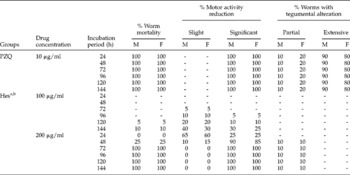
The mortality of male and female S. mansoni worms following in vitro exposure to a range of concentrations of hesperidin was calculated on a daily basis over 6 days. By 72 h, all male and female worms exposed to 200 μg/ml were killed, whereas in the 100 μg/ml concentration only 10% of worms had died after 144 h of incubation. No mortality was observed in either male or female worms incubated with 50 μg/ml up to the end of the study. In contrast, worms remained viable in the negative control group until the end of the incubation period (144 h). Meanwhile, praziquantel at 10 μg/ml resulted in the mortality of all worms within 24 h (table 1).
In vitro effects of hesperidin (Hes) and praziquantel (PZQ) on mortality, motor activity and tegumental alterations in 56-day-old adult worms of Schistosoma mansoni.

a Adult worm pairs incubated in CM (RPMI 1640+1% DMSO) remained healthy and there was no tegumental alteration up to the end of the study.
b Only 10% of male and 5% of female worms showed a slight reduction in motor activity after 144 h of incubation with 50 μg/ml of hesperidin. No mortality or tegumental alterations were observed in either male or female worms up to the end of the experiment.
Subsequently, the effect of hesperidin on the motor activity of the worms was examined, and a dose-dependent reduction of the activity was observed. A significant decrease in the motor activity was seen in 25% of the adult parasites after 24 h of exposure to 200 μg/ml. This percentage increased to 90% and 85% of male and female worms, respectively, after 48 h of incubation. In the meantime, 5 and 10% significant reduction in the motor activity of the adult worms were observed after exposure to 100 μg/ml for 96 and 120 h, respectively. This percentage increased to 30% and 25% for male and female worms, respectively, after 144 h of exposure. In contrast, small reductions in the motor activity were observed in 5% female and 10% male worms following exposure to 50 μg/ml for 144 h. On the other hand, all adult worms incubated in a medium containing 10 μg/ml praziquantel became immediately contracted and their motor activities were significantly reduced (table 1).
In addition to the mortality rate and changes in the motor capacity of S. mansoni adults, the results highlighted the effect of hesperidin on the tegument. Only 10% of living worms showed partial morphological alterations of the tegument after exposure to 200 μg/ml for 48 h upwards. No extensive tegumental alterations were observed in worms incubated with any tested concentration of hesperidin. In contrast, adult worms incubated with 10 μg/ml praziquantel showed tegumental alterations, and severe tegumental alterations were more pronounced in male than in female adults. Meanwhile, no tegumental changes in adult worms were observed in the negative control group (table 1).
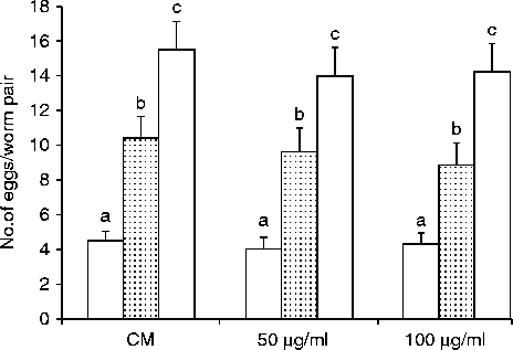
In order to evaluate the effect of hesperidin on oviposition, hesperidin was tested at 50 and 100 μg/ml. Hesperidin did not cause worm mortality after 72 h of incubation at these concentrations. There was no significant difference (P>0.05) in egg output of adult worms incubated with either 50 or 100 μg/ml for 24, 48 or 72 h compared to the corresponding negative control group (CM, fig. 1).
The effect of hesperidin on the fecundity of S. mansoni adult worms incubated for 24 h (white bars), 48 h (dotted bars) and 72 h (striped bars) in 0 (CM, culture medium), 50 or 100 μg/ml of hesperidin. Data are presented as mean eggs per worm pair ± standard deviation (SD) of ten worm pairs. For each time point, columns sharing common superscripts denote non-significant (P>0.05) differences with the corresponding control group (CM).

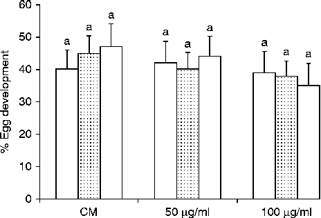
The effect of hesperidin on the development of eggs produced by the adult worms was further investigated based on the presence or absence of the miracidium. Tested concentrations (50 and 100 μg/ml) of hesperidin showed no significant (P>0.05) effect on deposited developed eggs after 24, 48 and 72 h of incubation in comparison to the negative control group (fig. 2).
The effect of hesperidin on egg development of S. mansoni adult worms incubated for 24 h (white bars), 48 h (dotted bars) and 72 h (striped bars) in 0 (CM, culture medium), 50 or 100 μg/ml of hesperidin. Data are expressed as mean developed eggs percentage ± standard deviation (SD) of ten worm pairs. Columns of each time point sharing common superscripts denote non-significant (P>0.05) differences.

The effect of hesperidin on worm and egg burden in vivo
A total number of 30 mice were each exposed to 60 cercariae for 2 h. After the exposure time, four mice were found with more than 5% of unpenetrated cercariae; therefore, they were excluded from the experiment. The remaining 26 mice were divided into three groups: 8 mice for the infected untreated group, 8 mice treated with 600 mg hesperidin/kg bw and 10 mice treated with 1000 mg praziquantel/kg bw.
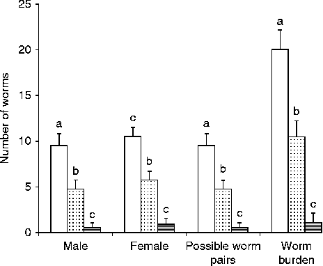
Hesperidin treatment significantly (P< 0.001) reduced male, female, possible worm pairs and total worm burden counts by 50, 45.2, 50 and 47.5%, respectively, compared to infected untreated mice. On the other hand, mice treated with praziquantel (two successive doses each of 500 mg/kg bw) showed 94.7, 91.7, 94.7 and 94.7% reduction (P< 0.001) in male, female, possible worm pairs and total worm burden counts, respectively, compared to infected untreated mice (fig. 3).
The effect of hesperidin treatment on worm burden. Mice were treated with either hesperidin (dotted bars), praziquantel (striped bars) or remained as untreated control mice (white bars). Data are expressed as mean number of worms ± standard deviation (SD) of eight mice per group. For each parameter, columns not sharing common superscripts denote significant (P< 0.001) differences.

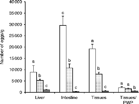
Hesperidin treatment significantly (P< 0.01) reduced eggs/g liver, eggs/g intestine and eggs/g tissues by 41.5, 63.7 and 58.6%, respectively, compared to infected untreated mice. However, hesperidin treatment did not significantly (P>0.05) alter eggs/g tissues per possible worm pairs; whereas praziquantel treatment significantly (P< 0.001) decreased eggs/g liver, intestine, tissues and eggs/g tissues per possible worm pairs by 88.1, 99.3, 96.7 and 75.6%, respectively, compared to the control group (fig. 4).
The effect of hesperidin treatment on egg burden. Mice were treated with either hesperidin (dotted bars), praziquantel (striped bars) or remained as untreated control mice (white bars). Schistosoma mansoni eggs were estimated in both liver and intestine tissues, and eggs/g tissues (liver + intestine) and eggs/g tissues per possible worm pairs (PWP) were calculated. Data are expressed as mean number of eggs ± standard deviation (SD) of eight mice per group. For each organ, columns not sharing common superscripts denote significant (P< 0.01) differences.

The effect of hesperidin on antibody responses to worm antigens
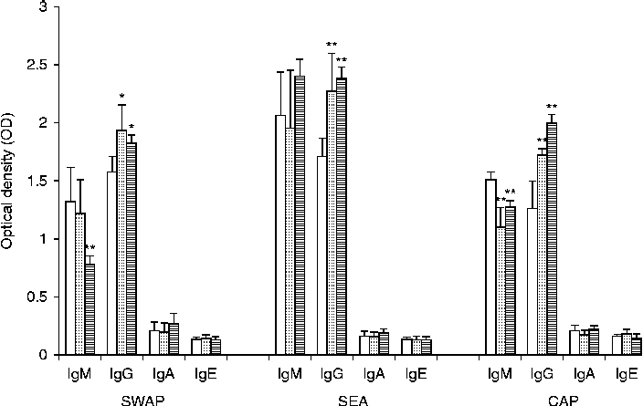
As hesperidin-treated mice demonstrated a higher reduction in parasite burden than in vitro studies, we decided to determine the specific humoral immune response induced in infected animals following treatment. Therefore, we measured the IgM, IgG, IgA and IgE levels in response to S. mansoni antigens at week 9 following infection, using indirect ELISA. Our data showed that anti-soluble worm antigen protein and anti-soluble egg antigen IgM levels were not significantly different (P>0.05), whereas the level of anti-cercarial antigen preparation IgM significantly decreased (P< 0.01) in hesperidin-treated mice compared with untreated mice. However, anti-soluble worm antigen protein (P< 0.05), anti-soluble egg antigen (P< 0.01) and anti-cercarial antigen preparation (P< 0.01) IgG levels were all significantly higher in the hesperidin-treated groups compared to the control group. On the other hand, praziquantel treatment significantly decreased (P< 0.01) IgM levels in response to both soluble worm antigen protein and cercarial antigen preparation, but did not significantly affect (P>0.05) IgM levels in response to soluble egg antigen. Similar to hesperidin-treated mice, praziquantel-treated mice showed a significant effect on IgG in response to soluble worm antigen protein (P< 0.05), soluble egg antigen (P< 0.01) and cercarial antigen preparation (P< 0.01) compared to infected untreated mice. Meanwhile, there was no significant difference between the IgA and IgE levels in response to different S. mansoni antigens (P>0.05) in the different groups (fig. 5).
The effect of hesperidin (dotted bars) and praziquantel treatment (striped bars) on specific antibody response to soluble worm antigen protein (SWAP), soluble egg antigen (SEA) and cercarial antigen preparation (CAP) of S. mansoni. Data reported as mean optical density (OD) ± standard deviation (SD) of eight mice per group. * P< 0.05, ** P< 0.01 versus the corresponding value of untreated control mice (white bars).

Discussion
The control of schistosomiasis depends almost completely on praziquantel, and there are no other reliable alternative drugs in sight. Oxamniquine, the only alternative commercially available drug, is expensive and active against only one of the three main schistosome species that are capable of infecting humans (Cioli et al., Reference Cioli, Valle, Angelucci and Miele2008). Recently, the development of new drugs from natural products for schistosomiasis has been highly encouraged (Mølgaard et al., Reference Mølgaard, Nielsen, Rasmussen, Drummond, Makaza and Andreassen2001; Sanderson et al., Reference Sanderson, Bartlett and Whitfield2002; Kayser et al., Reference Kayser, Kiderlen and Croft2003; Magalhães et al., Reference Magalhães, Machado, Morais, Moreira, Soares, da Silva, Da Silva Filho and Rodrigues2009, Reference Magalhães, Kapadia, da Silva Tonuci, Caixeta, Parreira, Rodrigues and Da Silva Filho2010; Parreira et al., Reference Parreira, Magalhães, Morais, Caixeta, de Sousa, Bastos, Cunha, Silva, Nanayakkara, Rodrigues and da Silva Filho2010). In this context, secondary plant metabolites may provide lead structures for the development of new anti-schistosomal drugs. In the present study, anti-schistosomal activity of hesperidin was assessed in vitro and in vivo. Data of the current study have demonstrated that hesperidin at 200 μg/ml was very effective against adult worms. However, lower concentrations showed little or no schistosomicidal effect. These results indicated that hesperidin was effective against adult worms only at higher concentrations. The exact mechanism by which hesperidin exerted its in vitro schistosomicidal effect is not clear and needs further investigation. However, it has been demonstrated that hesperidin inhibited lipase activity from porcine pancreas and Pseudomonas, where the IC50 was 32 and 132 μg/ml, respectively (Kawaguchi et al., Reference Kawaguchi, Mizuno, Aida and Uchino1997). Meanwile, Zhang et al. (Reference Zhang, Lu, Tao, Tao, Su and Wei2007) reported that hesperidin inhibited tyrosinase diphenolase activity with IC50 equal to 16.08 mm. Therefore, hesperidin might exert its anti-schistosomal activity through inhibition of vital enzymes of adult worms.
In the in vivo experiment, hesperidin was administrated i.p. as absorption proved to be higher by this route than after oral administration (Pires Das Neves et al., Reference Pires Das Neves, Carvalho, Carvalho, Fernandes, Soares, Bastos and Pereira2004; Hosseinimehr & Nemati, Reference Hosseinimehr and Nemati2006). The total dose of hesperidin (600 mg/kg bw) used in the current study was equal to 1/10 of the LD50. Many studies have used more than or equal to 10% of the LD50 for the experimental treatment with different drugs (Cantilena & Klaassen, Reference Cantilena and Klaassen1982; Biała & Langwiński, 1996). The total dose was divided into six injections (two times per week) each of 60 mg/kg bw to reduce any possible toxic effect of such a dose on S. mansoni-infected mice. Interestingly, Hosseinimehr & Nemati (Reference Hosseinimehr and Nemati2006) demonstrated that hesperidin did not show any genotoxic or toxic effects at doses up to 80 mg/kg bw in mice bone marrow cells when administrated i.p. Moreover, El-Sayed et al. (Reference El-Sayed, Abo-Salem, Abd-Ellah and Abd-Alla2007) reported that the pretreatment of rats with hesperidin once daily at a dose of 200 mg/kg bw i.p. for 28 days (a total dose of 5.6 g/kg bw) ameliorated acrylonitrile toxic volatile liquid-induced alterations in brain lipid peroxidation.
Data from the in vivo study showed that hesperidin treatment at a total dose of 600 mg/kg bw reduced both male and female worm burdens, by 50 and 45.2%, respectively. It appeared that this compound was active against both male and female worms, unlike other drugs which act preferentially against one sex rather than the other (Sanderson et al., Reference Sanderson, Bartlett and Whitfield2002). There are two, not mutually exclusive explanations for this reduction in worm burden. First, hesperidin possesses a direct schistosomicidal effect on adult worms, this appeared clear in data of the in vitro study. Second, hesperidin may induce an effective immune response against adult S. mansoni. The latter explanation is supported by the relatively greater humoral immune response elicited in vivo against S. mansoni antigens. Data of the present study clearly showed that hesperidin treatment augmented the mouse IgG response against soluble worm antigen protein, soluble egg antigen and cercarial antigen preparations of S. mansoni. It has been found that hesperidin significantly increased the contents of IgG in weaned piglets (Ying et al., Reference Ying, ZhiSheng and AnGuo2009). The absolute requirement for antibody response in protection against murine and human schistosomiasis has been amply documented (Hoffmann et al., Reference Hoffmann, James, Cheever and Wynn1999; Jankovic et al., Reference Jankovic, Wynn, Kullberg, Hieny, Caspar, James, Cheever and Sher1999; Dunne & Mountford, 2001). The effective factors in protective humoral immune response appeared to be dependent on IgG antibodies (Mangold & Dean, Reference Mangold and Dean1986). Moreover, the chemotherapeutic effect of praziquantel against S. mansoni was dependent mainly on IgG response (Brindley & Sher, Reference Brindley and Sher1987). Interestingly, we found that both hesperidin- and praziquantel-treated mice showed a similar pattern of IgG response to different S. mansoni antigens. Such antibodies may impair worm viability by interaction with critical sites in surface membrane-exposed molecules of importance for the parasite, such as the glucose transporter, the amino acid permease and enzymes necessary for the generation of energy and membrane repair. Such ‘neutralizing’ antibodies might interfere with the welfare of adult worms in the liver (El Ridi, Reference El Ridi2002). Moreover, it has been shown that in rat, primate and human schistosomiasis, antibody-dependent cell-mediated cytotoxicity, which mainly depends on IgG isotype, is the main mechanism involved in killing parasite larvae (Capron & Capron, Reference Capron and Capron1994). Despite the above-discussed mechanisms, the in vivo worm burden reductions were moderate. These limitations could not be explained at the moment because the exact mechanism by which hesperidin affected adult worms, either in vitro or in vivo, was not clear and needs further investigation.
Eggs laid by mature worms can become trapped in the liver and other tissues and lead to a severe syndrome including inflammation, granuloma and tissue fibrosis. Therefore, reducing egg load in the tissues can significantly relieve the morbidity caused by schistosomiasis (Huang et al., Reference Huang, Li, Qin, Cao, Yao, Liu, Li, Cai, Li and Shi2005). The current study demonstrated that hesperidin administration decreased tissue egg load by 58.6%. This decrease in egg burden could be attributed to the reduction in possible worm pairs and not to impaired worm fecundity, as eggs/g tissue per worm pairs was not significantly different from that of infected untreated mice. This result, supported by the in vitro study, showed that egg output of worm pairs was not altered by incubation with hesperidin. Therefore, hesperidin has no effect on the reproductive fitness of S. mansoni adult worms in vitro or in vivo. The effect of hesperidin on egg hatchability and juveniles was not studied and needs further investigation.
In conclusion, we demonstrated that hesperidin is an effective compound against S. mansoni both in vitro (especially in high concentrations) and in vivo. Hesperidin is a promising compound that may be used for the development of new schistosomicidal agents. Considering the results obtained, further biological studies are required in order to elucidate the mechanism of schistosomicidal action and its effect on immunity against adult S. mansoni.
Acknowledgements
This work was supported by grant number 1-433–1571 from Taif University, Ministry of Higher Education, Saudi Arabia.