




No CrossRef data available.
Article contents
Tumor necrosis factor receptor-mediated apoptosis in Trichinella spiralis-infected muscle cells
Published online by Cambridge University Press: 21 April 2005
Abstract
In order to reveal the mechanisms underlying nurse cell formation during Trichinella spiralis infection, the expression of the factors of tumor necrosis factor-alpha (TNF-α)/TNF receptor 1 (TNFR-1) signalling pathway mediating apoptosis was investigated. The analysed factors included TNF-α, TNFR-1, TNF receptor-associated death-domain (TRADD), caspase 3, caspase 8, TNF receptor associated factor-2 (TRAF2) and receptor interactive protein (RIP), all of which are involved in the TNF-α/TNFR-1 signalling pathway-mediated apoptosis. The quantitative RT-PCR indicated that the infected muscle tissues up-regulate the expression of pro-apoptosis genes (TNF-α, TNFR-1 and TRADD, caspase 3 and caspase 8), and anti-apoptosis genes (TRAF2 and RIP) at the beginning of cyst formation. The expression returned to the normal level after cyst formation. The quantitative RT-PCR analysis of mRNA from tissue samples isolated by laser capture micro-dissection confirmed that the up-regulation of these genes was restricted in infected muscle cells, was not in the inflammation cells around infected muscle cells nor in normal muscle cells. The in situ localization study of pro-apoptosis (TRADD, caspase 3) and anti-apoptosis gene products (TRAF2) indicated that these were expressed in the basophilic cytoplasm (infected muscle cell origin) of the nurse cells. Thus the present study suggests that the TNF-α/TNFR-1 signalling pathway is involved in nurse cell formation.
Information
- Type
- Research Article
- Information
- Copyright
- © 2005 Cambridge University Press
INTRODUCTION
Nematode Trichinella is a unique helminth that lives and develops in striated skeletal muscle cells. Unlike many other intracellular parasites, this parasite occupies the host cells for years without killing them. After invasion, larvae initiate a series of infected muscle cell changes including (1) re-entry of cell cycle and arrest in apparent G2/M of cell cycle (Jasmer, 1993), (2) enlarged nuclei which reflects a elevated host transcription (Despommier, Symmans & Dell, 1991; Yao & Jasmer, 2001), and (3) the degenerative and regenerative responses of muscle cells leading to nurse cell formation (Matsuo et al. 2000), which is completed around 18 days post-infection (Despommier, 1975).
Although little has been known about the underlying mechanisms of host cell remodelling by the invaded newborn larvae, a series of recent studies have reported that Trichinella infection induced apoptosis in infected muscle cells (Matsuo et al. 2000; Boonmars et al. 2004b), suggesting that apoptosis may play an essential role in this unique phenomenon, namely, muscle cell transformation leading to nurse cell formation.
It is well established that there are two principal pathways for apoptosis initiation, the mitochondrial pathway and the death receptor pathway (Gupta, 2003). In the mitochondrial pathway, Bax (Bcl-2 associated protein X) facilitates the mitochondrial release of cytochrome c that binds to Apaf 1 (apoptotic protease activating factor 1), leading to successive activation of effector caspases (Cosulich, Savory & Clarke, 1999; Vousden & Woude, 2000; Bratton et al. 2001).
In the death receptor signalling pathway, the signal is provided by the interaction between the ligand and the death receptor. Upon binding with the ligand, TNF-α (tumor necrosis factor), TNFR-1 (TNF receptor 1) recruits TRADD (TNF receptor-associated death-domain) which then recruits FADD-caspase 8 to initiate the pro-apoptotic signalling (see the review by Dempsey et al. 2003; Wajant, Pfizenmaier & Scheurich, 2003). On the other hand, if TRADD recruits TRAF2 (TNF receptor associated factor-2) and RIP (receptor interactive protein), it can initiate the reverse function, that is, anti-apoptotic signalling (Hsu, Xiong & Goeddel, 1995; Beg & Baltimore, 1996; Liu et al. 1996).
Our previous experiments have indicated that mitochondria-mediated apoptosis occurs in the T. spiralis-infected muscle cells (Boonmars et al. 2004b). T. spiralis-infected muscle cells showed morphological characteristic of apoptosis in basophilic cytoplasm (Matsuo et al. 2000). In favour of this observation, the expression of the genes associated with mitochondria-mediated apoptosis (Bax, Apaf-1; Caspase 9 and serine/threonine protein kinase, PKB) were elevated in the T. spiralis-infected muscles cells during cyst formation (Boonmars et al. 2004b).
From another line of our experiments, in cDNA microarray analysis, expression of TNFR-1 was highly up-regulated in T. spiralis-infected muscle (Wu et al. 2005). To expand these data, the present study investigated the kinetics of expression of several key genes; TNF-α, TNFR-1 and TRADD which initiate the TNF-α/TNFR-1 signaling pathway, caspase 8 and caspase 3 which mediates apoptosis signalling, and RIP and TRAF2 which mediates anti-apoptosis signalling. The results suggested that the TNF-α/TNFR-1 signalling pathway is induced and that both apoptosis and anti-apoptosis mechanisms are involved in nurse cell formation after T. spiralis infection.
MATERIALS AND METHODS
Parasite and infection
Nude mice were orally infected with 600 larvae of T. spiralis (ISS413). The muscle samples of hind limbs were collected at day 0, 8, 13, 18, 23, 28, 33, 38 and 48 days post-infection (p.i.).
Quantitative RT-PCR
Total RNA was isolated from the muscle sample using TRIZOL (GIBCOBRL, Life Technologies, Inc., CA, USA) according to the manufacturer's instructions. To diminish DNA contamination, the isolated RNA was treated with DNase.
Reverse transcription was performed using a Ready-To-Go Kit (Amersham Pharmacia Biotech Inc, NJ, USA) according to the manufacturer's instructions. In brief, 3 μg of the sample RNA and 1 μl of 0·5 μg/μl Oligo (dT) 12–18 were added to a Ready-To-Go tube, followed by RNase-free water to give a final volume of 33 μl. The tube was incubated at 37 °C for 60 min, and then at 90 °C for 5 min. The PCR reaction mixture was comprised of 3 μl of 1[ratio ]10 diluted reverse transcription products, 3 μl of 10× PCR buffer, 3 μl of dNTP (2·5 mM each), 6 μl of sense and anti-sense primers (5 μM), 0·12 μl of EX Taq polymerase (5 U/μl, Takara Shuzo, Co., Ltd, Kyoto, Japan), and distilled water to give the final volume of 30 μl. PCR amplification was performed under the condition of 1 cycle at 95 °C for 3 min; 25–35 cycles of 95 °C for 30 sec, 52–56 °C (depending on primers) for 30 sec and 72 °C for 1 min; and a final extension cycle at 72 °C for 10 min.
The expression levels of 7 kinds of genes related to TNF-α-induced apoptosis were investigated. PCR primers were developed using the published sequence, including TNF-α (Accession number: XM_123058, ggcctccctctcatcagttc/ccattcccttcacagagcaa), TNFR-1 (Accession number: L35302, gttccaacgctacctgagtg/gcatacagaatcgcaaggtc), TRADD (Accession number: U43184, agagtggcctggacctgtt/gtcagccaccagattcagt), caspase 8 (Accession number: XM_129752; gctggcagaaaataacttgg/gcaagcctgaatgaaaaaga), caspase 3 (Accession number: BC038825 gctctggtacggatgtggac/taacgcgagtgagaatgtgc), TRAF2 (Accession number: XM_130119, actgtgctgcctgtgtctat/cctctctacttcacggttca), and RIP (Accession number: U25995, tggagcaaactgactaagga/ggaggaagaaaaccaggact).
To compare the levels of expression of the genes, a semi-quantitative RT-PCR was performed as described previously (Boonmars et al. 2004b). A pair of primers was developed to amplify housekeeping gene G3PDH (glyceraldehyde 3-phosphate dehydrogenase) of mouse (Accession number: M32599, cccgtagacaaaatggtgaagg/gacacattgggggtaggaacac) was used as a constitutively expressed standard gene. The PCR product was electrophoresed in a 1·5% agarose gel, stained with 1 μg/ml ethidium bromide, and photographed under UV light. The photographs were analysed densitometrically using NIH Image 1.95 software (developed at the U.S. National Institutes of Health and available on the Internet at http://rsb.info.nih.gov/nih-image/). The optical intensities of the bands of the sample gene and G3PDH gene were measured, and the ratio of both the intensities represented a relative level of expression. Three independent experiments were performed and the values were expressed as mean±S.D.
To confirm that the PCR product with each gene primer was from muscle cells but not from the larva, cDNA of larva was used as a template and PCR was performed with the same conditions as described above.
Laser capture micro-dissection (LCM) and RT-PCR
To identify the origin of TNF-α-induced apoptosis genes, LCM was adapted to isolate RNA from defined cells as described previously (Boonmars et al. 2004b). In brief, 8 μm cryosections of muscles from day 23 p.i. were cut and placed onto glass slides pre-cooled, and dried immediately using a conventional dryer. The infected cells, normal cells (from the non-infected area of the same sample) and inflammation cells (from the area around the infected cell) were micro-dissected out using the laser micro-beam of the Robot-MicroBeam system (P.A.L.M. GmbH, Bernried, Germany), as shown in Fig. 3A. Samples with the same dimension of square μm were collected, which included about 200 infected muscle cells (1×10⁶/μm2 of tissue section).
The dissected tissues were treated with a lysis buffer and incubated at 37 °C for 15 min. After vigorous vortex for 30 min, the samples were subjected to RNA isolation using RNeasy Fibrous Tissue Mini Kit (QIAGEN, Maryland, USA). The isolated RNA sample was reverse-transcripted into cDNA using 1 μl of 0·5 μg/μl oligo(dT) 12–18 primers (Amersham Pharmacia Biotech) and a Ready-to-Go First-Strands beads kit (Amersham Pharmacia Biotech). The tube was incubated at 37 °C for 60 min and then at 95 °C for 10 min. The expression levels of each gene were determined as described above.
Immunohistochemistry
To examine the tissue localization of the investigated gene products in muscle cells, immunostaining was performed as previously described (Wu et al. 2001). In brief, cryosections of tissues from 18, 23 and 48 days p.i. were fixed with 4% paraformaldehyde (PFA) in 0·1 M phosphate buffered saline (PBS) for 5 min, washed with PBS for 15 min 3 times and then blocked with 5% skim milk (DIFCO Laboratories, Detroit, MI, USA) in PBS for 30 min. The primary antibodies included the rabbit polyclonal antibodies to TRADD (Santa Cruz Biotechnology Inc., CA, USA), to caspase 3 (Santa Cruz Biotechnology), and to TRAF2 (Santa Cruz Biotechnology), which represent the signal initiating factor, the apoptosis factor and the anti-apoptosis factor, respectively. The sections were incubated with the primary antibody and further processed using the Histostain SP (ZYMED Laboratories Inc., CA, USA) according to the manufacture's instructions. Slides were photographed and then stained with H&E for histological orientation. The negative control study was performed using PBS instead of the primary antibody.
TUNEL assay of chromatin fragmentation
The TUNEL assay was performed with an ApopTag Plasu Peroxidase In Situ Apoptosis Detection Kit (Intergen, NY, USA) according to the manufacturers instructions. In brief, the deparaffinized sections of T. spiralis-infected muscle tissue were treated with proteinase K. Endogenous peroxidase activity was quenched using 3% hydrogen peroxide in PBS. Sections were then incubated with digoxigenin-dNTP in the presence of terminal deoxynucleotidyl transferase (TdT), which incorporates the digoxigenin-dNTP into the 3′-OH ends of fragmented DNA. Finally, the digoxigenin was detected using anti-digoxigenin peroxidase conjugate, and the colour reaction was developed using diaminobenzidine substrate. Methyl green was used as a nuclear counter-stain.
RESULTS
Expression of TNF-α, TNFR-1 and TRADD
The mRNA expression level was indicated by the ratio with that of the control gene (G3PD). As shown in Fig. 1 and Fig. 2A, there was increased expression of TNF-α, TNFR-1 and TRADD. The expression of TNF-α rapidly increased from 0·30 (before the infection) to 1·16 (at 13 days p.i.). The expression remained high at about this level until 28 days p.i., and then decreased to 0·65 at 48 days p.i. The time- course of TNFR-1 and TRADD expressions were similar to that of TNF-α expression. Expression of TNFR-1 increased to 1·27 (13 days p.i.) −1·41 (28 days p.i.), and then decreased to 0·56 (48 days p.i.). Expression of TRADD was increased to 1·16 (13 days p.i.) to 1·55 (28 days p.i.), and then decreased to 0·81 (48 days p.i.).

Fig. 1. RT-PCR analysis of the expression of apoptosis-related genes. (A) G3PD; (B) TNF; (C) TNFR-1; (D) TRADD; (E) caspase 8; (F) caspase 3; (G) TRAF2; (H) RIP) in Trichinella spiralis-infected muscle tissue.

Fig. 2. Kinetics of the expression levels of apoptosis-related genes. (A) TNF, TNFR-1 and TRADD; (B) caspase 8 and caspase 3; (C) TRAF2 and RIP in Trichinella spiralis-infected muscle tissue. Total RNA was isolated from the infected muscle tissue at various stages (0, 8, 13, 18, 23, 28, 33, 38 and 48 days) post-infection. RT-PCR products were analysed by agarose gel electrophoresis. The band intensity was analyzed densitometrically using NIH Image 1.95 software. The expression level was represented by the ratio of the intensity of the sample gene relative to the intensity of the constitutively-expressed standard gene (G3PD) in the same sample. Data represent means±S.D. for 3 mice per group.
Expression of apoptosis signalling factor C8 and C3
As shown in Fig. 1 and Fig. 2B, at an early stage of infection, the expression of caspase 8 and caspase 3 increased rapidly from 0·35 and 0·26 (before the infection) to 0·87 and 1·15 (13 days p.i.), respectively. This was followed by a gradual increase reaching a peak at 28 days p.i. and a decrease down to 0·57 and 0·47 (48 days p.i.), respectively.
Expression of anti-apoptosis signalling factor TRAF2 and RIP
As shown in Fig. 1 and Fig. 2C, the expression of TRAF2 and RIP had slightly increased expression from 0 days p.i. to 13 days p.i. (from 0·16 and 0·30 to 0·44 and 0·78, respectively). After 13 days p.i., the expressions of both factors rapidly increased to 1·13 and 1·24 at 23 days p.i., respectively. The expression level was maintained during 23–28 days p.i., and then began to decrease slowly to 0·50 and 0·91 at 48 days p.i., respectively.
Detection of apoptosis related-genes in the infected cell by LCM
The apoptosis related-genes were detected with combination of LCM and subsequent RT-PCR. As shown in Fig. 3, the specific band of G3PD was observed and the expression levels were nearly the same as those in the 3 samples (normal, infected and inflammation cells). The expression levels of every gene (TNF-α, TNFR-1, TRADD, caspase 8, caspase 3, TRAF2 and RIP) were highly up-regulated in infected cells. The expression of the gene TNF-α, TRADD, TRAF2 and RIP were undetectable in normal cells and inflammation cells. Although the faint bands of TNFR-1, caspase 8 and caspase 3 appeared in normal cell samples, the bands were significantly dense in infected cell samples. The inflammation cell samples showed a similar result to the normal samples in the expression level of the three genes, that is, the expression levels of TNFR-1, caspase 8 and caspase 3 were nearly the same as those in normal cell samples.

Fig. 3. The expression levels of apoptosis-related genes (TNF, TNFR-1, TRADD, caspase 8, caspase 3, TRAF2 and RIP) in Trichinella spiralis-infected muscle cell (IMC), normal muscle cell (N) or inflammation cell (IC). The samples were collected using laser micro-dissection capture. Panel A: (a) an infected muscle cell at 23 days p.i. in cryosection; (b) shows that the inflammation cells around the infected muscle cell were dissected; (c) shows that the larva was removed; (d) shows that the infected muscle cell was dissected. Total RNA was isolated from the laser-micro-dissected samples, and RT-PCR products of various genes were analysed by agarose electrophoresis (Panel B). The band intensity was analysed densitometrically using NIH Image 1.95 software. The expression level was represented by the ratio of the intensity of the sample gene relative to the intensity of the constitutively expressed standard gene (G3PD) in the same sample (Panel C). Data represent means±S.D. for 3 samples per group.
In situ localization of apoptosis related-factors
To determine the tissue localization of these factors (TRADD, caspase 3 and TRAF2 proteins) in the infected muscle tissue, immunostaining was performed. As shown in Fig. 4, immunoreactivity of the 3 factors was present within the infected muscle cells, and a more intense positive immunoreaction was located at the cytoplasm. The direct comparison of the immunostaining (Fig. 4A–C for TRADD, Fig. 4G–I for caspase 3, and Fig. 4M–O for TRAF2) and H&E staining (Fig. 4D–F for TRADD, Fig. 4J–L for caspase 3, and Fig. 4P–R for TRAF2) slides confirmed that the cytoplasm of the positive staining is predominantly basophilic cytoplasm of the infected muscle cell. The positive staining was found at the early stage of infection such as 18 and 23 days p.i., not at the late stage of infection such as 48 days p.i. There was no positive staining in the enlarged nuclei within infected muscle cells. There was no detectable staining for these 3 kinds of proteins in uninfected muscle cells (as shown by ‘nm’ in Fig. 4) and the filtrating inflammatory cells around the cysts.

Fig. 4. Immunohistochemical staining for TRADD (A–F), caspase 3 (G–L) and TRAF2 (M–R). Cryosections of Trichinella spiralis-infected muscle tissues at 18 (A, G and M), 23 (B, H and N) and 48 (C, I and O) days post-infection were reacted with anti-TRADD, anti-caspase 3 and anti-TRAF2 antibodies. The brown colour indicates the positive reaction. After antibody staining, the same section was subjected to H&E staining for histological orientation (TRADD: D, E and F; caspase 3: J, K and L; TRAF2: P, Q and O). L: larva; b: basophilic cytoplasm; e: eosinophilic cytoplasm; nm: normal muscle cell.
DNA fragmentation
As shown in Fig. 5, positive staining for DNA fragmentation was observed in some of the enlarged nuclei within infected muscle cells at 18, 23 and 28 days p.i., as revealed by TUNEL method. There was no positive staining in the nuclei of normal muscle cells.

Fig. 5. TUNEL staining for DNA fragmentation in Trichinella spiralis-infected muscle cells. The sections at 18 (A), 23 (B) and 28 (C) days p.i. were stained using TUNEL method and counterstained with methyl green. The enlarged nuclei within infected muscle cells were positively stained (brown colour as indicated by arrows). The normal muscle nuclei were negatively stained (green colour).
DISCUSSION
The current study first clarified the kinetics and location of the TNF-α/TNFR-1 signalling pathway associated factors in T. spiralis-infected muscle tissues, which included TNF-α, TNFR-1, TRADD, caspase 8, caspase 3, TRAF2 and RIP.
The TNF-α/TNFR-1 signaling pathway is known to mediate apoptosis through TNF-R1/FRADD/FADD/caspase 8/caspase 3 signaling pathway (see the review by Dempsey et al. 2003; Wajant et al. 2003). This present kinetic study indicated that there is a rapid increase and decrease in TNF-α, TNFR-1 and TRADD expression levels in infected muscles (Fig. 2A). The timing of this temporal increase corresponds to the cyst formation which occurs by 18 days post infection. The effectors in the TNF pathway mediated-apoptosis (caspase 8 and caspase 3) also followed the same expression kinetics. In fact, the apoptosis is likely to be ongoing because the nurse cell transformation during the cyst formation accompanies some pathological changes similar to those seen in apoptosis, such examples include condensation, hypertrophy and/or fragmentation of infected muscle cell nucleus (Matsuo et al. 2000; Boonmars et al. 2004b). Our present study showed that there is occurrence of DNA fragmentation in some of the enlarged nuclei within infected muscle cells, which reconfirms that T. spiralis infection induces the apoptosis in muscle cells.
Jasmer and coworkers reported the interesting phenomenon after Trichinella infection in muscles such as reentry of cell cycle and arrest in G2/M of the cell cycle (Jasmer, 1993; Yao & Jasmer, 2001). So far no data are available about the origin of cells they observed, that is, satellite cell origin or infected muscle cell origin. If these cells are of muscle cell origin, they also can undergo apoptosis through the process described in the present study.
The infected muscle tissue contains numerous inflammation cells around the cyst. Therefore, it is necessary to confirm whether the up-regulated expressions of those genes were from infected muscle cells or inflammation cells. We adapted LCM to isolate RNA from infected muscle cells and the infiltrated cells separately. The quantitative RT-PCR confirmed that the elevated expression of the detected genes was from infected muscle cells, not from inflammation cells or normal muscle cells.
Moreover, the immunostaining confirmed that both pro-apoptotic (TRADD and caspase 3) and anti-apoptotic factors (TRAF2) are expressed predominantly in the basophilic cytoplasm of infected muscle cells.
The TNF-α/TNFR-1 signalling pathway is unique because it not only mediates apoptosis, but also mediates the opposing cellular response, anti-apoptosis, within the same cell. The anti-apoptosis response is mediated through TNF-R1/FRADD/TRAF2/RIP signaling pathway, which down regulates apoptosis response by activating NF-kB (see the review by Dempsey et al. 2003; Wajant et al. 2003). In Trichinella infection, there was an increased and decreased expression of TRAF2 and RIP during the course of cyst formation. The expression kinetics were similar to those of apoptosis gene expressions, which suggests the co-existence of apoptosis and anti-apoptosis in infected muscle cells. This co-existing condition may play an important role in nurse cell formation.
Our previous study indicated that the mitochondrial pathway mediates both apoptosis and anti-apoptosis in Trichinella infected muscle cell (Boonmars et al. 2004b). The present study provided evidence that the TNF-α/TNFR-1 signalling pathway also mediates both apoptosis and anti-apoptosis function during the infection.
Interestingly, there seems to be a difference in the location of these gene expressions between the mitochondrial pathway and the TNF-α/TNFR-1 signalling pathway mediating apoptosis (as summarized in Table 1). It has been known that there are two kinds of cytoplasm in T. spiralis-infected muscle cell, basophilic and eosinophilic cytoplasm (Matsuo et al. 2000; Wu et al. 2001; Boonmars et al. 2004a,b). The basophilic cytoplasm is derived from the infected muscle cell and is dominant in the early stage of infection. At the late stage of infection, the basophilic cytoplasm is replaced by the eosinophilic cytoplasm that is derived from satellite cells. In the mitochondrial signalling pathway, pro-apoptosis factors occur in basophilic cytoplasm and anti-apoptosis factors occur in eosinophilic cytoplasm. While in the TNF-α/TNFR-1 signalling pathway, the apoptosis and anti-apoptosis factors occurred in the same cytoplasm, basophilic cytoplasm, not in eosinophlic cytoplasm. Thus the basophilic cytoplasm is a place where the two factors with opposite functions coexist. The pro-apoptotic factors seems to be dominant because the basophilic cytoplasm is subjected to disappearance following the completion of the cyst formation (Boonmars et al. 2004b).

TNF-α has been traditionally considered as a secretory product of the immune system, a majority of activated leukocytes, in response to infection or inflammation (Tracey & Cerami, 1990; Vilcek & Lee, 1991; Korner & Sedgwick, 1996). The secreted TNF-α combines with TNFR-1 on the surface of cells to initiate apoptosis signals. However, recent studies have provided surprising evidence that the stressed or injured myocytes produce TNF which functions as a local autocrine or paracrine mediator (Tews & Goebel, 1996; De Bleecker et al. 1999; Warren et al. 2002; Li, 2003). Despite its potential importance, the effects and mechanisms of TNF-α on skeletal muscle remain largely undefined. The present study showed that there is up-regulated expression of TNF-α in infected muscle cells during the early stage of infection. A quantitative analysis of mRNA isolated from infected muscle cells by laser capture microdissection indicated that the increased expression of TNF-α is confined to infected muscle cells, not to the inflammation cells (the area around infected cells) nor to normal muscle cells because they gave undetectable results. These results further support the previous observation by other authors, who reported that endogenous TNF-α is expressed by the injured myofibres during the course of regeneration (Collins & Grounds, 2001; Zador et al. 2001; Warren et al. 2002), dystrophic mdx myofibers (Tews & Goebel, 1996), and inflammatory myopathies (De Bleecker et al. 1999). Since TNF-α is multi-functional, it likely plays many roles in the nurse cell formation, but the involvement in apoptosis and anti-apoptosis signalling may be one of the functions of the TNF-α autocrined in infected muscle cells.
Thus the over-expression of endogenous TNF-α in the T. spiralis-infected muscle cells is likely involved in the opposite function, that is, apoptosis and anti-apoptosis in the infected muscle, which is one of potential mechanisms of the nurse cell formation.
This study was partially supported by a Grant-in-Aid for Scientific Research (15590366) from the Ministry of Education, Culture, Sports, Science and Technology of Japan.
References
REFERENCES
Beg, A. A. and
Baltimore, D. (
1996).
An essential role for NF-kappaB in preventing TNF-alpha-induced cell death.
Science
274,
782–784.CrossRefGoogle Scholar
Boonmars, T.,
Wu, Z.,
Nagano, I.,
Nakada, T. and
Takahashi, Y. (
2004 a).
Differences and similarities of nurse cells in cysts of T. spiralis and T. pseudospiralis.
Journal Helminthology
78,
7–16.Google Scholar
Boonmars, T.,
Wu, Z.,
Nagano, I. and
Takahashi, Y. (
2004 b).
Expression of apoptosis-related factors in muscles infected with T. spiralis.
Parasitology
128,
323–332.Google Scholar
Bratton, S. B.,
Walker, G.,
Srinivasula, S. M.,
Sun, X. M.,
Butterworth, M.,
Alnemri, E. S. &
Cohen, G. M. (
2001).
Recruitment, activation and retention of caspases-9 and -3 by Apaf-1 apoptosome and associated XIAP complexes.
EMBO Journal
20,
998–1009.CrossRefGoogle Scholar
Collins, R. A. and
Grounds, M. D. (
2001).
The role of tumor necrosis factor-alpha (TNF-alpha) in skeletal muscle regeneration. Studies in TNF-alpha(−/−) and TNF-alpha(−/−)/LT-alpha(−/−) mice.
Journal of Histochemistry and Cytochemistry
49,
989–1001.CrossRefGoogle Scholar
Cosulich, S. C.,
Savory, P. J. and
Clarke, P. R. (
1999).
Bcl-2 regulates amplification of caspase activation by cytochrome c.
Current Biology
9,
147–150.CrossRefGoogle Scholar
De Bleecker, J. L.,
Meire, V. I.,
Declercq, W. and
Van Aken, E. H. (
1999).
Immunolocalization of tumor necrosis factor-alpha and its receptors in inflammatory myopathies.
Neuromuscular Disorders
9,
239–246.Google Scholar
Dempsey, P. W.,
Doyle, S. E.,
He, J. Q. and
Cheng, G. (
2003).
The signaling adaptors and pathways activated by TNF superfamily.
Cytokine and Growth Factor Review
14,
193–209.CrossRefGoogle Scholar
Despommier, D. D. (
1975).
Adaptive changes in muscle fibers infected with Trichinella spiralis.
American Journal of Pathology
78,
477–496.Google Scholar
Despommier, D. D.,
Symmans, W. F. and
Dell, R. (
1991).
Changes in nurse cell nuclei during synchronous infection with Trichinella spiralis.
Journal of Parasitology
77,
290–295.CrossRefGoogle Scholar
Gupta, S. (
2003).
Molecular signaling in death receptor and mitochondrial pathways of apoptosis (Review).
International Journal of Oncology
22,
15–20.CrossRefGoogle Scholar
Hsu, H.,
Xiong, J. and
Goeddel, D. V. (
1995).
The TNF receptor-1 associated protein TRADD signal cell death and NF-kB activation.
Cell
81,
495–504.CrossRefGoogle Scholar
Jasmer, D. P. (
1993).
Trichinella spiralis infected skeletal muscle cells arrest in G2/M and cease muscle gene expression.
Journal of Cell Biology
121,
785–793.CrossRefGoogle Scholar
Korner, H. and
Sedgwick, J. D. (
1996).
Tumour necrosis factor and lymphotoxin: molecular aspects and role in tissue-specific autoimmunity.
Immunology and Cell Biology
74,
465–472.CrossRefGoogle Scholar
Li, Y. (
2003).
TNF-α is a mitogen in skeletal muscle.
American Journal of Physiology: Cell Physiology
285,
C370–C376.Google Scholar
Liu, Z. G.,
Hsu, H.,
Goeddel, D. V. and
Karin, M. (
1996).
Dissection of TNF receptor 1 effector functions: JNK activation is not linked to apoptosis while NF-kappaB activation prevents cell death.
Cell
87,
565–576.CrossRefGoogle Scholar
Matsuo, A.,
Wu, Z.,
Nagano, I. and
Takahashi, Y. (
2000).
Five types of nuclei present in the capsule of Trichinella spiralis.
Parasitology
121,
203–210.CrossRefGoogle Scholar
Tews, D. S. and
Goebel, H. H. (
1996).
Cytokine expression profile in idiopathic inflammatory myopathies.
Journal of Neuropathology and Experimental Neurology
55,
342–347.CrossRefGoogle Scholar
Tracey, K. J. and
Cerami, A. (
1990).
Metabolic responses to cachectin/TNF. A brief review.
Annals of the New York Academy of Sciences
587,
325–331.CrossRefGoogle Scholar
Vilcek, J. and
Lee, T. H. (
1991).
Tumor necrosis factor: New insights into the molecular mechanisms of its multiple actions.
Journal of Biological Chemistry
266,
7313–7316.Google Scholar
Vousden, K. H. and
Woude, G. F. (
2000).
The ins and outs of p53.
Nature, Cell Biology
2,
E178–E180.CrossRefGoogle Scholar
Wajant, H.,
Pfizenmaier, K. and
Scheurich, P. (
2003).
Tumor necrosis factor signaling.
Cell Death and Differentiation
10,
45–65.CrossRefGoogle Scholar
Warren, G. L.,
Hulderman, T.,
Jensen, N.,
Mckinstry, M.,
Mishra, M.,
Luster, M. I. and
Simeonova, P. P. (
2002).
Physiological role of tumor necrosis factor alpha in traumatic muscle injury.
FASEB Journal
16,
1630–1632.CrossRefGoogle Scholar
Wu, Z.,
Matsuo, A.,
Nakada, T.,
Nagano, I. and
Takahashi, Y. (
2001).
Different response of satellite cells in the kinetics of myogenic regulatory factors and ultrastructural pathology after Trichinella spiralis and T. pseudospiralis infection.
Parasitology
123,
85–94.CrossRefGoogle Scholar
Wu, Z.,
Nagano, I.,
Boonmars, T. and
Takahashi, Y. (
2005).
A spectrum of functional genes mobilized after Trichinella spiralis infection in skeletal muscle.
Parasitology
130.CrossRefGoogle Scholar
Yao, C. and
Jasmer, D. P. (
2001).
Trichinella spiralis-infected muscle cells: abundant RNA polymerase II in nuclear speckle domains colocalizes with nuclear antigens.
Infection and Immunity
69,
4065–4071.CrossRefGoogle Scholar
Zador, E.,
Mendler, L.,
Takacs, V.,
De Bleecke, R. J. and
Wuytack, F. (
2001).
Regenerating soleus and extensor digitorum longus muscles of the rat show elevated levels of TNF-alpha and its receptors, TNFR-60 and TNFR-80.
Muscle and Nerve
24,
1058–1067.CrossRefGoogle Scholar
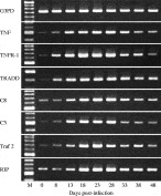
Fig. 1. RT-PCR analysis of the expression of apoptosis-related genes. (A) G3PD; (B) TNF; (C) TNFR-1; (D) TRADD; (E) caspase 8; (F) caspase 3; (G) TRAF2; (H) RIP) in Trichinella spiralis-infected muscle tissue.
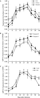
Fig. 2. Kinetics of the expression levels of apoptosis-related genes. (A) TNF, TNFR-1 and TRADD; (B) caspase 8 and caspase 3; (C) TRAF2 and RIP in Trichinella spiralis-infected muscle tissue. Total RNA was isolated from the infected muscle tissue at various stages (0, 8, 13, 18, 23, 28, 33, 38 and 48 days) post-infection. RT-PCR products were analysed by agarose gel electrophoresis. The band intensity was analyzed densitometrically using NIH Image 1.95 software. The expression level was represented by the ratio of the intensity of the sample gene relative to the intensity of the constitutively-expressed standard gene (G3PD) in the same sample. Data represent means±S.D. for 3 mice per group.
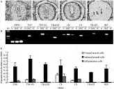
Fig. 3. The expression levels of apoptosis-related genes (TNF, TNFR-1, TRADD, caspase 8, caspase 3, TRAF2 and RIP) in Trichinella spiralis-infected muscle cell (IMC), normal muscle cell (N) or inflammation cell (IC). The samples were collected using laser micro-dissection capture. Panel A: (a) an infected muscle cell at 23 days p.i. in cryosection; (b) shows that the inflammation cells around the infected muscle cell were dissected; (c) shows that the larva was removed; (d) shows that the infected muscle cell was dissected. Total RNA was isolated from the laser-micro-dissected samples, and RT-PCR products of various genes were analysed by agarose electrophoresis (Panel B). The band intensity was analysed densitometrically using NIH Image 1.95 software. The expression level was represented by the ratio of the intensity of the sample gene relative to the intensity of the constitutively expressed standard gene (G3PD) in the same sample (Panel C). Data represent means±S.D. for 3 samples per group.
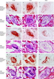
Fig. 4. Immunohistochemical staining for TRADD (A–F), caspase 3 (G–L) and TRAF2 (M–R). Cryosections of Trichinella spiralis-infected muscle tissues at 18 (A, G and M), 23 (B, H and N) and 48 (C, I and O) days post-infection were reacted with anti-TRADD, anti-caspase 3 and anti-TRAF2 antibodies. The brown colour indicates the positive reaction. After antibody staining, the same section was subjected to H&E staining for histological orientation (TRADD: D, E and F; caspase 3: J, K and L; TRAF2: P, Q and O). L: larva; b: basophilic cytoplasm; e: eosinophilic cytoplasm; nm: normal muscle cell.

Fig. 5. TUNEL staining for DNA fragmentation in Trichinella spiralis-infected muscle cells. The sections at 18 (A), 23 (B) and 28 (C) days p.i. were stained using TUNEL method and counterstained with methyl green. The enlarged nuclei within infected muscle cells were positively stained (brown colour as indicated by arrows). The normal muscle nuclei were negatively stained (green colour).

Table 1. Comparison of mitochondria-mediated and death receptor-mediated apoptosis in Trichinella spiralis-infected muscle cells
 You have
Access
- 24
- Cited by