Introduction
ATD studies used to examine the effects of a rapid reduction in serotonin function have investigated the role of serotonin function in AD (Porter et al., Reference Porter, Lunn, Walker, Gray, Ballard and O'Brien2000; Reference Porter, Lunn and O'Brien2003), PD (Scholtissen et al., Reference Scholtissen, Verhey, Adam, Prickaerts and Leentjens2006; Mace et al., Reference Mace, Porter, Dalrymple-Alford and Anderson2010a; Reference Mace, Porter, Dalrymple-Alford, Wesnes and Anderson2010b), and recovered depression in the elderly (Porter et al., Reference Porter, Phipps, Gallagher, Scott, Stevenson and O'Brien2005; Reference Porter, Bourke and Gallagher2007). These studies have found that ATD generally decreased global cognitive status (as measured by the MMSE) in patients compared with healthy controls. We have suggested that this may be a result of a sudden reduction in serotonin function compounding an impaired cholinergic system in these conditions.
There is evidence that cholinergic impairment is comparatively greater in PDD and DLB (Tiraboschi et al., Reference Tiraboschi2002; Bohnen et al., Reference Bohnen2003); thus, we predict that the effect of ATD on global cognitive status would be greater in dementia associated with Lewy body pathology than in AD.
Methods
PDD and DLB patients, as defined by the Movement Disorders Society task force (Emre et al., Reference Emre2007) and the consensus guidelines for dementia with Lewy bodies (McKeith et al., Reference McKeith1996), were recruited by a neurologist (TA) and old age psychiatrist (CC) in Christchurch, New Zealand. Patients had a recent physical and neurological examination. Exclusion criteria included a history of affective or other psychiatric disorder, serious medical disease, and serotonergic medication. All patients participated voluntarily and written informed consent was obtained. The study received ethical approval from the Upper South B Regional Ethics Committee, Christchurch. The design, procedure, and assessments have been described elsewhere (Mace, Reference Mace, Porter, Dalrymple-Alford and Anderson2010a; Reference Mace, Porter, Dalrymple-Alford, Wesnes and Anderson2010b). Cognitive status was measured using the Modified Mini-Mental State test (3MS) (Teng and Chui, Reference Teng and Chui1991). Practice effects were attenuated by using three different items for each registration and recall category as per the manual. Paired samples t-tests were used to compare results between placebo and depletion using SPSS for Windows Release 18 (SPSS, Chicago, Illinois).
Results
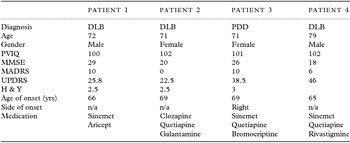
The study was designed to recruit 20 patients. However, the trial was terminated because of adverse events such that only four patients completed testing. Of the six patients recruited to the study, one female PDD patient, taking lisuride, sinemet, selegiline, procyclidine, and ranitidine, vomited at noon on the first test day and was taken home before testing. One female patient with DLB, taking rivastigmine, became obviously very confused during the late afternoon of the second visit (depletion) and meaningful testing could not be carried out. Confusion was not, in the weeks prior to the study, a significant part of her presentation. At follow-up, her nursing staff reported she had hallucinated on her way home. The characteristics of the four patients who completed two test days are presented in Table 1.
Patient characteristics at baseline

Note. DLB = dementia with Lewy bodies; H & Y = Hoehn and Yahr (Reference Hoehn and Yahr1967); MADRS = Montgomery–Asberg Depression Rating Scale; MMSE = Mini-Mental State Examination; PDD = Parkinson's disease and dementia; PVIQ = Predicted Verbal IQ (Nelson, Reference Nelson1982); UPDRS = United Parkinson's Disease Rating Scale.
Assessments
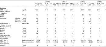
Data for all patients were limited and only complete data sets are shown in Table 2 and reported in text.
Individual results for complete assessments

Note. ATD = Acute Tryptophan Depletion; COWA = Controlled Oral Word Association test; CRT = Choice Reaction Time; DigitsB = Digit Span Backward; DigitsF = Digit Span Forward; DLB = dementia with Lewy bodies; 3MSE = Modified Mini-Mental State Examination; ODFAS = One Day Fluctuation Assessment Scale; PDD = Parkinson's disease and dementia; TRP = plasma tryptophan; VOSP = Visual Object and Space Perception test.
*p < 0.05.
There was a significant difference in TRP at 4 hours when levels were lower after depletion (M = 775.00, SD = 427.20) and higher after the placebo (M = 5800.00, SD = 3496.67; t3 = 3.25, p = 0.047).
3MSE scores were lower after depletion (M = 65, SD = 17.98) compared with placebo (M = 71, SD = 15.17; t3 = −2.64, p = 0.08). The effect size (d) with all four patients was 1.38 (CI = −0.03 to 2.80). When restricted to the three DLB patients, d = 1.84 (CI = −0.22 to 3.91).
There was no significant difference in the change scores on Montgomery–Asberg Depression Rating Scale (MADRS), that is, the difference between baseline and 4.5 hours, after depletion (M = 5.5, SD = 4.51) compared to placebo (M = 2.25, SD = 3.60; t3 = 1.01, p = 0.39).
Discussion
In the four patients who tolerated ATD, the procedure significantly lowered tryptophan levels by an average of 61% at 4 hours, which is in keeping with previous studies.
The patients in this study found the protocol difficult to tolerate and a decision was made to terminate the study early. This is in contrast to previous studies in the healthy elderly, including a group aged between 70 and 90 (Mace et al., Reference Mace, Porter, Dalrymple-Alford, Wesnes and Anderson2011), and studies mentioned previously in Alzheimer's and Parkinson's diseases in which ATD has been found to be relatively tolerable. The poor tolerability of ATD in this group could relate to the marked impairment in the cholinergic system found in DLB and PDD.
In particular, one patient became very confused and hallucinated during depletion. Although potentially related to ATD, we note the scale we used to assess fluctuations in consciousness during the day − which was designed to assess fluctuations in DLB (Walker et al., Reference Walker2000) − did not show a consistent difference between ATD and placebo in the other patients. In this patient, there was uncertainty regarding the nursing home's administration of her usual rivastigmine on that morning and, although clinically significant confusion is not generally a sequela of missing a dose of a cholinesterase inhibitor, the confusion could possibly have been secondary to this omission. This patient was only taking rivastigmine, making interactions between current therapy and the effects of ATD less complicated than in the other patients. Confusion had not been a particular clinical problem in the weeks prior to depletion.
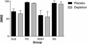
Based on previous studies and the marked cholinergic deficit in DLB/PDD, we hypothesized that ATD would reduce global cognitive status in DLB/PDD. Given the small sample size, it is not surprising that results were not significant. However, three of the four patients did have a lower 3MSE during depletion, while in the fourth patient it was unchanged. A reduction in 3MSE is in line with the results of previous studies as illustrated in Figure 1 and is compatible with the explanation that an overall cholinergic deficit, combined with a profound reduction in serotonin function caused by ATD, gives rise to a global cognitive impairment. One study referred to in Figure 1 was conducted in recovered elderly depressed subjects in whom cholinergic deficit has not been well described. However in PD at least, depression does seem to be associated with greater cholinergic deficit (Bohnen et al., Reference Bohnen, Kaufer, Hendrickson, Constantine, Mathis and Moore2007). The magnitude of the change in 3MSE obviously depends on the severity of cognitive impairment at baseline. In unimpaired groups, the 3MSE is likely to be relatively insensitive to change because subjects are operating at close to ceiling. As can be seen from Figure 1, the magnitude of change is greater in the DLB/PDD and the Alzheimer's (senile dementia of the Alzheimer's type) groups, both of whom have a much lower baseline. Variability of scores is of course also much greater in these groups and it is important to note once again the low number of patients tested in the current study and the variability that this introduces. The small number of subjects with variable test results also makes it very difficult to draw any further conclusions from the more detailed neuropsychological testing. The results of more detailed testing have not been consistent in the previous studies described.
Effects of ATD on global cognitive function in DLB/PDD (current data), PD (Mace et al., Reference Mace, Porter, Dalrymple-Alford, Wesnes and Anderson2010b), senile dementia of the Alzheimer's type (SDAT) (Porter et al., Reference Porter, Lunn, Walker, Gray, Ballard and O'Brien2000) and recovered depression in the elderly (RD) (Porter et al., Reference Porter, Phipps, Gallagher, Scott, Stevenson and O'Brien2005).

Surprisingly, none of the studies described have shown a significant differential effect on mood in the patient groups despite all being more vulnerable to depression than healthy subjects. In this small group of patients with DLB/PDD, there was no clear effect on MADRS scores.
The changes in cognitive status which appear to have occurred in this study, in response to ATD, may not be a direct result of reduced central serotonin function. Young et al. (Reference Young2013) review a number of other effects of ATD to which the patients in this study, by virtue of their neurodegenerative brain impairment, may be more vulnerable.
A further complicating factor in this study is that patients were on a range of medication including three patients who were taking a combination of L-dopa and a peripheral decarboxylase inhibitor and three who were on quetiapine. It is beyond the scope of this paper to review all the possible implications of this especially given the small number of patients. However, of particular note may be the data suggesting that the upregulation of 5HT2 receptors during ATD is important (Price et al., Reference Price, Malison, McDougle, McCance-Katz, Owen and Heninger1997). The 5HT2 antagonist effect of quetiapine could significantly alter this effect.
The adverse effects seen in this study raise further concern regarding the ethics of administering compounds that may cause even a temporary deterioration in a patient group. The possible information gained, given uncertainty regarding the specificity of the method, must be weighed against the possible side effects. If, following this assessment, symptom provocation studies such as this one are contemplated, we agree with the recommendation of Young et al. (Reference Young2013) that vulnerable participants be carefully observed (including a post-test follow-up) and administered 1 gram of tryptophan at the end of the study day to restore tryptophan levels.
The small number of patients completing the current study and uncertainties regarding the ATD method makes it impossible to draw firm conclusions. However, important results are that the protocol was poorly tolerated in this group and that there was a trend towards a reduction in global cognitive status during ATD possibly suggesting that, as hypothesized, this group is particularly susceptible to a sudden reduction in serotonin function. Clinically, it is difficult to draw conclusions from a small experimental study such as this. In our practice however, this study has increased our caution and degree of monitoring when withdrawing SSRI medication in this group of patients.
Acknowledgments
This research received no specific grant from any funding agency in the public, commercial, or not-for-profit sectors.
Conflict of interest
None.
Description of authors’ roles
Design of study – all authors. Data collection – J Mace. Supervision of data collection – R Porter, T Anderson. Patient recruitment – C Collins, T Anderson. Statistical design and analysis – J Mace, R Porter. Writing of article – all authors.