Introduction
The term dementia does not denote a disease (American Psychiatric Association, 2013), but a syndrome characterized by the progressive deterioration of the cognitive function, more commonly the memory, but also of other domains such as speech, praxis, visual perception, and executive functions (Waldemar et al., Reference Waldemar2007). The medical perspective regarding dementia evolved drastically in the 20th century, as there was not a specific diagnosis before 1900. However, after much effort and with the aid of pathological, neuroimaging, and genetic studies, it is now possible to classify dementia both clinically and pathologically (Neary et al., Reference Neary2002).
Due to the variations in sensitivity and specificity of the diagnostic criteria, and due to the growing need for accurate diagnostics for clinical research and appropriate orientations, more accurate diagnoses are expected in order to provide improvements in the quality of life – objective and subjective well-being – of patients and caregivers (Weyerer et al., Reference Weyerer2003; Alzheimer's Association, 2016). Moreover, supplementary exams are more and more expected to assist with and confirm diagnostic suspicions, as well as to exclude reversible diseases that might simulate them – the so-called reversible dementia, such as depression, normal pressure hydrocephalus (NPH) or neurosyphilis (Weytingh et al., Reference Weytingh1995).
With this purpose in mind, the last revision by the American Academy of Neurology (Knopman et al., Reference Knopman2001) on the parameters used to diagnose dementia was published in 2001, which instructs that all patients should obtain a Cranial Computed Tomography (CT) scan or Magnetic Resonance Imaging (MRI), a depression triage scale such as the “Geriatric Depression Scale” (GDS), dosing of vitamin B12, thyroid-stimulating hormone (TSH), and Venereal Disease Research Laboratory test (VDRL) for those with a risk factor, or residing in an endemic area. In endemic areas, the European guideline also recommends the serology for human immunodeficiency virus (HIV) (British HIV Association, 2008; Nightingale et al., Reference Nightingale2013) and, more recently, the systematic sleep analysis, particularly in the search for the Obstructive Sleep Apnea syndrome (OSA) (Waldemar et al., Reference Waldemar2007; Verstraeten et al., Reference Verstraeten2007). The recommendation made by the Brazilian Academy of Neurology is even broader, as it also includes a complete blood count, blood urea concentrations, creatinine, albumin, hepatic enzymes, and calcium, in order to discard misleading clinical and psychiatric causes (Nitrini et al., Reference Nitrini2005).
Considering the significant increase in the occurrence and prevalence of patients with dementia in the last few years, particularly in developing countries (Prince et al., Reference Prince2013) and considering the progressive increase in the requests for supplementary exams available, added to the progressive reduction in the time given for clinical assessment (Cooper et al., Reference Cooper and Huppert1994), and the scarcity of longitudinal publication related to the topic (Koepsell et al., Reference Koepsell2013), it is important to demonstrate which clinical, radiological, and laboratorial variables are relevant for the diagnosis and its stability of demential syndromes.
Methods
Subjects
A survey was carried out with all medical records from the Service of Cognitive and Behavioral Neurology of the Department of Neurology and Neurosurgery of the Federal University of São Paulo, between 2000 and 2015. The evaluations of each patient and the completion of each medical record were made by neurologists, geriatricians, psychiatrists, or residents in each of these fields, accompanied by neuropsychologists from that service. All medical records were evaluated by a single researcher.
Criteria for inclusion were as follows:
-
1. At least one full year of follow-up with at least two medical visits.
-
2. CDR (Clinical Dementia Rating) ≥1.0 and ≤2.0.
-
3. Diagnosis defined by the end of the follow-up period.
-
4. Medical file duly completed.
-
5. Presence of a family member or companion.
Criteria for exclusion were as follows:
-
1. Less than one year of follow-up.
-
2. CDR <1.0 or >2.0.
-
3. Diagnosis not defined by the end of the follow-up period.
-
4. Incomplete data on the file.
-
5. Absence of a family member at the appointments.
Assessment and instruments
A data collection file was elaborated to include social-demographic data. Then, the reported complaints from patients (self-report) and family or informants were assessed and divided to the following: memory, speech, executive function, behavior, visual-spatial, perception, and gait. The neurological exam was classified according to the changes that contribute to the differential diagnosis of dementia (Larner, Reference Larner2014).The time since the commencement of the symptoms as well as the psychiatric, vascular, and non-vascular background was described. The Clinical Dementia Rating (CDR) (Montaño et al., Reference Montaño2005) and the Neuropsychiatric Inventory (NPI) (Camozzato et al., Reference Camozzato2008) were included based on informant's descriptions.
All patients were subject to neuropsychological tests in the first evaluation, which include, the Mini-Mental State Exam (MMSE) (Bertolucci et al., Reference Bertolucci1994), the Consortium to Establish a Registry for Alzheimer's Disease (CERAD) battery test (Bertolucci et al., Reference Bertolucci2001), the verbal fluency within 1 min with fruits and animals (Brucki et al., Reference Brucki1997), the clock drawing test (CDT) (Fuzikawa et al., Reference Fuzikawa2003), Digit-Span (Zimmermann et al., Reference Zimmermann2015), drawing of the Rey complex figure and figure recall (Oliveira et al., Reference Oliveira2004) from which it was possible to determine the cognitive domains indicated as altered in the tests (short-term memory, long-term memory, speech, executive function, and visual-spatial function). The MMSE and the CDR of the first evaluation were recorded for comparison with these same data collected after one year of follow-up.
The neuropsychological tests were standardized, accordingly to their validity in the Brazilian population and on their intra- and inter-rater reliability (Bertolucci et al., Reference Bertolucci1994; Brucki et al., Reference Brucki1997; Bertolucci et al., Reference Bertolucci2001; Fuzikawa et al., Reference Fuzikawa2003; Oliveira et al., Reference Oliveira2004; Zimmermann et al., Reference Zimmermann2015). The cut-off values and interpretations were based accordingly to the original publications controlling specially for the education bias, so important in our population due to its great variability. It is important to cite that the population in our sample is from the same center that published most of the Brazilian data about the tests applied.
According to the specific criteria of each disorder, the diagnoses were defined in the first medical appointment and one year after the supplementary exams. The depressive disorder was triaged with the application of the Brazilian version of the GDS, with 15 items (GDS 15) (Almeida et al., Reference Almeida1999). The mandatory exams for the differential diagnosis of dementia were considered relevant (TSH, B12, VDRL, HIV, Cranial CT Scan, or MRI, and when prudent, single-photon emission computerized tomography, polysomnography, liquor, and electroencephalogram (EEG). The study was approved by the Research Ethics Committee of the Federal University of São Paulo.
Statistical analysis
In the description of data, the qualitative variables were summarized to absolute frequencies (n) and relative frequencies (%); the quantitative variables were expressed as the average ± standard deviation, median, minimum, and maximum values. In order to identify the factors associated with the primary objective related to the diagnostic change, binary logistic regression models were used. At first, the isolated effect of each variable was investigated by simple logistic regression (single-variate analysis). The variables with p < 0.10 in the single-variate analysis were included in a multiple logistic regression (multivariate analysis). Based on the complete multivariate model (with all variables selected by the single-variate model), a reduced model was adjusted to consider only the variables with p ≤ 0.05. The significance level considered was 0.05. The results were presented in odds ratio and respective 95% confidence intervals. The patients with diagnostic change were evaluated individually in order to identify the exam related to such development. The statistical software used in all of the analyses was SPSS 19.0 for Windows.
Results
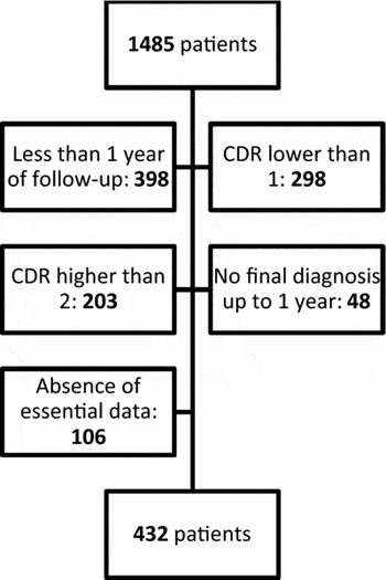
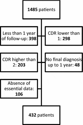
From a total of 1,485 patients evaluated by the Service of Cognitive and Behavioral Neurology of the Department of Neurology and Neurosurgery of the Federal University of São Paulo between 2000 and 2015, 432 patients were selected in accordance with the inclusion and exclusion criteria (Figure 1).
Description of patients selected by the criteria for inclusion and exclusion.
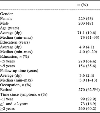
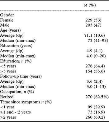
The demographic and clinical characteristics are described in Table 1.
Demographic and clinical characteristics
Source: Author's personal files.
In the initial evaluation, 263 patients (60.9%) presented mild dementia (CDR 1.0) and 169 patients (39.1%) presented moderate dementia (CDR 2.0). After at least one year of follow-up, 204 (47.2%) presented CDR 1.0, 160 (37%) presented CDR 2.0, 52 (12%) presented CDR 03, and 16 (3.7%) presented CDR 0.5. The initial MMSE average was 18.2 ± 5.4 with a median of 18.0, evolving to 17.1 ± 6.7 with a median of 18.0 after one year.
Regarding the neuropsychological domains, all patients had at least one domain affected. Most patients (76%) had a significant loss in executive function measured by the CDT and verbal fluency, followed by long term memory (69%) measured by the CERAD list, MEEM, and Rey figure. Language was affected in 60%, especially anomia in the Boston Test. The least affected domain was visuo-spatial abilities in 24.1% of the patients based on the CDT and Rey figure. Behavior abnormalities were observed in 35% of the patients during anamnesis, physical examination, and NPI.
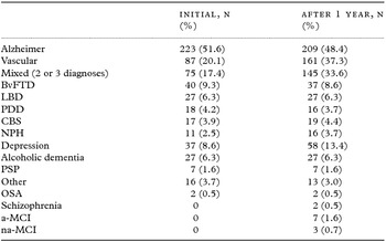
Table 2 discriminates the initial and final diagnosis of the sample. There was a decrease in the number of patients with only with diagnosis after the minimum one-year follow-up from 82.6% at first to 66.4% after one year. In the end, 140 patients (32.4%) received two diagnoses, and 05 patients (1.2%) received three diagnoses.
Distribution of diagnoses after the follow-up period (%)
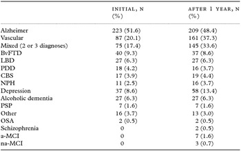
Note: BvFTD – Behavioral variant of Fronto-temporal Dementia, LBD – Lewy Body Dementia, PDD – Parkinson's Disease Dementia, CBS – Corticobasal degeneration, NPH – Normal Pressure Hydrocephalus, PSP – Progressive Supranuclear Palsy, OSA – Obstructive Sleep Apnea, a-MCI – amnestic Minor Cognitive Impairment, na-MCI – non-amnestic Minor Cognitive Impairment.
Source: Author's personal files.
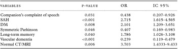
Table 3 shows the final result of the reduced multivariate model regarding all the clinical, laboratorial, and radiologic variables for diagnostic change.
Factors associated with the diagnostic change – results from the multi-variant analysis (reduced model)
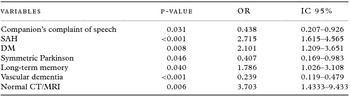
Note: SAH – Systemic Arterial Hypertension, DM – Diabetes Mellitus, CT– Computed Tomography, MRI – Magnetic Resonance Imaging
Source: Author's personal files.
The diagnostic change occurred in 113 of the 432 patients (26.6%). Among these patients, the main reason was the addition of the vascular dementia or vascular cognitive impairment (VCI) as a contributor, or even as a final diagnosis, passing from 87 patients (20.1%) to 161 patients (37.3%), leading to an 85% increase due to the neuroimaging analysis. The CT/MRI also played a role in the increase in the diagnosis of NPH that rose from 11 to 16 patients. The depression triage was also relevant in the diagnostic change, as depression was initially understood as comorbidity or even ignored, but later showed to be a significant cause of the cognitive loss demonstrated by the diagnostic change after proper treatment. The increase value was 62%, from 36 patients to 58 patients (8.3% to 13.4%).On the other hand, vitamin B12, VDRL, TSH, HIV did not contribute to the change in the diagnoses of patients. Other three patients demonstrated change regardless of the exams.
Discussion
This work assessed the clinical, laboratorial, and radiologic variables related to the clinical stability of demential syndromes in an expressive sampling of 432 patients in a Brazilian reference hospital. The longitudinal assessment was relevant for the completion of the ancillary exams and diagnostic review after at least two medical evaluations. Based on the results, it is clear that all of the evaluation phases were relevant, including anamnesis, physical exam, neuropsychological tests, and supplementary exams. Except for neuroimaging and depression triage, none of the other exams considered to be mandatory (Knopman et al., Reference Knopman2001; Nitrini et al., Reference Nitrini2005; NICE guidelines, 2006), had a direct relationship with the diagnostic stability.
Assigning an underlying pathological process to a patient with dementia is essential for clinical practice and research. However, few works have discussed so far the diagnostic evolution of patients with dementia in a longitudinal manner. To this date, we have identified only one study related to the diagnostic stability of patients with dementia (Koepsell et al., Reference Koepsell2013). Other studies are related to early-onset dementias (Panegyres et al., Reference Panegyres2007), minor cognitive impairment (MCI) (Han et al., Reference Han2012), or the clinical-pathological correlations (Mok et al., Reference Mok2004).
Koepsell et al. (Reference Koepsell2013) performed a prospective evaluation of 4,141 patients from 32 centers in the USA, from 2005 to 2009, who presented a clinical diagnosis of dementia or MCI. The patients were evaluated at least twice, with an interval of at least one year, as with our sample. The objective of the study was to evaluate the stability of the diagnosis of dementia and evaluate the factors related to such stability. Most of the patients (90%) had the same diagnosis in at least two medical appointments. Older patients (older than 70 years), non-white patients, patients with milder conditions (CDR 0.5 or 1.0), lower education level (less than 08 years), patients with more than one initially associated comorbid diagnosis, patients not accompanied by close family members, and patients evaluated by different physicians in the two medical appointments presented a higher chance of change. The main changes were caused by vascular disease, depression, and clinical change independent of exams, which is exactly the data found in our sample.
Considering the variants used in our studies, it is possible to identify many congruencies. In both studies, the presence of comorbidities, particularly vascular comorbidities such as hypertension and diabetes, increased the possibility of changes. The education level also played an important role in our study, particularly in the subgroup of patients with Alzheimer's Disease (AD) and, likewise, patients with lower education had a higher chance of diagnostic change. The relationship between education and dementia is complex However, the concept of cognitive reserve is critical to understand this relationship (Meng et al., Reference Meng and D'Arcy2012). The cognitive reserve refers to the ability to tolerate changes related to age and to the pathology related to the disease without developing signs or symptoms of the disease. Patients with lower education have higher risks both of incidence (OR: 1.88) and in prevalence (OR: 2.61) of dementia. Therefore, higher education protects against AD and VCI. However, in the higher education sub-group, once the diseases onsets, there is faster cognitive decline associated with the higher pathological load than other patients with the same impairment but lower education (Meng et al., Reference Meng and D'Arcy2012) leaving a narrow margin for confounding factors such as depression.
Some significant variants would not be possible in our work, such as the comparison between patients evaluated by the same physician or by another professional, as the operation of the University service makes it unlikely that a same physician could evaluate the patients in all of the sessions. Moreover, even though our interview form included the family relationship between the patient and the companion, the proximity of their daily lives was not mentioned, and was therefore not included in the analysis. However, these two factors are interesting because they are preventable causes of diagnostic instability, reinforcing the need for close relatives and multidisciplinary teams in the patient's attendances (Verhey et al., Reference Verhey1993; Carr et al., Reference Carr2000). Another variable not included in our analysis was race. The concept of race in Brazil is not clear sociologically or genetically, unlike the USA (Travassos et al., Reference Travassos2004).
Koepsell (Reference Koepsell2013) describes that the diagnoses that presented the highest concordance (κ) were those of atypical Parkinsonism (Progressive Supranuclear Palsy – PSP 0.78, Corticobasal syndrome – CBS 0.79, and Lewy Body Disease – LBD 0.77) and Primary Progressive Aphasia – PPA (0.76). On the other hand, diseases with lower concordance between the initial and final diagnoses were depression (0.46) and Vascular Dementia (0.69), while the latter presented a 35.5% increase in diagnoses as a final comorbid disease. Likewise, in our sample, atypical Parkinsonism had the highest diagnostic stability (κ) (PSP 1.0; Lewy 0.96; CBS 0.94), while vascular dementia and depression had the lowest correlations between the initial and final diagnoses (0.53 and 0.75). Therefore, many similarities between the studied populations are made evident, even though they are located in different countries.
More than just match previous results, we positively added significant variables, such as the type of complaint, impaired domains in the neuropsychological testing, changes in the neurological exam, and radiologic alterations. In our sample, the variables that reduced the chance of diagnostic change were the companion's complaint about speech, the presence of symmetric Parkinsonism in the physical exam, and the initial diagnosis of Vascular Dementia. On the other hand, the variables that increased the chance of diagnostic change were the presence of systemic arterial hypertension, type-II diabetes, long-term memory impairment, and normal CT or MRI (Table 3). It is worth noting that all stages of the medical evaluation presented at least one relevant variable.
The importance of the vascular and depressive cognitive impairment was evident for the diagnostic stability. However, a small group of patients presented diagnostic changes independent of the exams. Three patients had the initial diagnosis of PPA and evolved to CBS or Behavioral variant of Fronto-Temporal Dementia (BvFTD). What may initially seem random may hide a profound and paradigmatic change in the study of dementia. This is the assumption that the definition of PPA (and all other dementias) can only affirm the initial neural circuitry impaired, and not the underlying pathological process or the final progression. The new approach that attempts to unify the circuitry and the molecular pathology is referred to as Molecular Nexopathies (Warren, Reference Warren2013).
In interpreting results of this study, it should be kept in mind that this work involves a single reference center that receives patients previously evaluated, which reduces the clinical and neurosurgical causes of dementia typically described by articles done in primary services (Weytingh et al., Reference Weytingh1995). For the same reason, random and systematic errors are likely to decrease with the reproduction of this kind of study in a multicenter environment including different settings and tests. One example is that a reasonable percentage of supplementary exams were not carried out. For instance, 15% of patients were not evaluated with the depression scale and 13% of patients did not have their b12 vitamin serum level measured. Besides, a selection bias occurred considering the inclusion criteria of follow-up and closed diagnosis. And finally, that the diagnosis was based on the clinical diagnosis (possible or probable) and not pathological diagnosis (definite) (Mok et al., Reference Mok2004).
In conclusion, with the advances in neuroimaging and biomarkers, new frontiers are going to be crossed and new opportunities for research will be available for dementia. However, in order to add value to new technologies, we must base our practices on a solid practice based on evidence. Given the complexity of the clinical presentation, the changes in the neurological physical exam and neuropsychological tests, the variations in sensitivity and specificity of diagnostic criteria and supplementary exams, we can affirm that the diagnosis of demential syndromes is a dynamic process which benefits from an open minded and multi-professional health service.
Conflict of interest
None.
Description of author's roles
Moraes, FM was responsible for collecting the data, carrying out the statistical analysis and writing the paper and Bertolucci PHF designed the study, supervised the data collection and the paper.