Marfan syndrome is a genetic disorder of connective tissue affecting several organ systems including the skeleton, lungs, eyes, heart, and blood vessels.Reference Dietz 1 Increased mortality risk in Marfan syndrome is attributed to progressive aortic root dilatation leading to aortic root dissection or rupture, requiring careful serial monitoring by echocardiography.Reference Loeys, Dietz and Braverman 2 A 12-lead electrocardiogram is often included as part of the routine follow-up evaluation of paediatric Marfan patients.Reference Yetman, Bornemeier and McCrindle 3 However, the utility of electrocardiograms in paediatric and adolescent Marfan patients remains unclear. There are few studies in the literature that report an association between Marfan syndrome and significant electrocardiographic findings, including a higher prevalence of premature atrial and ventricular contractions, first-degree atrioventricular block, left atrial enlargement, and repolarisation abnormalities correlating with mitral valve prolapse or mitral valve regurgitation and left ventricular dilation by echocardiography.Reference Yetman, Bornemeier and McCrindle 3 – Reference Phornphutkul, Rosenthal and Nadas 6 The objective of this study was to describe the frequency and type of electrocardiographic abnormalities in paediatric and adolescent Marfan patients, and to assess whether these electrocardiographic abnormalities were confirmed by echocardiography.
Materials and methods
Patient population
Children followed up at Stanford Children’s Health with a diagnosis of Marfan syndrome (January, 2003–July, 2017) were identified retrospectively via review of electronic medical records. Patients were included if they were ⩽18 years of age, met revised Ghent criteria for Marfan syndrome,Reference Loeys, Dietz and Braverman 2 and had a 12-lead electrocardiogram and echocardiogram performed within a 3-month period before any cardiac or pectus surgery. Patients who underwent cardiothoracic surgery including pectus repair, aortic root replacement, aortic valve repair, and/or mitral valve repair or replacement were excluded. Medical records were reviewed for demographic data, diagnostic criteria for Marfan syndrome, and medical therapy.
Age, body surface area, gender, race, and ethnicity-matched control patients were selected retrospectively from electronic medical records and consisted of patients seen in the outpatient cardiology clinic for evaluation of a murmur, chest pain, palpitations, or syncope requiring performance of a 12-lead electrocardiogram, and discharged from clinic with a normal cardiac evaluation.
Data collected
Echocardiograms were reviewed for aortic root diameters at the sinuses of Valsalva, aortic valve regurgitation, mitral valve prolapse, and mitral valve regurgitation. Left ventricular size, shortening fraction (by M-mode), and ejection fraction (by 5/6 area×length) were measured by a blinded investigator (C.T.N.).Reference Wyatt, Meerbaum, Heng, Gueret and Corday 7 z-Scores were calculated when applicable.Reference Sluysmans and Colan 8 , Reference Colan 9 Left atrial volume was measured by single plane in the apical view and indexed to body surface area by two independent blinded investigators – C.T.N. and E.S.S.T..Reference Vieira-Filho, Mancuso and Oliveira 10 Left atrial dilation was defined as a left atrial volume ⩾29 ml/m2 or z-score ⩾2.Reference Lang, Bierig and Devereux 11 , Reference Bhatla, Nielsen, Ko, Doucette, Lytrivi and Srivastava 12
Two investigators, C.T.N. and S.R.C., blinded to diagnosis – Marfan syndrome or control – reviewed 12-lead electrocardiograms in all patients for evidence of rhythm abnormalities, atrioventricular conduction delay, chamber enlargement, hypertrophy, or ischaemia.Reference Davignon, Rautaharju, Boise, Soumis, Megelas and Choquette 13 – Reference Towbin, Bricker and Garson 15 PR, QRS, and QTc intervals were compared with published age-adjusted normative values.Reference Davignon, Rautaharju, Boise, Soumis, Megelas and Choquette 13 QTc prolongation was defined as >450 ms for males and ⩾460 ms for females.Reference Rautaharju, Surawicz and Gettes 16 , Reference Schwartz, Moss, Vincent and Crampton 17 More than 1 mm ST segment elevation or depression present in at least two contiguous leads was defined as abnormal.Reference Towbin, Bricker and Garson 15 , Reference Wagner, Macfarlane and Wellens 18 Abnormal Q waves were defined as wider than 35 ms or greater than 4 mm in leads I, aVL, II, III, aVF, V3, V5, or V6.Reference Towbin, Bricker and Garson 15 The electrocardiograms were examined for atrial and ventricular enlargement and ventricular hypertrophy based on published criteria for children.Reference Davignon, Rautaharju, Boise, Soumis, Megelas and Choquette 13 , Reference Hancock, Deal and Mirvis 14
Statistical analysis
Descriptive statistics were calculated, with continuous data presented as mean±standard deviation, medians (range), and 95% confidence intervals. Parametric testing was used to compare data with normal distributions, such as age, body surface area, and echocardiographic measurements. All unpaired comparisons were performed using Student’s t-test. Non-parametric testing was used to compare data with non-normal distributions. Categorical variables were compared between study groups using Fischer’s exact test.
Intra-class correlation analysis was used to compare echocardiographic measurements of the left atrial and ventricular size, as well as to calculate inter-observer reproducibility for ventricular systolic function measurements between two investigators – C.T.N. and E.S.S.T. Intra-class correlation analysis was also used to evaluate inter-observer reproducibility for identification of abnormal Q waves, ST segment changes, atrial or ventricular enlargement and ventricular hypertrophy, and QTc intervals on 12-lead electrocardiograms between two investigators – C.T.N. and S.R.C.
All analyses were performed using IBM SPSS Statistics version 23.0 (IBM Corporation, Armonk, New York, United States of America). A two-tailed p-value of ⩽0.05 was considered statistically significant. The study protocol was approved by the Stanford University Institutional Review Board.
Results
Patient population
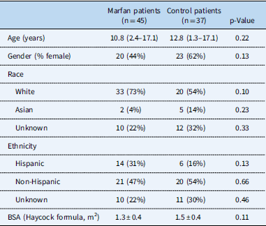
In total, 45 patients with a diagnosis of Marfan syndrome met the inclusion criteria (10.8 [2.4–17.1] years). There were no significant differences in gender, race, ethnicity, or body surface area between Marfan patients and age-matched controls (Table 1).
Patient demographics.

BSA=body surface area
Data are presented as mean±standard deviation, median (range), and number (%)
In total, 20 (44%) patients had a family history of Marfan syndrome, 12 (27%) patients had ectopia lentis, 13 (29%) patients had positive genetic testing for Marfan syndrome (FBN1 mutation), and 21 (47%) patients had a systemic score ⩾7 according to revised Ghent criteria.Reference Loeys, Dietz and Braverman 2 In the absence of family history of Marfan syndrome, nine (20%) patients had an aortic root z-score ⩾2 and ectopia lentis, nine (20%) patients had an aortic root z-score ⩾2 and FBN1 mutation, 14 (31%) patients had an aortic root z-score ⩾2 and systemic score ⩾7, and two (4%) patients had ectopia lentis and FBN1 mutation with known aortic root dilation, with patients meeting diagnostic criteria by more than one possible combination. With a family history of Marfan syndrome, three (7%) patients had ectopia lentis, seven (16%) patients had a systemic score of ⩾7, and 16 (36%) patients met criteria for aortic root dilation, again with patients meeting diagnostic criteria by more than one possible combination.
In all, 26 (58%) patients had a pectus abnormality, including pectus excavatum or carinatum. The majority of Marfan patients (34, 76%) were on medication therapy – beta blocker, angiotensin receptor blocker, or angiotensin-converting enzyme inhibitor. In total, 37 control patients were predominantly referred for a history of dizziness or vasovagal syncope (24, 65%). All control patients had a 12-lead electrocardiogram at the time of the clinic visit, and 31 (84%) patients received an echocardiogram.
Electrocardiographic findings
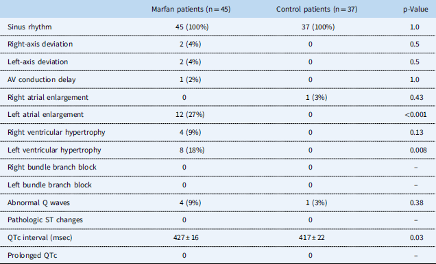
The electrocardiographic findings and their respective frequencies among Marfan and control patients are summarised in Table 2. A total of 33 electrocardiographic abnormalities were identified in 17 Marfan patients. Sinus rhythm was noted in all Marfan patients. One Marfan patient demonstrated first-degree atrioventricular block with a PR interval of 200 ms. No Marfan patient demonstrated bundle branch block or pathologic ST segment changes. No ventricular (or atrial) ectopy on resting 12-lead electrocardiogram was noted in either the Marfan group or the control group.
Electrocardiographic findings in Marfan and control patients

AV=atrioventricular
Data are presented as mean±standard deviation and number (%)
Two observers – C.T.N. and S.R.C. – had 100% agreement on identifying abnormal Q waves, pathologic ST segment changes, left and right atrial and ventricular enlargement, and ventricular hypertrophy. The inter-observer agreement was excellent for QTc interval measurements with an intra-class correlation coefficient of 0.86 (95% CI: 0.51–0.97).
Overall, electrocardiographic abnormalities were detected in 38% of Marfan patients compared with 5% in the control group (p<0.001) (Table 2). In total, 12 (27%) Marfan patients demonstrated electrocardiographic evidence of left atrial enlargement compared with none in the control group (p<0.001). Eight (18%) Marfan patients met electrocardiographic criteria for left ventricular hypertrophy compared with none in the control group (p=0.008). Four (9%) Marfan patients demonstrated abnormal Q waves compared with 1 (3%) patient in the control group (p=0.38). Left or right-axis deviation was seen in a small subset of Marfan patients, both at a rate of 4%, whereas axis deviation was not demonstrated in any control patient (p=0.50). QTc interval was noted to be longer in Marfan patients compared with controls (427±16 versus 417±22 ms, p=0.03), with four (9%) Marfan patients demonstrating borderline prolonged QTc intervals – three males with a QTc of 450 ms; one female with a QTc of 459 ms. Of these four patients with borderline prolonged QTc interval, none complained of palpitations or syncope during clinic visits.
When Marfan patients with and without pectus deformity were compared, there was no significant difference in the frequency of abnormal electrocardiographic findings including left atrial enlargement, left ventricular hypertrophy, or abnormal Q waves (12/26 (46%) versus 5/19 (26%), p=0.22). When Marfan patients with and without severe aortic root dilation (z-score ⩾4.5) were compared, there was no significant difference in the frequency of left atrial enlargement, left ventricular hypertrophy, or abnormal Q waves on 12-lead electrocardiogram (4/10 (40%) versus 11/35 (31%), p=0.71). There was also no significant difference in the frequency of left atrial enlargement, left ventricular hypertrophy, or abnormal Q waves on electrocardiograms in Marfan patients who were on medical therapy compared with Marfan patients not on medical therapy (12/34 (35%) versus 6/12 (50%), p=0.49).
Echocardiographic findings
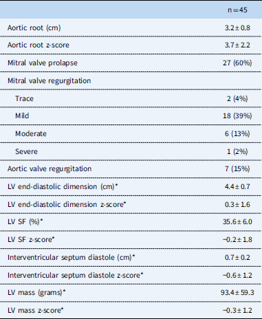
Echocardiographic findings in Marfan patients are summarised in Table 3. In total, 38 (84%) Marfan patients had aortic root dilation (z-score ⩾2), of whom 10 (22%) patients had severe aortic root dilation (z-score ⩾4.5). In total, 27 (60%) Marfan patients demonstrated mitral valve prolapse, with seven (16%) patients with more than mild mitral regurgitation. Left ventricular dilation, with a left ventricular end-diastolic dimension z-score ⩾2, was seen in eight (18%) Marfan patients, and left ventricular systolic dysfunction, with a shortening fraction z-score ⩽−2, was seen in five (11%) Marfan patients. Inter-observer variability of echocardiographic measurements of left atrial and left ventricular size and function were good with intra-class correlation coefficients ranging from 0.7 to 0.9.
Echocardiographic findings in Marfan patients

LV=left ventricle; SF=shortening fraction
Data are presented as mean±standard deviation and number (%)
* Represents M-mode measurements
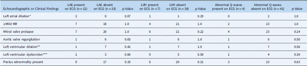
Two of 12 patients with left atrial enlargement by electrocardiogram were confirmed to have left atrial enlargement by echocardiogram, one of whom had moderate mitral regurgitation related to mitral valve prolapse, and the other had moderate left ventricular systolic dysfunction (Table 4). The patient with moderate mitral regurgitation also met criteria for left ventricular hypertrophy by electrocardiogram, not confirmed on echocardiogram.
Abnormalities on electrocardiogram and echocardiographic and clinical findings in Marfan patients

ECG=electrocardiogram; LAE=left atrial enlargement; LVH=left ventricular hypertrophy; MR=mitral regurgitation
* Defined by diastolic volume >28 ml/m2 or z-score ⩾2
** Defined by end-diastolic dimension z-score ⩾2
*** Defined by shortening fraction z-score ⩽−2
Of the seven patients meeting criteria for left ventricular hypertrophy by electrocardiogram, none demonstrated left ventricular hypertrophy on echocardiogram; however, one patient with mild concentric left ventricular hypertrophy by echocardiogram was not captured on electrocardiogram. Of the four patients with abnormal Q waves on electrocardiogram, one patient demonstrated mild left ventricular systolic dysfunction.
In subgroup analyses, comparisons were made between Marfan patients with and without electrocardiographic evidence of left atrial enlargement, left ventricular hypertrophy, or abnormal Q waves (Table 4). There were no significant differences in the frequency of any echocardiographic findings including left atrial enlargement by echocardiography, mild or greater mitral regurgitation, mitral valve prolapse, aortic valve regurgitation of any degree, left ventricular dilation, and left ventricular dysfunction.
Discussion
In this retrospective review of the utility of serial outpatient electrocardiograms in children with Marfan syndrome, we demonstrated the following: there was a higher frequency of left atrial enlargement and left ventricular hypertrophy by electrocardiogram in Marfan patients compared with controls, not confirmed by echocardiogram; and QTc interval was longer in Marfan patients compared with controls (p=0.03), with four Marfan patients demonstrating borderline prolonged QTc intervals for gender.
Our findings are consistent with the limited data published on electrocardiographic abnormalities in paediatric Marfan patients. These prior studies reported conduction abnormalities, ventricular arrhythmias, and arrhythmia-induced sudden cardiac death – in up to 4% of patients – associated with prolonged QTc and mitral valve prolapse in Marfan patients.Reference Yetman, Bornemeier and McCrindle 3 – Reference Chen, Fagan, Nouri and Donahoe 5 In a study of 36 children with Marfan syndrome, six patients had first-degree atrioventricular block and three patients had prolonged QTc (>450 ms), whereas a study of 24 paediatric Marfan patients showed ventricular ectopy on resting electrocardiograms in eight patients (33%), with five of eight demonstrating a baseline prolonged QTc interval.Reference Chen, Fagan, Nouri and Donahoe 5 , Reference Phornphutkul, Rosenthal and Nadas 6 The investigators reported that delayed repolarisation, when combined with mitral valve prolapse, mitral valve regurgitation, and left ventricular dilation, was associated with ventricular ectopy.
Pectus deformity is a common finding in Marfan syndrome observed in approximately two-thirds of patients and, as expected, was present in 58% of our Marfan syndrome population.Reference Pyeritz and McKusick 19 Pectus abnormalities have been reported to be associated with electrocardiographic abnormalities in Marfan patients, specifically with complete P wave inversion in lead V1;Reference Martins De Oliveira, Sambhi and Zimmerman 20 however, in our study there was no significant difference in the frequency of abnormal electrocardiographic findings in Marfan patients with and without pectus deformity.
There are limitations in this study inherent to its retrospective nature and small sample size. Marfan patients have poor acoustic windows often prohibiting accurate assessment of cardiac structures by echocardiography. Although inter-observer reproducibility of echocardiographic measurements was good, quantitative echocardiographic assessment may be less accurate in Marfan patients compared with other children populations. In addition, in our clinical centre, we do not routinely obtain 24-hour Holter recordings on Marfan patients, which may have enhanced this data set.
In conclusion, although Marfan patients exhibited a higher frequency of left atrial enlargement and left ventricular hypertrophy on 12-lead electrocardiograms compared with controls, these findings were not supported by echocardiography. Thus, serial 12-lead electrocardiograms in routine follow-up of asymptomatic paediatric Marfan patients may be more appropriate for a subgroup of Marfan patients only, specifically those with prolonged QTc at their baseline visit. Further investigation is needed to determine best practice in routine electrocardiographic screening in the paediatric Marfan population, as this is an important consideration in the era of appropriate resource utilisation.
Acknowledgements
None.
Financial Support
This research received no specific grant from any funding agency or from commercial or not-for-profit sectors.
Conflicts of Interest
None.
Ethical Standards
The authors assert that all procedures contributing to this work comply with the Helsinki Declaration of 1975, as revised in 2008, and has been approved by the institutional committee (Stanford University Institutional Review Board).