INTRODUCTION
The Mediterranean bumblebee shrimp Gnathophyllum elegans (Risso, 1816) is widely distributed throughout the Mediterranean Sea (Adriatic Sea, Aegean Sea, Alboran Sea, Ionian Sea and Israelean coast) and adjacent Atlantic regions (Azores, Canary Islands, Madeira and Moroccan Atlantic) (d'Udekem d'Acoz, Reference d'Udekem d'Acoz1999; Türkay, Reference Türkay, Costello, Emblow and White2001). As a nocturnal species it can be found in shallow coastal waters (0–35 m) (Denitto et al., Reference Denitto, Moscatello and Belmonte2009; Pipitone & Vaccaro, Reference Pipitone, Vaccaro, Pessani, Tirelli and Froglia2011), hidden under stones and pebbles at daytime. Zariquiey Álvarez (Reference Zariquiey Álvarez1955) mentioned that at night the species can be dredged in Posidonia oceanica Delile, 1813 meadows. Occasionally it is associated with the sea anemone Telmatactis cricoides (Duchassaing, 1850) (Wirtz, Reference Wirtz1997). In our sampling area we did not observe any kind of association and found specimens obviously free living. However, when transferred in a community aquarium we monitored the bumblebee shrimp associated with different echinoderms, e.g. Echinaster (Echinaster) sepositus (Retzius, 1783) and Arbacia lixula (Linnaeus, 1758).
The family Gnathophyllidae includes five genera with a total of 14 species (De Grave & Fransen, Reference De Grave and Fransen2011). The larval development of this family is poorly documented; only the first stage zoea in Gnathophyllum americanum Guérin-Méneville, 1855 (in Guérin-Méneville, 1855–1856) is described so far (Bruce, Reference Bruce1986). Also the systematic classification of the genera is under discussion. There are notions of a close relationship between the Gnathophyllidae Dana, 1852, Hymenoceridae Ortmann, 1890 and members of the palaemonid subfamily Pontoniinae Kingsley, 1879. To put further arguments into this question, in the present study we describe and illustrate the first zoea in G. elegans and compare the first zoeal characteristics of G. elegans with those of G. americanum, Hymenocera picta Dana, 1852 and Periclimenes amethysteus (Risso, 1827).
MATERIALS AND METHODS
On May 2012, one ovigerous female of Gnathophyllum elegans was collected in a depth of 2 m while night-snorkelling west of the isle of Šolta (43°23′00′′N16°13′47′′E), Croatia. The specimen was identified according to Zariquiey Álvarez (Reference Zariquiey Álvarez1968) and kept in an aquarium with a temperature of 20–23°C and salinity of 35 psu at the Zoologische Staatssammlung München (Germany) till larvae hatched. About 200 first zoeas were released on 20 June 2012. Few larvae were observed and photographed in the egg integument and instantly after hatching in order to document natural pigmentation. Larvae were fixed immediately in a graded ethanol series (30%, 50%, 70% for 10 min each, see Meyer & Melzer (Reference Meyer and Melzer2004)) and then dissected using a stereomicroscope and thin tungsten wires. Of each type of appendage about 20 pieces were dissected and kept separately in small glass vials containing 70% ethanol. For scanning electron microscopy (SEM) dissected appendages and entire larvae were dehydrated in a graded acetone series (70%, 80%, 90%, 2 × 100%, 10 min each) and then critical-point-dried in a Baltec CPD 030. After mounting on SEM stubs with self-adhesive carbon stickers, specimens were coated with gold on a Polaron ‘Sputter Coater’ (Quorum Technologies, UK) and studied with a LEO 1430VP (Zeiss, Germany) SEM at 10–15 kV. Several appendages were scanned and compared to identify the exact number of setae. For classification of the different types of setae the terminology of Ingle (Reference Ingle1992) was followed.
Measurements of the first zoeas were made using LEO's SEM-User-Interface-Software (N = 4). Total length (TL) was measured from the base of the rostrum to the posterior end of the telson, carapace length (CL) from the base of the rostrum to the posterior margin of the carapace and carapace width (CW) between the lateral margins of carapace directly behind the eyes. Furthermore the rostrum length (RL) measured from the tip of the rostrum to the antennule peduncle and the antenna endopod length (AEL) measured to the scale of the antenna are given in this description.
Complete larvae and dissected maxillipeds were also studied with light microscopy (LM) using the Leica DM 5000 B microscope (Leica, Germany) equipped with the CCD Camera ProgRes SpeedXTcore 5 (Jenaoptik, Germany). Drawings of the mandibles and the telson were prepared by interpretation of several SEM and LM images.
The female and the zoea larvae are deposited at the Zoologische Staatssammlung München under the registration numbers ZSMA 20120316 and ZSMA 20120317. Additionally the DNA barcode (cytochrome oxidase I sequence) of a different specimen of Gnathophyllum elegans sampled in the same region (ZSMA 20111534) is available on the Boldsystems website (www.boldsystems.org) under the number CFAD190-11.
RESULTS
First zoea of Gnathophyllum elegans
Dimension (mm): TL = 1.860 + 0.044; CL = 0.446 + 0.020; CW = 0.459 + 0.038; RL = 0.185 + 0.016; AEL = 0.173 + 0.023.
Carapace (Figure 1A, B): with short, slender and unarmed rostral process; epigastric tubercle with dorsal organ present; carapace armed with pterygostomial spine, otherwise absence of spines. Compound eyes sessile.
Gnathophyllum elegans, first zoea: (A) LM image, lateral view, natural pigmentation visible; insert showing egg just before hatching; (B) SEM image, lateral view, arrowhead points to dorsal organ; insert showing detail of dorsal organ. Abbreviations: I–VI, abdominal segments; AB, abdomen; AN, antenna; AU, antennule; C, carapace; E, eye; LAB, labrum; MXP 1–3, first to third maxillipeds; PER1&2, pereiopods 1 and 2; PT, pterygostomial spine; R, rostrum; T, telson. Scale bars: A, 200 µm; B, 200 µm, insert 20 µm.

Anterior sensory dorsal organ (SDO) (Figure 1B): small protrusion composed of four small cuticular depressions disposed as the corners of a square with the centre occupied by a pore.
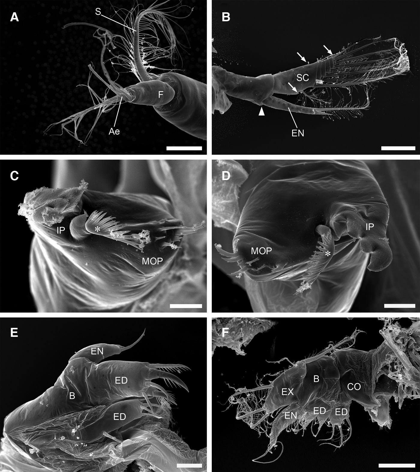
Antennule (Figures 1A, B & 2A): subcylindrical, unsegmented peduncle with distal plumose seta; flagellum with four aesthetascs and plumose seta.
Gnathophyllum elegans, Zoea I, SEM images showing appendages of cephalothorax and mouthparts: (A) antennule; (B) antenna, arrowhead = small spine; arrows = small setae and tubercle; (C) right mandible, asterisk = ‘lacinia mobilis’; (D) left mandible, asterisk = ‘lacinia mobilis’; (E) maxillule; (F) maxilla. Abbreviations: AE, aesthetasc; B, basis; CO, coxa; ED, endite; EN, endopod; EX, exopod; F, flagellum; IP, incisor process; MOP, molar process; S, seta; SC, scaphocerite. Scale bars: A, 40 µm; B, 100 µm; C, 10 µm; D, 10 µm; E, 20 µm; F, 40 µm.
Antenna (Figures 1A, B & 2B): biramous; protopod unsegmented, with medio-terminal small spine; endopod apically with plumose seta and short spine; scaphocerite with four segmentations distally and ten plumose setae plus simple seta distomedially, first and last reduced, with small tubercle proximally on medial border; small seta on the outer side of the proximal article and small distal plumose seta, small simple seta on the apex.
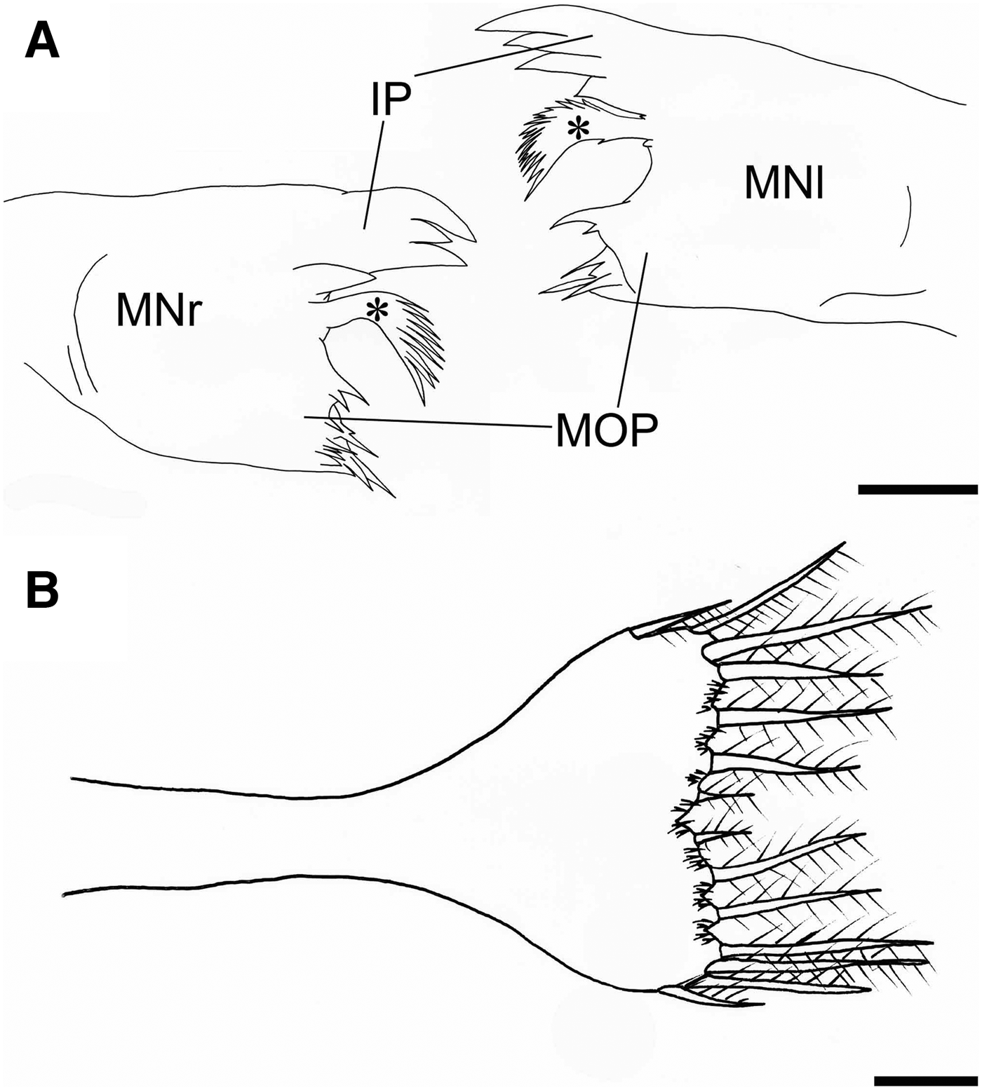
Mandibles (Figures 2C, D & 5A): right and left mandibles almost identical; mandibular palps absent. Incisor process with marginal protrusion bearing row of three acute spines ventrally and serrated ‘lacinia mobilis’, molar process slender, bearing group of small spines. Right mandible with clearly separate submarginal spine between incisor process and ‘lacinia mobili’. Left mandible with submarginal spine grown together with ventral row of spines.
Maxillule (Figure 2E): coxal endite with four simple distal setae and slightly serrated proximal located seta; basal endite with two stout and slender plumose setae and two simple setae distally; endopod compressed, stout, distally acute with preterminal simple seta; exopod absent.
Maxilla (Figure 2F): coxal endite with three simple setae, basal endite bilobed, proximal lobe with simple seta, and distal lobe with two simple setae, endopod unsegmented with long simple seta and microtrichia on inner margin. Exopod with five marginal plumose setae.
First maxilliped (Figure 3A, B): basis with three simple setae. Endopod 3-segmented with 0, 1, 1 + 3 setae; exopod partially crossed by six incisions, the last one armed laterally with simple seta, distally four long plumose setae arranged in a row.
Gnathophyllum elegans, first zoea, SEM (A, C, D, G) and LM (B, E, F) images showing maxillipeds: (A) endopod and exopod of first maxilliped, arrow = seta; (B) first maxilliped; (C) endopod of second maxilliped; (D) endopod of third maxilliped; (E) second maxilliped; (F) third maxilliped; (G) exopod of third maxilliped, arrow = seta. Abbreviations: EN, endopod; EX, exopod. Scale bars: A, 20 µm; B, 150 µm; C, 20 µm; D, 40 µm; E, 200 µm; F, 200 µm; G, 6 µm.
Second maxilliped (Figure 3C, E): basis with two simple setae. Endopod 3-segmented, proximal segment largest and unarmed, intermediate segment with distomedial serrated and distolateral simple seta distally, distal segment with small simple seta located proximally and three simple setae plus strong serrate seta distally. Exopod partially crossed by nine incisions, setae arranged as in first maxilliped.
Third maxilliped (Figure 3D, F, G): basis with two simple setae and small proximomedial tubercle. Endopod 3-segmented; proximal segment with 1 + 1 simple setae; intermediate segment with two strong serrate distal setae, distal segment armed with proximolateral simple and three simple and strong serrate distally located setae. Exopod partially crossed by eight incisions, setae arranged as in first maxilliped.
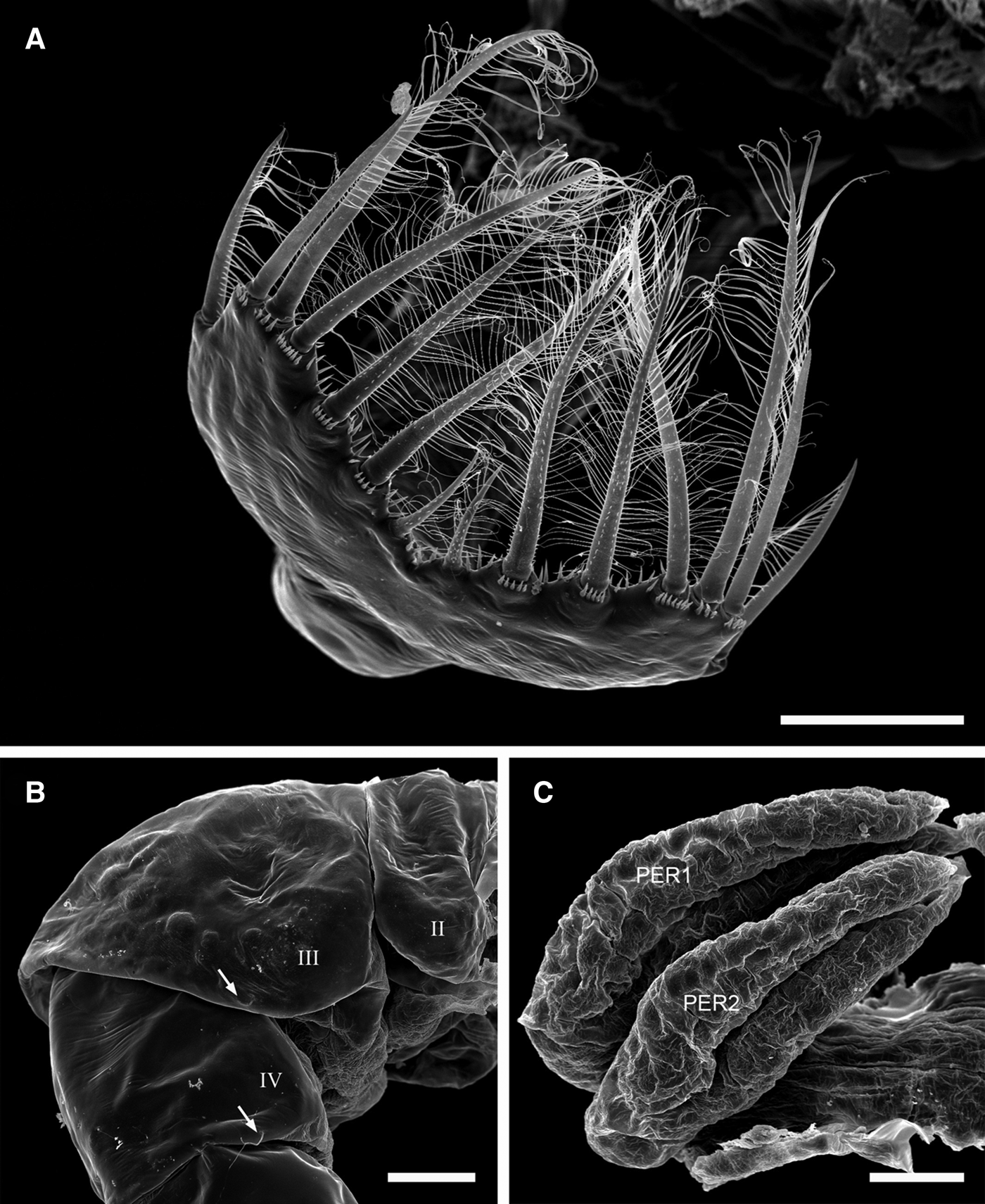
First and second pereiopods (Figure 4C): present as biramous rudiments.
Gnathophyllum elegans, first zoea, SEM images: (A) telson; (B) detail of abdomen segments II–IV, arrows = setae; (C) pereiopods 1 and 2. Abbreviations: II–IV, abdominal segments; PER1&2, pereiopods 1 and 2. Scale bars: A, 100 µm; B, 60 µm; C, 30 µm.
Third to fifth pereiopods: not differented.
Abdomen (Figures 1A, B & 4B): 6-segmented; strongly flexed between third and fourth segments; small simple seta on the posterior-lateral margin of the third and fourth somite present.
Pleopods and uropods: absent.
Telson (Figures 4A & 5B): fused with sixth abdominal segment; broadly triangular; 7 + 7 plumodenticulate setae on the posterior margin; the inner- and outermost smaller, the two outermost on each side with setules medially; minute spines only between four inner setae; setal bases armed with minute spines except inner and outermost.
Gnathophyllum elegans, first zoea, drawing: (A) posterio-ventral view of mandibles; (B) dorsal view of telson. Abbreviations: asterisk, ‘lacinia mobilis’; IP, incisor process; MNl, left mandible; MNr, right mandible; MOP, molar process; Scale bars: A, 20 µm; B, 100 µm.
DISCUSSION
The present description of the first zoea in Gnathophyllum elegans is based on a combination of SEM and LM techniques applied to fixed larvae and dissected appendages. Using the high resolution power of the SEM even minute structures such as setules, spines or the anterior sensory dorsal organ can be located and described. We also could analyse the gnathal edge of the mandibles in great detail, a feature often omitted in larval descriptions. Ingle (Reference Ingle1992) mentioned that left and right mandibles in zoea-larvae are usually slightly dissimilar and that details are not easy to resolve by LM due to their gross three-dimensional structure. In the case of G. elegans this difference is marginal and can only be observed in the position of the submarginal spine on the incisor process.
The family Gnathophyllidae (superfamily Palaemonoidea Rafinesque, 1815) consists of five genera: Gnathophylleptum d'Udekem d'Acoz, 2001, Gnathophylloides Schmitt, 1933, Gnathophyllum Latreille, 1819, Levicaris Bruce, 1973, and Pycnocaris Bruce, 1972 (Martin & Davis, Reference Martin and Davis2001; De Grave & Fransen, Reference De Grave and Fransen2011). Until now the description of the first zoeal stage in Gnathophyllum americanum was the only one present for a representative of this family (Bruce, Reference Bruce1986).
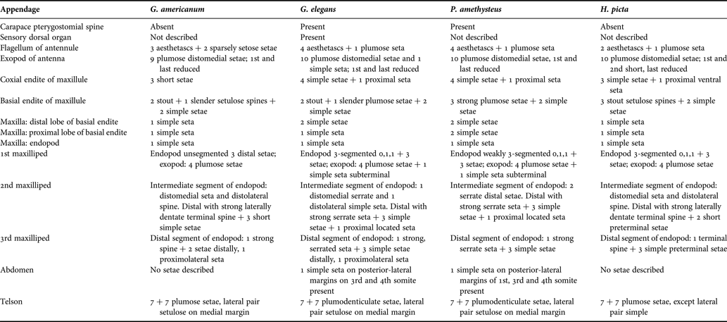
Differential diagnosis between the larval characters of the first zoea in G. elegans and G. americanum shows morphological differences in the carapace shape and the arrangement and number of setae on different appendages, e.g. the antennule, antenna, maxillule, maxilla, first to third maxillipeds and the telson (Table 1). The two Gnathophyllum-species can thus be readily distinguished from each other. However, some of the differences like the presence or absence of the pterygostomial spine and the segmentation of the endopod of the first maxilliped may also be a result from the different analysing techniques used in both examinations, since the SEM's resolution power is much higher than that of the LM.
Comparison of morphological features of the first zoeas Zoea-I larvae of Gnathophyllum americanum, G. elegans, Periclimenes amethysteus and Hymenocera picta. Data on P. amethysteus are taken from Geiselbrecht & Melzer (2010), Gnathophyllum americanum and Hymenocera picta from Bruce Reference Bruce1986, Reference Bruce1988. Bold marked features show similarity between G. elegans and P. amethysteus.
The absence of the distinct basal lobe on the endopod of the maxilla and the branchiostegal spines as observed in the G. elegans-zoea are morphological characteristics thought to be specific for larvae of the palaemonid subfamily Pontoniinae as well as the Gnathophyllidae and the Hymenoceridae (Mitsuhashi et al., Reference Mitsuhashi, Sin, Lei, Chan and Chu2007).
Thus, with respect to species specific features and above-family systematics our results fit well with what could be expected. However, the search for features that allow us to distinguish the studied gnathophyllid zoeae from pontoniids and hymenocerids proved to be more difficult. In Table 1 we summarize characters of selected representatives of these taxa for detailed comparison. Periclimenes amethysteus was selected as a Pontoniine ‘model’ because the larval description is also based on a combined technique using LM and SEM and thus is most suitable for comparison with our results (Geiselbrecht & Melzer, Reference Geiselbrecht and Melzer2009). A comparison of 14 zoeal characteristics in G. elegans and P. amethysteus shows a high degree of correspondence, though they are placed in different families. Seven of these features are identical, and only six differences are shown, i.e. setation of exopod of antenna, basal endite of maxillule, proximal lobe of basal endite of maxilla, second maxilliped, third maxilliped and the abdomen. It was not possible to compare the structure of the sensory dorsal organ, because an adequate description of this feature was lacking in P. amethysteus. This is surprising, since the two studied gnathophyllids, G. elegans and G. americanum, belonging to the same genus, show differences in eight conspicuous features. Moreover, G. elegans and Hymenocera picta, a representative of Hymenoceridae, the putative sister taxon of the Gnathophyllidae, show 11 differences (Table 1).
What could be explanations for these partly contradictory results? As mentioned above, our SEM technique used for the analysis of G. elegans and P. amethysteus shows minute details that are probably not detected with LM alone, viz., the technique used for description of G. americanum and H. picta. Differences in meticulousness could thus hinder comparison of zoeae and suggest false similarities. For example the structure of the sensory dorsal organ or the mandibles can only be described by use of the SEM. Nonetheless our study clearly supports a notion already put forward in several other differential diagnoses of pontoniine and gnathophyllid zoeas, i.e. their high degree of correspondence (Bruce, Reference Bruce1986, Bruce, Reference Bruce1988; Yang & Ko, Reference Yang and Ko2002; Yang & Ko, Reference Yang and Ko2004; Mitsuhashi et al., Reference Mitsuhashi, Sin, Lei, Chan and Chu2007).
Another explanation for the character distribution shown here might lie in the not-well-understood relationships between the Gnathophyllidae and the Pontoniinae. Holthuis (Reference Holthuis1955) erected the family Gnathophyllidae based on adult mouthpart morphology (shape of the third maxilliped) by excision of genera Gnathophylloides, Gnathophyllum, Levicaris and Pycnocaris from the Pontoniinae, and thereby producing probably paraphyletic Pontoniinae as a ‘leftover’ group. With the first description of the larval features of G. americanum and their high degree of similarity to Pontoniinae zoeae, Bruce (Reference Bruce1986) synonymized the Gnathophyllidae with the former, but Chace & Bruce (Reference Chace and Bruce1993) concluded tentatively that the unique mouthparts of the Gnathophyllidae discard the possibility of synonymy. Moreover, Williamson & Rice (Reference Williamson and Rice1996) and Williamson (Reference Williamson2001) proposed that highly similar larvae do not necessarily signify related adults and, therefore, the Pontoniinae and the Gnathophyllidae may be distinct. This would suggest that some of the zoeal features presented in the present study might be plesiomorphies and thus not useful for phylogenetic considerations.
Recent molecular systematic analyses based on nuclear rDNA and mitochondrial sequences (Mitsuhashi et al., Reference Mitsuhashi, Sin, Lei, Chan and Chu2007; Bracken et al., Reference Bracken, De Grave, Felder, Martin, Crandall and Felder2009; Li et al., Reference Li, De Grave, Chan, Lei and Chu2011) show that the Gnathophyllidae are placed in a cluster of lineages including the Hymenoceridae as the sister group of Gnathophyllidae, plus several lineages belonging to Palaemoninae Rafinesque, 1815, Pontoniinae and Anchistioididae Borradaile, 1915 supporting the idea of pontoniine paraphyly.
It seems that the above described unclear character distribution between gnathophyllid and related zoeae is caused by a combination of still valid traditional taxonomic acts, lack of consequent cladistic analyses including molecular trees and insufficient knowledge of larval morphology. Further investigations combining molecular methods and classical morphological analyses (larval and adult) involving more gnathophyllid genera are therefore required.
ACKNOWLEDGEMENTS
We thank Stefan Friedrich (Zoologische Staatssammlung München, Germany) for expert technical assistance and collection management. Furthermore we would like to thank the captain Bernd Meyer and the crew of the SY ‘Janu’ for their support.
FINANCIAL SUPPORT
This study was partly supported by the Sea Life Center München, Germany.