INTRODUCTION
Ostertagia ostertagi, often called the brown stomach worm of cattle, damages the abomasum during larval development and during the tissue-feeding adult stage. Ostertagia is among the most pathogenic of this parasite group. Ingested larvae (L3) enter the gastric glands, moult (L4) and remain there during the pre-patent period. After a final moult, they emerge to the lumen of the abomasum as young adults. Infections cause depression in appetite and impair digestion resulting in severe morbidity and mortality. Understanding the host-parasite relationship and in particular the effects of L4 on the host is important for developing strategies to attenuate the infection and its consequence on animal health.
Nucleotide-metabolizing enzymes, and in particular those capable of hydrolyzing nucleoside di- and triphosphates into mononucleotides and organophosphate i.e. apyrases (EC 3.6.1.5), have been described in arthropods (e.g. Hamasaki et al. Reference Hamasaki, Kato, Terayama, Iwata and Valenzuela2009), plants (Handa and Guidotti, Reference Handa and Guidotti1996), and vertebrates (e.g. Leal et al. Reference Leal, Streher, Neu, Bittencourt, Leal, Da Silva, Morsch and Schetinger2005). Inasmuch as similar activities are encoded by a number of seemingly unrelated proteins such as heat shock proteins, actin and sugar kinases, it has been proposed that these proteins are the product of convergent evolutionary events (Valenzuela et al. Reference Valenzuela, Belkaid, Rowton and Ribeiro2001). In general, nucleotide-metabolizing enzymes can be categorized into 3 broad groups predicated upon their amino acid sequences and substrate specificities; the ectonucleoside triphosphate diphosphohydrolyases (E-type NTPDases), the 5′ nucleotidases belonging to the CD73 lymphocyte protein family, and those predominantly associated with haematophagous arthropods (Valenzuela et al. Reference Valenzuela, Charlab, Galperin and Ribeiro1998, Reference Valenzuela, Belkaid, Rowton and Ribeiro2001). In general, the E-NTPDases are specific for di- and triphosphates, function well in the presence of Mg2+ or Ca2+ and are characterized by up to 5 (Handa and Guidotti, Reference Handa and Guidotti1996; Vasconcelos et al. Reference Vasconcelos, Ferreira, Carvalho, Souza, Kettlun, Mancilla, Valenzuela and Verjovski-Almeida1996) distinct and highly conserved regions where regions 1, 4 and 5 have been specifically linked to biochemical activity (Schulte am Esch et al. Reference Schulte am Esch, Sévigny, Kaczmarek, Siegel, Imai, Koziak, Beaudoin and Robson1999). The 5′-nucleotidases tend to be well conserved across species and occur as glycosyl phophatidylinositol (GPI) – anchored membrane proteins that can circulate as soluble proteins once released from the membrane. Their primary activity is on nucleoside 5′monophosphates. Those belonging to the ‘haematophagous arthropod’ group are Ca2+-dependent and also have conserved motifs but these are not shared with the E-NTPDases. They rapidly degrade di- and triphosphates to monophosphates and their relation to functionality has been investigated in a human-derived homologue (Dai et al. Reference Dai, Liu, Deng, Smith and Lu2004). Beyond substrate activities, the function of nucleotide-metabolizing enzymes as it relates to parasitism is unresolved; however, blood-feeding insects are known to secrete apyrases to facilitate feeding and prevent platelet activation through the depletion of adenosine diphosphates (ADP) (Valenzuela et al. Reference Valenzuela, Charlab, Galperin and Ribeiro1998, Reference Valenzuela, Belkaid, Rowton and Ribeiro2001). Furthermore, regulation of extracellular ATP levels has a direct impact on purinergic signalling and regulating the host immune responses, and may be influenced directly by nucleotide-metabolizing enzymes secreted by the parasite (Bhardwaj and Skelly, Reference Bhardwaj and Skelly2009). This suggests that some parasites have evolved methods to influence this signalling event by controlling the concentration of interactive nucleotides within their local environment.
Apyrases have been identified in helminths. The gene encoding ATPDase 1 of Schistosoma mansoni, which belongs to the human CD39 family, is present in 5 developmental stages but predominantly in the adults where the protein is localized to the parasite surface (DeMarco et al. Reference DeMarco, Kowaltowski, Mortara and Verjovski-Almeida2003). A unique 5′ nucleotidase was identified in Trichinella spiralis which hydrolyses 5′ diphosphates and 5′ monophosphates but not 5′ triphosphates (Gounaris et al. Reference Gounaris, Selkirk and Sadeghi2004). An ATP-diphosphohydrolase was discovered in the tegument surface of Taenia crassiceps cysticerci which is also a member of the human CD39 family (Guevara-Flores et al. Reference Guevara-Flores, Olvera-Sánchez, Gómez-Concha, Juárez, Esparza-Perusquía, Pardo, Mendoza-Hernández, Martínez and Flores-Herrera2008). Common to these classes of apyrases is a requirement for Mg2+ or Ca2+; however, the newest class of soluble apyrases found predominantly among the haematophagous arthropods has an absolute requirement for Ca2+ over Mg2+ and has not yet been identified in parasitic nematodes. One such protein has been found in Caenorhabditis elegans (Uccelletti et al. Reference Uccelletti, Pascoli, Farina, Alberti, Mancini, Hirschberg and Palleschi2008) but with notable differences to the Ostertagia-derived protein in substrate specificity, ubiquitous stage expression and its localization to membrane tegument. Herein, we identified a gene from an immunologically-screened cDNA library that encodes a functional apyrase present exclusively in Ostertagia ostertagi L4 that conforms in sequence and function to the apyrases identified in haematophagous arthropods and, in like manner, is Ca2+ dependent.
MATERIALS AND METHODS
Animals and parasite strain
Holstein-Friesen calves acquired within 48 h of birth were maintained on concrete to preclude unwanted infections. Calves were weaned at 60 days of age and maintained on a 16% protein diet supplemented with alfalfa and orchard grass hay; water was provided ad libitum. The strain of O. ostertagi was originally isolated at the USDA Regional Parasite Laboratory in Auburn, AL and has been maintained by serial passage through helminth-free calves for over 40 years. Parasites were obtained by infecting donor calves with 2×10⁵ O. ostertagi L3. Animals were killed 7–10 days p.i. to obtain L4 from the abomasum, or at 21 days p.i. to recover adult worms also from the abomasum which were picked microscopically. Infective L3 were acquired from 14-day-old sphagnum moss cultures of fertilized eggs, enriched using the Baermann funnel technique, then stored at 4°C in tap water until use. For kinetic studies, 2 animals were infected as described above and 2 were left as uninfected controls. Animals were serially bled prior to infection then at 1-week intervals for 4 weeks and the sera (4-week) used for immunologically screening a cDNA library and Western blots. All animal protocols and treatments were approved through the ARS Animal Care and Use Committee.
Library construction and immunological screening
Total RNA was isolated from L4 by homogenizing in Trizol™ as recommended by the manufacturer (Invitrogen). Isolated RNA was treated with RNAse-free DNAse and solubilization in DEPC-treated water to a final concentration of 7·8 μg/μl determined spectrophotometrically at 257 nm. The cDNA library was constructed using 1·2 μg of total RNA and the SMART™ I PCR cDNA Library Construction Kit (Clontech). Following PCR amplification and adaptor ligation, the amplified cDNA was purified through a Sephacryl 400 spin column to remove unligated adaptors and small PCR fragments that might interfere with vector ligation. The double-stranded cDNA was ligated overnight into EcoRI-digested and dephosphorylated λgt11 arms using T4 DNA ligase. The following day the ligated sample was packaged and used to transform Y1090r-cells.
Approximately 50 000 clones were transferred to isopropyl β-D-1-thiogalactopyranoside (IPTG) impregnated Nytran membranes and immunologically screened with bovine infection serum essentially as described under the heading Western blots. Primary bovine antibodies (1:200; obtained 4 weeks p.i.) were incubated overnight at 4°C with blocked filters (PBS containing 0·05% Tween-20 and 5% nonfat dried milk). The following day, the filters were washed 5X in PBS-Tween solution, incubated for 2 h in the same solution containing a 1:1000 dilution of peroxidase-labelled rabbit anti-bovine IgG, then washed again; the final wash was in distilled water. The filters were developed in 4-chloro-1-naphthol (0·06%) and hydrogen peroxide (0·02%) (Wang and Esen, Reference Wang and Esen1985). Positive plaques (10) were selected and purified to homogeneity by re-screening according to the methods described above.
Subcloning, protein expression and purification
One positive clone identified from the immunological screening and temporarily designated Oo7-64 was subcloned into pUC19 and subjected to automated sequencing using M13 forward and reverse primers. A full-length sequence was identified by rescreening the cDNA library with an Oo7-64 DNA probe radio-isotope labelled by nick translation (Rigby et al. Reference Rigby, Dieckmann, Rhodes and Berg1977). Positive plaques were validated for size by colony PCR (Nishikawa et al. Reference Nishikawa, Fowlkes and Kay1989) using reverse Oo7-64 PCR primer #625 (5′ gcctttgatgttcatcgacgatgtc), forward or reverse λgt11 primers, and Taq polymerase (Fisher Scientific). The full-length open reading frame was validated by amplifying and sequencing the gene directly from cDNA using forward and reverse primers 1014 (5′ aggatggttctctacatcctttcactg) and 1015 (5′ tcaaatgaagtggagtccctc), respectively. Alignment with known apyrases was performed by Clustal W (Thompson et al. Reference Thompson, Higgins and Gibson1994) using default parameters. Alignments included both Ca2+-dependent and non-dependent nucleotide-metabolizing enzymes. Phylogenetic analyses were performed with MEGA 4.1 (Tamura et al. Reference Tamura, Dudley, Nei and Kumar2007) using algorithms for Neighbor-joining. Bootstrap interior branch-test (1000 replicates) was used to estimate internal branch node support.
The mature protein, with the putative secretory signal removed (determined using Signal P (Bendtsen et al. Reference Bendtsen, Nielsen, Von Heijne and Brunak2004)), was cloned into the BamHI site (underlined) of the pSUMO expression vector (Life Sensors) using forward and reverse PCR primers #1064 (5′ aaaggatccacttcctcctggatatccagacagc) and #1058 (5′ aaaggatccaatggcttttattcgtgtgaatatc), respectively. The ligated plasmid, designated rOos-apy-1, was first transformed into DH5α cells for plasmid purification and sequence validation, followed by transformation into BL21Star cells for protein expression. Protein expression was performed in 500 ml of LB broth at 37°C containing 100 μg/ml ampicillin and induced with IPTG (1 mm final concentration) for 5 h. The cells were harvested and lysed in 0·50 mg/ml of lysozyme followed by overnight freezing at −80°C. The next day, the cells were sonicated, and the mixture was treated with 50 μg/ml DNAse for 30 min on ice. The recombinant protein (rOos-APY-1) was purified by affinity column chromatography on a TALON™ resin (BD Biosciences Clontech), eluted in 300 mm imidazole, pH 7·0 and quantified spectrophotometrically (Stoschek, Reference Stoschek1990).
Antisera, Western blots and immunohistochemical staining
Multiple attempts to generate antibodies against rOos-APY-1 were unsuccessful using conventional immunization periods. Consequently, 5 mice were subjected to long-term immunization over a period of 22 weeks. A 1·1 ml solution containing 585 μg of rOos-APY-1 and 1 mg of Alhydrogel in 1X SSC (150 mm NaCl, 15 mm NaCitrate, pH 7·0) was prepared and subject to gentle rocking for 1 h. Mice were injected intraperitoneally with 200 μl of prepared antigen; a second immunization was given 2 weeks later. All subsequent injections were performed at 1-month intervals over the next 4 months. Mice immunized with adjuvant only were used as negative controls. All mice were bled 2 weeks after the final injection.
For Western blots, somatic extracts from L3 were prepared by grinding snap-frozen parasites to a powder in liquid nitrogen and re-constituting in PBS. The suspension was centrifuged (15 000 g at 4 °C for 20 min) and the supernatant stored at −80 °C. Soluble extracts (S1) from L4 (7 days p.i.) and adult (21 days p.i.) O. ostertagi were prepared as described (Smith et al. Reference Smith, Smith and Pettit2000). All protein concentrations were determined using the bicinchoninic acid (BCA) protocol (Pierce) against bovine serum albumin (BSA) standards. Ostertagia ostertagi somatic extracts from L3, L4 and adult worms (8 μg each) along with purified recombinant apyrase were subjected to electrophoresis on a NuPAGE® Bis-Tris 4–12% gels under reducing conditions employing NuPAGE® MES SDS running buffer (Invitrogen). Separated proteins were transferred to nitrocellulose membranes (Invitrogen) which were washed briefly in TNTT (10 mm Tris pH 7·4, 0·5 m NaCl, 0·05% Tween 20, 0·01% thiomersal), then blocked in TNTT overnight at 4°C. The blots were incubated with hyperimmune mouse serum (M α rOos-APY-1) or control serum diluted 1:500 in TNTT, for 1 h at RT with constant rocking then washed (3×10 min/wash) in TNTT. Membranes were then incubated for 1 h at room temperature (RT) with a 1:1000 dilution of rabbit anti-mouse Ig horseradish peroxidase (HRP) conjugate (DakoCytomation) in TNTT. The blots were subsequently washed 5 times in TNTT and antibody binding visualized by peroxidase activity using ECL™ detection (GE Healthcare). Western blots using bovine experimental infection sera obtained 4 weeks post-infection to screen rOos-APY-1 were screened as described for the cDNA library.
Immunohistochemical staining was performed on Ostertagia L4. Larvae were fixed in 4% paraformaldehyde, 0·4 m Na cacodylate, pH 7·0 for 15 min at 55°C, for 15 min with rocking at RT, then an additional 15 min at 55°C. The paraformaldehyde was removed by extensive washing in PBS containing 0·01% Tween-20 followed by 3 washes and overnight soaking in 70% ethanol prior to embedding in paraffin. Sectioned slides (5 μm) were deparaffinized in xylene followed by multiple washes in decreasing amounts of ethanol ranging from 100% to 70% and soaking in deionized water. Slides were subjected to antigen retrieval in citrate buffer (0·01 m, pH 6·0) by microwaving on high for 5 min, resting for 5 min then microwaving an additional 5 min. The slides were cooled to RT, rinsed in 1X TBS buffer (150 mm NaCl, 50 mm Tris, pH 7·4) and blocked for 30 min in 5% normal goat serum (NGS) in TBS. Mouse primary antibodies were diluted 1:1000 in 5% NGS-TBS and incubated overnight at 4°C. The next day, the slides were washed 3×5 min in TBS, then incubated for 30 min at RT in TBS containing a 1:400 dilution of Alexa Fluor® 488 goat anti-mouse secondary antibody (Molecular Probes), 1% NGS and 0·5 μg/ml DAPI as counterstain. After washing, the slides were coated with Prolong Gold anti-fade mounting medium and coverslips. Images were acquired with a Zeiss Axioskop (Carl Zeiss) fitted with a Spot digital camera (Diagnostic Instruments, Sterling Heights, Mich., USA). Fluorochromes were viewed with the following filter sets: a G 365-nm excitation filter, FT 395-nm dichroic beamsplitter and LP 420-nm emission filter were used for DAPI detection, and a BP 450-to 490-nm excitation filter, FT 510 dichroic beamsplitter and LP 515 emission filter were used for Alexa Fluor 488 detection.
Apyrase activity and inorganic phosphate determination
Apyrase activity was determined by assaying inorganic phosphate levels generated from soluble nucleotides using malachite green (Baykov et al. Reference Baykov, Evtushenko and Avaeva1988). Modifications included dissolving 0·60 g malachite green in 410 ml of 5·0 N sulfuric acid and making the colour reagent (fresh daily) consisting of 10 ml of dye solution, 0·2 ml Tween-20 and 2·5 ml of 7·5% ammonium molybdate in 5 N sulfuric acid rather than in water. Enzymatic reactions were performed in a buffer consisting of 50 mm Tris, pH 6·5, 150 mm NaCl, 5 mm MgCl2 or CaCl2, 2·5 mm DTT, 0·5–1·0 mm each nucleotide and 10 ng of rOos-APY-1. All reactions were carried out in 80 μl for 20 min at 37°C after which 20 μl of colour reagent were added and the solutions read 10 min later at OD650. For studies evaluating the pH optimum, reactions were carried out in Bis-Tris (pH 5·0–7·0) and Tris-HCl (pH 7·0–9·5) buffers in increments of 0·5 pH units where ATP was used as substrate. Results for pH determination are presented as OD650; substrate specificity is presented as % OD650 relative to ADP.
RT-PCR
Semiquantitative RT-PCR was performed on cDNA using total RNA from L3, L4 and adult parasites. For each sample, approximately 1 μg of Trizol™ isolated RNA was subjected to SuperScript™ First Strand Synthesis (Invitrogen) using an oligo dT primer according to manufacturer recommendations. For PCR amplification, forward #716 (5′ tcggaaacacagggacattggtacttcc) and reverse #1048 (5′ ctggcggccgcaaatgaagtggagtccctcgaa) primers were synthesized to amplify a 358 bp fragment of Oos-apy-1. Two Ostertagia genes were used for normalizing the PCR amplifications; forward #675 (5′ aactgcctggctcccctggccaaggtcat) and reverse #676 (5′ ggtgggaacacggaaagccat) primers were generated to a 249 bp fragment of Ostertagia glyceraldehyde 3-phosphate dehydrogenase (Oos-gapdh) and a second set of forward (#988-5′ agcgcaagcagcatcatcgtaac) and reverse (#1071-5′ aaaggatccttagcgacacccgcaagcttcgac) primers were synthesized to a 357 bp region of a Tgf-β homologue, Ostertagia Tgh-1 (Oos-tgh-1; unpublished data). Underlined segments of primers #1048 and #1071 represent inserted tags containing restriction enzyme sites. All amplifications were carried out in 25 μl using 0·25 μl cDNA and Taq polymerase as described (Zarlenga et al. Reference Zarlenga, Chute, Gasbarre and Boyd2001), and cycled at 94°C for 30 sec, 55°C for 30 sec, and 72°C for 2 min with a final extension for 7 min at 72 °C. Amplifications of Oos-apy-1, Oos-gapdh and Oos-tgh-1 were performed for 25, 28 and 29 cycles, respectively, so as not to exceed the linear range of the respective amplification curves. All products were separated on 2% NuSieve agarose gels and visualized by ethidium bromide staining.
RESULTS
The cDNA library consisted of 7×10⁵ primary clones. The efficiency of cloning was 95% based on the ratio of blue and white colonies on IPTG/X-gal agar plates. This was confirmed by colony PCR which indicated the size of the clones to range between 450 and 2000 bp. The length of the Oos-apy-1 open reading frame is 1020 bp (GenBank Accession no. GU936595) and encodes a 339 amino acid protein approximately 39 kDa in molecular mass. As predicted by SignalP (Bendtsen et al. Reference Bendtsen, Nielsen, Von Heijne and Brunak2004) via the eukaryotic hidden Markov model and the neural networks, rOos-APY-1 protein contains a putative secretory signal encompassing the first 14 amino acids with a signal peptide probability of 0·977; no signal anchor was predicted.
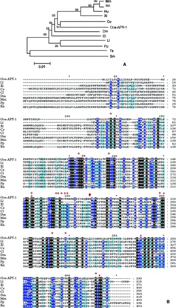
Clustal W sequence alignment with known apyrases including those of the CD39 family (Schistosoma mansoni) and other nematode derived 5′ nucleotidases (Trichinella spiralis) indicates that rOos-APY-1 belongs to a Ca2+-dependent apyrase class of proteins with strongest sequence similarity to that found in C. elegans (Fig. 1A). Bootstrap data indicate that all nodes were well supported in the NJ-generated tree. The S. mansoni and T. spiralis enzymes formed a clade basal to the Ca2+-dependent group. The sandfly apyrases from Lutzomyia longipalpis and Phlebotomus papatasi, and the collection of apyrases from mammals each formed well supported clades as well. Amino acid alignment of only the Ca2+-dependent apyrases generated noticeable regions of consensus (Fig. 1B). Among the 306 aa constituting the body of the alignment (aa115-aa421), 58% similarity and 16% identity were observed among the 8 species examined. Absolute congruence occurred between the 5 sites previously defined as essential for Ca2+ binding in the human apyrase homologue (Dai et al. Reference Dai, Liu, Deng, Smith and Lu2004) and these same loci in rOos-APY-1; however, only 62% of those amino acids defined as critical for nucleotide binding (Dai et al. Reference Dai, Liu, Deng, Smith and Lu2004) were congruent among these two apyrases (Fig. 1B). The rOos-APY-1 protein sequence exhibited 93% identity with an EST sequence (AM744030) from Teladorsagia circumcincta (Smith et al. Reference Smith, Nisbet, Meikle, Inglis, Sales, Beynon and Matthews2009) that encodes an L4 protein secreted 5 days post-infection.
(A) Phylogenetic tree of apyrases representing nematodes, arthropods and vertebrates. The tree was generated from protein sequences aligned using Clustal W followed by Neighbor-joining software using Mega 4.1. Numbers represent branch confidence levels using the interior branch test (1000 samplings) excluding gaps. GenBank™ Accession numbers are as follows: Mus musculus (Mm, AAH20003); Rattus norvegicus (Rn, NP_653355); Homo sapiens (Hu; Q8NG05); Xenopus laevis (Xl, Q8AVX8); Caenorhabditis elegans (Ce, AAA68725); Ostertagia ostertagi (rOos-APY-1,GU936595); Drosophila melanogaster (Dm, AAF54638); Cimex lectularius (Cl, AAD09177); Lutzomyia longipalpis (Ll, AAD33513); Phlebotomus papatasi (Pp, AAG17637); Trichinella spiralis (Ts, AY127571); Schistosoma mansoni (Sm, AY323529). Scale bar is equivalent to 0·01 substitutions per site. (B) Clustal W alignment of apyrases presented in Fig. 1A excluding nucleotide-metabolizing enzyme sequences from Trichinella spiralis and Schistosoma mansoni. Amino acids highlighted in black=100% identity; dark blue=minimum 80% identity; light blue=minimum 60% identity. Red stars (*) and plusses (+) define Ca2+ and nucleotide binding sites, respectively (from Dai et al. Reference Dai, Liu, Deng, Smith and Lu2004). The locus defined by a diamond (◆⧫) reflects the site important for ADP/ATP substrate identification.

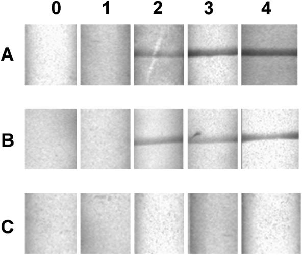
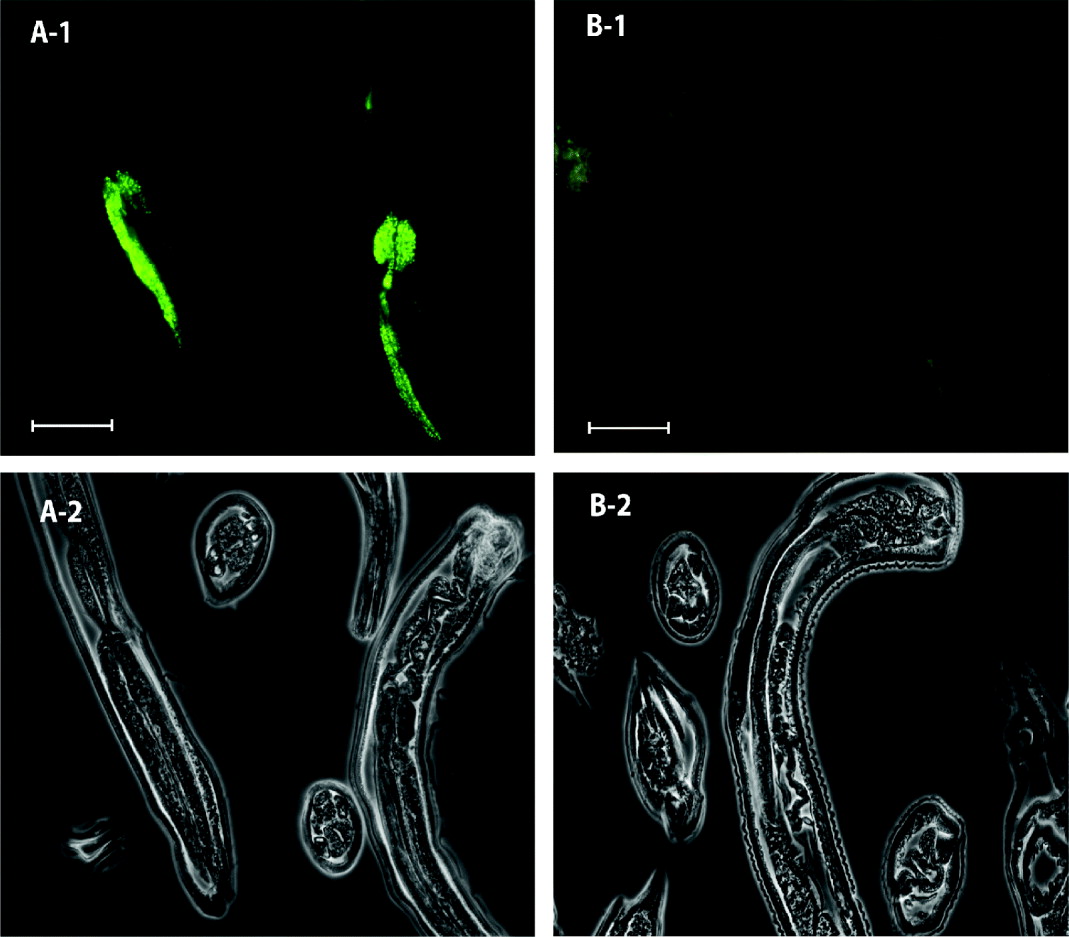
Western blots using bovine infection serum to screen rOos-APY-1 showed that host antibodies to the Ostertagia apyrase surfaced as early as 2 weeks post-infection and remained elevated throughout the course of the study (4 weeks) (Fig. 2) long after maturation of the L4 to adult parasites. Data from RT-PCR showed that the predominant stage for gene expression is the L4 with substantially less RNA transcript present in adults (Fig. 3A). These results were confirmed by Western blots using mouse anti-rOos-APY-1 to screen whole worm extracts from Ostertagia L3, L4 and adult parasites which exhibited specific and intense binding to L4 extracts (Fig. 3B); however, contrary to the RT-PCR data, no antibody binding was observed in extracts of adult parasites. Immunohistochemical staining of paraformaldehyde-fixed L4 revealed strong and specific interactions between mouse anti-rOos-APY-1 antibodies and the glandular bulb of the oesophagus at the oesophageal/intestinal junction (Fig. 4). No binding was observed using control serum.
Western Blot of rOos-APY-1 immunoscreened with serum from experimentally-infected animals. A, animal # 329; B, animal #330; C, uninfected control. Numbers above each lane represent weeks post-infection.

(A) RT-PCR analysis of RNA isolated from Ostertagia ostertagi L3, L4 and adult parasites. Oos-tgh-1 and Oos-gapdh represent constitutively produced Tgf-β and glyceraldehyde-3-phosphate dehydrogenase homologues in Ostertagia, respectively. (B) Western blot of crude worm extract from O. ostertagia L3 (lane 2), L4 (lane 3) and adult (lane 4) parasites using polyclonal mouse antibodies to the recombinant protein. Affinity-purified recombinant Ostertagia apyrase fusion protein was used as a positive control (lane 1) where the fused sumoylation cassette altered the migration of the recombinant protein.

Fluorescence immunohistochemical staining of paraformaldehyde-fixed parasites. Fixed, paraffin-embedded Ostertagia ostertagi L4 were screened using M α rOos-APY-1 (A-1) or control serum (B-1) as defined in the Materials and Methods section. Figures A-2 and B-2 represent monochrome, phase-contrast micrographs of Figs A-1 and B-1, respectively. Scale bars=50 μm.

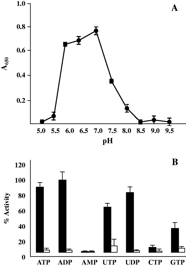
A biofunctional rOos-APY-1 enzyme was successfully expressed in a bacterial sumoylation cassette. Using this expressed fusion construct and the malachite green assay to monitor free phosphate, the rOos-APY-1 exhibited a strict requirement for Ca2+ relative to Mg2+ and an optimum pH range between 6·0 and 7·0 with a pH 7·0 maximum defined by the conditions and points used in this study (Fig. 5A). This coincides with pH optima found in the human homologue (Smith et al. Reference Smith, Hicks-Berger, Kim and Kirley2002; Murphy et al. Reference Murphy, Ivanenkov and Kirley2003). The recombinant protein showed highest relative activity with ADP as substrate followed by ATP, UDP, UTP and GTP. Little activity was observed with CTP; the monophosphate AMP was not further dephosphorylated (Fig. 5B).
Evaluation of pH optimum (A) and substrate specificity (B) of rOos-APY-1. A malachite green assay was used to monitor release of inorganic phosphate at OD650 after 10 min incubation with colour reagent. Results for pH determination are presented as OD650 (A); substrate specificity is presented as % OD650 relative to ADP (B). Reactions were carried out in triplicate. Error bars represent standard deviation. For pH determination, reactions were performed in buffered Bis-Tris (pH 5·0–6·5) or Tris-HCl (pH 7·0–9·5) where ATP was used as substrate. Studies on substrate specificity were performed in 50 mm Tris, pH 6·5, containing either 5 mm MgCl2 (□) or CaCl2 (▪).

DISCUSSION
Nucleotide-metabolizing enzymes are ubiquitous in nature; however, within this broad group of enzymes there exist subsets with more specialized functions. Some are capable of degrading tri-and diphosphates with no activity on monophosphates (apyrases), while others are capable of degrading monophosphates to nucleosides with little ability to remove phosphates from other nucleotides. One group of particular interest and recently discovered within haematophagous arthropods is a Ca2+-dependent family with little sequence overlap to the more common human CD39 family, but with broad similarity in substrate activity. Herein we describe an apyrase within 4th-stage O. ostertagi that by sequence and function corresponds with the Ca2+-dependent apyrases first identified in blood-sucking arthropods. Phylogenetic analysis showed support for a monophyletic lineage of the Ca2+ apyrases that is distinct from the CD39 family (S. mansoni) (DeMarco et al. Reference DeMarco, Kowaltowski, Mortara and Verjovski-Almeida2003) and the 5′nucleotidase in T. spiralis (Gounaris et al. Reference Gounaris, Selkirk and Sadeghi2004) which specifically dephosphorylates di- and mononucleotides. The phylogenetic tree is in general agreement with those presented elsewhere (Smith et al. Reference Smith, Hicks-Berger, Kim and Kirley2002; Anderson et al. Reference Anderson, Oliveira, Kamhawi, Mans, Reynoso, Seitz, Lawyer, Garfield, Pham and Valenzuela2006) showing a monophyletic lineage among nematodes and vertebrates relative to the apyrases derived from insects.
Elucidation of the tertiary structure of the human Ca2+-dependent apyrase (Dai et al. Reference Dai, Liu, Deng, Smith and Lu2004), in conjunction with functional analyses, identified conserved amino acids Ser182, Asp183 Glu230, Glu300, Ser363 and Glu416 as critical for Ca2+ binding. These amino acids reside within a central tunnel formed in the X-ray crystal structure that permits Ca2+ access to the 5 blades or regions of the protein and proximity to the nucleotide-binding site. These same amino acids were observed at the aligned loci in rOos-APY-1 confirming the functional dependency on Ca2+. Absolute conservation at Glu230 with respect to Ca2+ and nucleotide binding suggests that the Ca2+ molecule is directly involved with the active site (Dai et al. Reference Dai, Liu, Deng, Smith and Lu2004).
Amino acids defined as critical for nucleotide binding were somewhat less conserved (62%) but superficially located on the 3D model (Dai et al. Reference Dai, Liu, Deng, Smith and Lu2004) providing unabated access to the protein. Unlike the human homologue which has greatest activity on guanosine and uridine base analogues, rOos-APY-1 exhibits highest reactivity on ATP and ADP which is more in line with the substrate specificity observed in insects. Random mutations of the human homologue demonstrated that changing Glu261 to Met261 enhanced enzymatic activity 5 to 7-fold on both GDP and ADP (Dai et al. Reference Dai, Liu, Deng, Smith and Lu2004). The rOos-APY-1 possesses a Met at that locus lending support to the importance of this site in substrate recognition, activity and/or protein conformation. Apyrases from L. longipalpis and P. papatasi which are most active on ATP and ADP contain Leu at the 261 locus so it is difficult to fully ascribe this specific amino acid change to ADP functionality; however, prior work on L. longipalpis (Charlab et al. Reference Charlab, Valenzuela, Rowton and Ribeiro1999) and P. papatasi (Valenzuela et al. Reference Valenzuela, Belkaid, Rowton and Ribeiro2001) did not provide relative enzyme activities on other nucleotides to rule out this possibility. On the other hand, research findings on Phlebotomus duboscqi (Hamasaki et al. Reference Hamasaki, Kato, Terayama, Iwata and Valenzuela2009) which also possess a Leu261 and which shows no activity on substrates other than ADP, points to the replacement of Glu rather than the specific introduction of Met at this locus that creates a less sterically hindered enzyme with expanded substrate activity. Comparison to a C. elegans-derived protein (AAA68725) (Uccelletti et al. Reference Uccelletti, Pascoli, Farina, Alberti, Mancini, Hirschberg and Palleschi2008) exhibited notable differences. First, substrate specificity of the C. elegans protein was relegated to UDP and weakly to GDP and exhibited no activity on ATP or ADP as shown here. Secondly, the C. elegans-derived protein was expressed in all stages whereas the Ostertagia protein expression was restricted to the L4 only. Finally, sequence identity between the Ostertagia and C. elegans proteins is 44%, whereas identity with the secreted protein from T. circumcincta is 93% (Smith et al. Reference Smith, Nisbet, Meikle, Inglis, Sales, Beynon and Matthews2009) and shows 100% congruence in the first 15aa defined as a putative secretory signal (personal communication). The rapid rise in host antibody responses to the native parasite protein is consistent with this protein being secreted in the earlier stages of development.
Both RT-PCR and Western blot data demonstrated that the predominant stage of gene and protein expression is the L4 when rapid growth commences. Limited RT-PCR product was observed in adult parasites and this was confirmed by Northern blots (data not shown); however, adult parasites which also actively feed showed no signs of the protein by Western blot analysis. The native protein was localized to the more anterior region of the oesophagus which is predominantly muscle, so enzymatic activity probably emanates from the bulb. Inasmuch as the protein is not observed in mature adults that also actively feed, the function of the apyrase is most likely linked to regulating the microenvironment of the parasite where the enzyme is pumped anterior from the oesophageal bulb to interact with host extracellular nucleotides possibly in an effort to control inflammation during its explosive growth within the gastric glands. A similar proposal for controlling inflammation has been put forth for the family of ATP-catabolizing enzymes present on the tegumental surface of S. mansoni (Bhardwaj and Skelly, Reference Bhardwaj and Skelly2009) and the cascade of comparable enzymes present in secreted products from T. spiralis (Gounaris and Selkirk, Reference Gounaris and Selkirk2005).
The penetration of Ostertagia into and its emergence from the gastric glands causes substantial tissue damage. Infected glands as well as glands in the vicinity of those parasitized show a loss of parietal and zymogenic cells that are replaced by undifferentiated epithelium. Such tissue damage undoubtedly results in the release of cytosolic ATP into the surrounding milieu capable of priming cells of the innate immune system and initiating inflammatory response mechanisms. However, as long as viable L4 are present within the gastric glands, there is complete absence of inflammation (Snider et al. Reference Snider, Williams, Knox, Marbury, Crowder and Willis1988). Furthermore, inflammatory responses rapidly surface only after the L4 have shed their cuticles and emerge from the glands or after they have been killed in situ with anthelmintic treatment. In part, this relates to experiments showing that extracts of O. ostertagi contain products capable of inhibiting in vitro proliferation of bovine lymphocytes obtained from infected cattle (Gómez-Muñoz et al. Reference Gómez-Muñoz, Canals-Caballero, Almeria, Pasquali, Zarlenga and Gasbarre2004) and suppressing the local immune responses. These ‘suppressive factors’ have been identified predominantly in products secreted by L4 (De Marez et al. Reference De Marez, Cox, Claerebout, Vercruysse and Goddeeris1997), the principal stage for apyrase production. Interestingly, Li et al. (Reference Li, Sonstegard, Van Tassell and Gasbarre2007) demonstrated that cytokine types and levels coinciding with inflammatory responses were substantially higher in Angus heifers resistant to GI nematode infections relative to susceptible animals, and concluded that resistant animals can better maintain inflammatory responses at the site of infection. Similar conclusions have been drawn following the examination of sheep breeds resistant or susceptible to Haemonchus contortus infection (MacKinnon et al. Reference MacKinnon, Burton, Zajac and Notter2009). Although apyrases are likely not the only parasite product capable of modifying the host immune responses, these findings support a role for the apyrases in abating inflammation by reducing local ATP levels and exacerbating the infection.
Parasitic helminths produce a variety of host-immunomodulatory factors on their cuticular surface or in excretory and secretory (ES) products (reviewed by Maizels and Yazdanbakhsh, Reference Maizels and Yazdanbakhsh2003 and Johnston et al. Reference Johnston, MacDonald and McKay2009). Several studies have indicated that some parasitic nematodes produce macrophage migration-inhibitory factor (MIF) and TGF-β orthologues (Vermeire et al. Reference Vermeire, Cho, Lolis, Bucala and Cappello2008; McSorley et al. Reference McSorley, Grainger, Harcus, Murray, Nisbet, Knox and Maizels2010). Host immune responses can also be regulated by altering ATP levels in interstitial spaces and controlling the ATP:adenosine (Ado) ratios which directly affect purinergic signalling. Increases in levels of eATP affect numerous cell types in the mammalian immune system. ATP alters IL-8 secretion by eosinophils, maturation of dendritic cells (DC), inflammatory cytokine secretion by monocytes and DC, induction of TH17 cells, and T and B cell co-stimulation (reviewed by Trautmann, Reference Trautmann2009). Because of the pro-inflammatory role played by ATP, it has been classified as a Danger-Associated Molecular Pattern (DAMP) (Bours et al. Reference Bours, Swennen, Di Virgilio, Cronstein and Dagnelie2006) or a host-derived molecule that is released by tissues in response to some external perturbation i.e. pathogen or cell injury. Purinergic receptors on immune and non-immune cells have specificity either for Ado and inosine [P1 (A1, A2 and A3) receptors], or ATP [P2X and P2Y receptors], ADP, UTP and UDP [P2Y receptors] suggesting that the native Ostertagia apyrase functioning in interstitial spaces can directly affect the P2 purinergic signalling within the gastric glands given its substrate specificity. As such, it is clear that modulating host eATP can confer an advantage to nematode establishment and survival.
A study of Oos-APY-1 protein production within inhibited larvae may shed light on its ultimate function in the L4 and within the gastric glands. Such a molecule warrants targeting for vaccine development inasmuch as it emanates from the feeding end of the worm and interrupting this process could drastically affect the ability of the worm to metabolize nutrients or modulate the local immune responses. Inasmuch as the crystal structure of the human homologue is known, this could help in the design of compounds to specifically attenuate the function of the parasite derived enzymes.
ACKNOWLEDGEMENTS
Soluble extracts (S1) from adult and L4 O. ostertagi were kindly donated by Dr Stuart Smith, Moredun Research Institute. Authors express thanks to M.B. Chute, P. Boyd and D. Hebert for technical assistance. D.S.Z. is funded by the U.S. Department of Agriculture, Agricultural Research Service (ARS). A.J.N. is funded by the Scottish Government Rural and Environment Research and Analysis Directorate (RERAD).