

Introduction
The metabolic phenomenon known as the lactate paradox (LP) was first identified in the early 1930s and described as ‘Standard work performances, on first going to high altitudes, produce greater rises in blood lactic acid than at sea level. After acclimatization, lactic acid values similar to those at sea level are found for a given performance’Reference Edwards1. However, it was not until 1981, during a research expedition to Mount Everest (Mt. Everest), that the LP began to gain widespread attention from the scientific community. During this expedition to 6300 m above sea level (SL), subjects performed maximal aerobic exercise. The blood lactate concentration (blood [La]) attained during this maximal effort was found to be significantly lower than expectedReference West, Boyer, Graber, Hackett, Maret and Milledge2, Reference West3. The data obtained from this expedition spurred a number of following studies and reviews concerning lactate kinetics during exercise at high altitudes (ALTs). The studies confirmed the initial findings obtained from the Mt. Everest project, and researchers began to look for the mechanisms that would explain these perplexing resultsReference Edwards1, Reference Beidleman, Muza, Rock, Fulco, Lyons and Hoyt4–Reference Pronk, Tiemessen, Hupperets, Kennedy, Powell and Hopkins16.
New studies were designed to answer two fundamental questions: Why does peak blood [La] that is achieved through aerobic exercise decrease with increasing ALT? And, why does blood [La] decrease during acclimatization at ALT, despite there being no change in O2 consumption for a given workload? However, the studies performed to answer these questions created questions of their own and new hypotheses. Thus, the theories and explanations of the LP's two main components quickly changed as each new study revealed more biochemical details and nuances concerning the paradox. This pattern of the proposed mechanisms for the LP being replaced by new data and new proposals in the literature has continued to the present. To date, no single mechanism that explains the full extent of the LP has been identified.
The existence of the LP was strongly challenged at the turn of the century by a group of researchers in Denmark. Their studies suggested that when subjects were given enough time to acclimatize, blood [La] during exercise at ALT returns to values typically measured at SLReference Lundby, Saltin and Van Hall14, Reference Van Hall, Calbet, Sondergaard and Saltin17–Reference Van Hall, Lundby, Araoz, Calbet, Snader and Saltin19. However, the question as to why this return occurs without an increase in VO2max remained unanswered. The very existence of the LP was later discussed in a Point: Counterpoint debate between Dr John West and Dr Garret van Hall in the Journal of Applied Physiology Reference Van Hall20, Reference West21. Upon the conclusion of this debate, the overall consensus between the researchers was that the LP did indeed exist, even if only under a small set of parameters. However, no absolute parameters were specifiedReference Mazzeo, Secher, Rasmussen, Kayser, Wagner and Samaja22. Currently, the dominant questions regarding the LP are as follows: (1) What specific physiological, ambient conditions and workloads elicit the LP? (2) What are the specific physiological/biochemical mechanisms causing the LP? and (3) What are the specific physiological/biochemical mechanisms for sustaining the LP upon return to SL?
Several of the proposed LP mechanisms have been separately reviewed in previous literature. However, as a whole, the specific results and conclusions of nearly 30 years of research in this area have not been addressed in a single review. Our purpose is to present the prominent literature regarding the LP, and highlight the conditions or parameters under which the LP does or does not exist. In doing so, the intent is to provide a more directed source for future LP mechanism research.
Peak blood [La] declines as a function of increasing ALT
In 1981, Operation Everest provided strong insight into the human body's ability to cope with severe hypoxiaReference West, Boyer, Graber, Hackett, Maret and Milledge2. Though this was not the first time low blood [La] at ALT had been noted, this project became the focal point for ensuing research done on the LP. The peak elevation reached by the study was 6300 m, or approximately 20,500 ft. As part of this project, subjects performed incremental exercise on a portable cycle ergometer. The exercise protocols included maintaining 50 rpms for workloads up to 900 kg min− 1, and 75 rpms for workloads above that. At SL, workloads below 900 kg min− 1 had to be maintained for 5 min. Higher workloads had to be maintained for 3 min. At ALT, workloads above 600 kg min− 1 had to be maintained for 3 min. Subjects breathing low-O2 gas mixtures at ALT were required to maintain any given workload for up to 3 min.
Most of the tests performed during this study were conducted on subjects breathing ambient air. In order to simulate exercise at elevations of 8050 and 8848 m, West et al. Reference West, Boyer, Graber, Hackett, Maret and Milledge2 employed the use of low-oxygen gas mixtures: ‘a sophisticated laboratory was set up at an altitude (ALT) of 6300 m and at a barometric pressure of 351 Torr. Subjects were studied both while breathing ambient air and while inhaling low O2 mixtures containing 16 and 14% O2. These mixtures gave virtually the same inspired PO2 as that of climbers at altitudes of 8050 m (barometric pressure 283 Torr) and at the summit of Mt. Everest, altitude of 8848 m (barometric pressure 253 Torr)’.
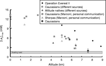
The partial pressure of oxygen (PO2) is a function of atmospheric pressure and the fractional composition of inspired air (FiO2). Despite the fractional composition of oxygen content in the air remaining constant at 20.93% from low to high ALTs, the large reduction in atmospheric pressure with increased elevation decreases the atmospheric PO2 drastically. Decreases in PO2 cause incomplete arterial oxygen saturation (SaO2) leading to a decrease in the total arterial oxygen content (CaO2). By using low-oxygen gas mixtures to decrease FiO2, West was able to simulate a PO2 similar to that expected to be found at the top of Mt. Everest. Among the data collected was the finding that during exhaustive exercise, peak blood [La] declines linearly as ALT increases.
The decline in peak blood [La] with increasing elevation during aerobic exercise to exhaustion is depicted in Fig. 1. As illustrated by the graph, and mentioned by West, peak blood [La] is not predicted to exceed resting blood [La] values when exercise is performed above approximately 7500 mReference West23. Hochachka et al. Reference Hochachka24 described this aspect of the LP thus: ‘The paradox is that anaerobic glycolysis works perfectly well when subjects acclimatized to high altitudes start from rest, and in fact it is well known that the anaerobic power output of muscles is unaffected by hypobaric hypoxia. Therefore, the problem arises as to why it seems to be impossible to activate the pathway in muscles brought to fatigue during aerobic work at high altitude, where the demands for glycolytic adenosine triphosphate (ATP) synthesis are normally exaggerated’.
The decline in peak blood [La] with increasing elevation during aerobic exercise to exhaustion, data borrowed from Cerretelli and Samaja 2003Reference Cerretelli and Samaja7. The black diamonds reflect the data recorded from Operation Everest II. This study took place at SL, and utilized a large decompression chamber in which the air pressure within the room was reduced to simulate the decreasing atmospheric pressure that is accompanied by increasing altitude.

This indicates that, starting from rest, lactate production during sub-maximal exercise, but not during all-out exercise, is affected by changes in oxygen availability to the working muscles. ATP production at high intensities is generated mostly by glycolysis and the phosphocreatine (PCr) system. Therefore, all-out exercise performance lasting up to 10 s is unaffected by changes in oxygen availability because it does not rely heavily on the electron transport chain (ETC) to produce ATP. Sub-maximal exercise ATP production does rely heavily on the ETC. Therefore, in order to maintain sub-maximal exercise performance with decreased oxygen availability, either oxygen utilization must be made more efficient or other metabolic processes must compensate.
La release from working muscles is decreased after acclimatization
It is well known that blood [La] reflects the balance between lactate flux from working muscles and lactate uptake by non-working muscles, the liver and, depending on the concentration, working muscles as well. In 1989, Bender et al. Reference Bender, Groves, McCullough, McCullough, Trad and Young5 investigated the LP by measuring lactate release from exercising muscles through the course of acclimatization. The study took place at 4300 m, and subjects remained at an ALT for 18 days. A steady-state exercise protocol on a cycle ergometer was performed. The subjects completed three bouts of exercise corresponding to 43, 70 and 95% of VO2max for 5, 5 and 3 min, respectively. Subjects were given 5 min rest between the first two exercise bouts and 10 min between the second and third. Lactate release was calculated by measuring leg blood flow and differences between arterial and venous blood [La]. Results showed that the net lactate release by working muscles at a given workload and/or O2 consumption were decreased after acclimatization. This finding was also confirmed in the 1991 study done by Brooks et al. Reference Brooks, Wolfel, Groves, Bender, Butterfield and Cymerman25, in which lactate release and uptake by working muscles were measured.
The results of these studies suggest that exposure to chronic hypoxia induces a metabolic adaptation within the muscles; in this case, an adaptation that affects the production of lactate. However, it cannot be assumed that decreased lactate production and release from working muscles are solely responsible for the LP. The effect of lactate uptake by non-working muscles must also be analysed for its possible contribution to the LP.
Effect of ALT exposure on lactate uptake
Brooks et al. Reference Brooks, Wolfel, Groves, Bender, Butterfield and Cymerman25 were among the earliest researchers to point out that blood [La] during exercise is not only a function of lactate production and release by working muscles, but is also dependent on lactate uptake by non-working muscles. They investigated the rate of lactate release and uptake by working muscles as well as uptake by inactive limbs during steady-state exercise on a cycle ergometer equating to approximately 50% of the individual's VO2max. The study took place at 4300 m, and the duration of ALT exposure was 3 weeks. Lactate release/uptake was measured by calculating the difference between arterial and venous blood [La]. Results demonstrated that during acute hypoxia, inactive limbs uptake more lactate than they release, but that after acclimatization, the rate of lactate flux and uptake is equal. In working limbs, lactate is both released and taken up, though the latter is dependent on the concentration of lactate within the blood. This study suggests that through acclimatization, the ability of non-working muscles to uptake and oxidize blood lactate is diminished.
Blood glucose and fatty acid utilization after acclimatization
Studies done by Roberts and Brooks et al. investigated changes in glucose and fatty acid metabolism with chronic exposure to ALT. The first study, led by Brooks et al. in 1991Reference Brooks, Butterfield, Wolfe, Groves, Mazzeo and Sutton26, investigated the changes in the rate of glucose appearance and disappearance within the blood during rest and exercise through the course of high-ALT acclimatization. Subjects spent 3 weeks at an ALT of 4300 m and performed a regularly scheduled steady-state exercise on a cycle ergometer at 50% of VO2max. Their findings revealed that when compared with SL values, the rate of both glucose appearance and disappearance increased significantly with chronic ALT exposure. These results led to the conclusion that glucose utilization by working muscles during exercise is increased after chronic ALT exposure.
In 1996, Roberts et al. Reference Roberts, Reeves, Butterfield, Mazzeo, Sutton and Wolfel27, Reference Roberts, Butterfield, Cymerman, Reeves, Wolfel and Brooks28 investigated the effect of β-blockade on fatty acid and glucose utilization during rest and exercise through the course of high-ALT acclimatization. This study employed the same timetable, elevation and exercise protocol used by Brooks et al. in 1991: 3 weeks at 4300 m and a steady-state exercise protocol at 50% of the individual's VO2max. The results of this study were the same as those obtained by Brooks et al. The use of glucose increases with acclimatization, while the use of fatty acids is diminished. These results added to the complexity of the LP. If glucose becomes the preferred substrate during exercise at ALT, it can be assumed that La production, and therefore blood [La], increase. The increase in glucose utilization after ALT exposure, however, may be due to its more efficient yield of ATP per volume of oxygen when fully metabolized. ‘A potential benefit of this adaptation relates to the fact that, during times when oxygen availability is limited, carbohydrates yield more energy per litre of oxygen than do fats and proteins. Thus, this would result in a more oxygen-efficient use of fuels’Reference Mazzeo29.
The more efficient use of oxygen by glucose can be calculated by totalling the amount of ATP created by complete combustion of 1 mol of glucose and fat divided by the oxygen (mol) required for the combustion. The exchange ratio of NADH/FADH (nicotinamide adenine dinucleotide–flavin adenine dinucleotide) to ATP is 2.5:1.5, respectivelyReference Nelson and Cox30. This is accomplished by the passing of electrons through the ETC and subsequent pumping of H+ from the mitochondrial matrix (negative space) to the inner mitochondrial membrane (positive space), thus creating a strong [H+] concentration difference between the two. The flow of H+ back into the mitochondrial matrix creates a proton-motive force which is used by complex 5 to turn ADP (adenosine diphosphate)+inorganic phosphate (Pi) into ATP. With 4 being ‘the most accepted number of H+ required to drive the synthesis of an ATP molecule’, the number of ATP created per NADH/FADH is dependent on where the electron transfer beginsReference Nelson and Cox30. NADH formed in the mitochondrial matrix enters at complex 1 and pumps a total of 10H+ (complex 1-4H+; complex 3-4H+; complex 4-2H+) across the inner mitochondrial membrane. FADH and cytosolic NADH (in mammalian skeletal muscles) enter the ETC at complex 3. Missing out on the first 4H+ pumped out at complex 1 results in a total of 6H+ being pumped across the inner mitochondrial membrane into the positive space (4H+ at complex 3 and 2H+ at complex 4). Therefore, we get 10H+/(4H+/ATP) = 2.5 ATP per NADH formed within the mitochondrial matrix, and 6H+/(4H+/ATP) = 1.5 ATP for FADH and cytosolic NADH.
The calculation of total ATP mol− 1 O2 from glucose and fat is as follows:




Total:
- 8NADHmatrix
= 20ATP
- 2NADHcytosol (enters ETC at complex 3)
= 3ATP
- 2FADHmatrix
= 3ATP
- 2GTP
= 2ATP
- 2ATP
= 2ATP
30ATP



Total:
- 31NADHmatrix
= 77.5ATP
- 15FADHmatrix
= 22.5ATP
- 8GTP
= 8ATP
108ATP


Carbohydrates are 7% more efficient in their use of oxygen when used to produce ATP. This, in conjunction with the studies done by Brooks and Roberts, indicates that carbohydrates are the preferred carbon source for oxidative metabolism, potentially explaining why glycolytic recruitment can increase for a given workload without increasing the production of lactate.
The LP as a function of VO2
The aggravating effect of hypoxia on the slow component of VO2 has been recordedReference Springer, Barstow, Wasserman and Cooper31, Reference Engelen, Porszasz, Riley, Wasserman, Maehara and Barstow32. However, what is undoubtedly the most important aspect of the LP is the finding that blood [La] and lactate release by working muscles decreases for a given workload or VO2 during acclimatizationReference Bender, Groves, McCullough, McCullough, Trad and Young5, Reference Brooks, Butterfield, Wolfe, Groves, Mazzeo and Sutton6, Reference Grassi, Ferretti, Marzorati, Kayser, Bordini and Colombini10. MazzeoReference Mazzeo29 described it perfectly: ‘This is considered paradoxical as the reduction in lactate occurs despite the findings that whole-body and working muscle VO2 do not differ between short- and long-term altitude exposure’.
In a 1988 study done by Bender et al., changes in VO2 during steady-state exercise were measured at three exercise intensities corresponding to 43, 70 and 95% VO2max. These three intensities were used at SL and at both an acute and chronic exposure to 4300 m ALTReference Bender, Groves, McCullough, McCullough, Huang and Hamilton33. Exercise during acute ALT exposure was carried out at SL in a hypobaric chamber (445 Torr). The exercise protocol employed a cycle ergometer, and the length of stay at ALT was 18 days. Results showed that whole-body and leg VO2 were maintained during sub-maximal exercise in acute versus chronic hypoxia, as well as in normoxia before and after ALT acclimatization. These findings have been confirmed elsewhereReference Brooks, Butterfield, Wolfe, Groves, Mazzeo and Sutton26, Reference Rowell, Saltin, Kiens and Christensen34.
Despite VO2max decreasing as elevation increases, in both acute and chronic hypoxia, whole-body and working muscle VO2 match SL values during sub-maximal exercise. During acute hypoxia, the decreases in oxygen saturation and content of arterial blood (SaO2 and CaO2) are offset by an increase in HR. During chronic hypoxia, increased haemoglobin density offsets the decrease in stroke volume. These changes allow oxygen delivery to working muscles to match demand. However, just as VO2max decreases as elevation increases, so too does the intensity of the sub-maximal exercise at which oxygen supply can meet demand.
LP persistence in de-acclimatization
Perhaps one of the most intriguing aspects of the LP is its lingering effect upon return to SL. Studies done by Grassi et al. Reference Grassi, Ferretti, Kayser, Marzorati, Colombini and Marconi9, Reference Grassi, Ferretti, Marzorati, Kayser, Bordini and Colombini10 investigated the changes in blood [La] during exercise at SL, after chronic exposure to ALT and during deacclimatization upon return to SL. The first study used intermittent bouts of all-out exercise on a cycle ergometer corresponding to approximately 200% of the individual's VO2maxReference Grassi, Ferretti, Kayser, Marzorati, Colombini and Marconi9. The exercise bouts lasted 5, 15, 25, 35 and 45 s (or until exhaustion). Tests were performed at SL, at weeks 1 and 4 of exposure to 5050 m ALT, and again 1 week after returning to SL. The subjects' average time to exhaustion under all test conditions was above 30 s. However, none of the average times were significantly different. Results did show that in addition to peak blood [La] being significantly reduced after 4 weeks at ALT, peak blood [La] after 1 week of de-acclimatization remained significantly lower than peak blood [La] at SL before exposure to ALT.
In a second study, Grassi et al. Reference Grassi, Ferretti, Marzorati, Kayser, Bordini and Colombini10 once again used a cycle ergometer, but this time they employed an incremental exercise protocol. The study took place again at 5050 m. Exercise began at 30 W and increased by 30 W every 4 min until exhaustion. Tests were performed at SL (preliminary test), during weeks 1, 3 and 5 of chronic ALT exposure and again at SL during weeks 1, 2, 3, 4 and 5 of de-acclimatization. Peak blood [La] was lower at ALT than at SL in all tests. However, peak blood [La] at SL was not significantly different from peak blood [La] at ALT during weeks 1 and 2 of de-acclimatization. Peak blood [La] did not reach preliminary SL values until the third week of return. Peak work rate was highest during the preliminary SL test and lowest during the ALT week 1 test. Peak work rate did improve slightly during the stay at ALT.
These studies indicate that the adaptation(s) the body goes through in response to prolonged ALT exposure affect blood [La] not only during chronic hypoxia, but also when returning to SL.
Grassi et al. Reference Grassi, Ferretti, Kayser, Marzorati, Colombini and Marconi9 also measured blood [La] during recovery from exhaustive exercise: ‘In all experimental conditions, blood [La] ([Lab]) increased during the first 4–6 min of recovery and, after reaching a peak ([Lab]p), it diminished progressively according to a monoexponential function of time, with half times of ~11–13 min’. The time course for lactate removal was not significantly different under any of the experimental conditions, despite the presence of the LP (i.e. peak blood [La] being lower after ALT acclimatization). Though these results indicated that recovery times are not affected by ALT acclimatization, no information was collected on recovery time for exercise at SL immediately following ALT acclimatization. Initial lactate tracer studies done by Brooks and GaesserReference Brooks and Gaesser35 showed that the fate of lactate following exercise is oxidation. Therefore, it seems that lactate recovery times at ALT should be prolonged. However, as described previously, oxygen delivery to working muscles is maintained for sub-maximal exercise. These findings indicate that despite VO2max being decreased at ALT, oxygen delivery to recovering muscles may still be matched. Recovery time after exercise at extreme ALTs of 6000 m or higher, both before and after ALT acclimatization, would make for a good study.
In summary, these studies reaffirm that oxygen availability plays an indirect role in La production, and that prolonged exposure to hypoxia induces a metabolic change within the working muscles. Therefore, the question becomes: to what extent does oxygen availability to working muscles have an effect on blood [La] at various exercise intensities?
Effect of exercise intensity on LP expression
Another intriguing aspect of the LP is that it is not expressed when employing certain exercise protocols. In another study carried out by Grassi et al., the LP was investigated under varying exercise intensities on a cycle ergometer. The study took place at 5050 m, and the length of ALT exposure lasted approximately 21 days. Results showed that when performing all-out exercise lasting 10 s, the LP was not present. However, the LP was present when performing incremental exercise to exhaustion or all-out exercise lasting at least 30 sReference Grassi, Mognoni, Marzorati, Mattiotti, Marconi and Cerretelli11.
The average power output measured during the 10 s all-out effort was not significantly different between tests at SL and ALT. The average power output, as a function of body mass, was not significantly different either. Consequently, peak blood [La] did not change with exposure to ALT with this short-duration, high-intensity exercise. This protocol showed that when the exercise intensity is high enough, the LP may not be elicited. However, this should be expected since all-out exercise lasting 10 s does not rely on oxidative metabolism.
The average power output and average power output as a function of body mass during 30 s of all-out effort were significantly lower at ALT than at SL. The peak blood [La] was significantly lower at ALT than at SL as well. This protocol showed that the LP can be elucidated if exercise is not intense enough to elicit failure in < 30 s.
As with the 30 s maximal effort, maximal power output and maximal power output as a function of body mass were significantly lower at ALT during the incremental exercise protocol than at SL. Consequently, peak blood [La] was also significantly lower. The LP was clearly evident in this scenario. These results demonstrate that chronic oxygen availability has some underlying effect on blood [La] and lactate production during sub-maximal exercise.
Peak blood [La] rebounding after sufficient acclimatization
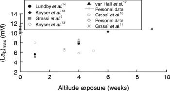
The peak blood [La] results of several independent acclimatization studies are summarized in Fig. 2. Recent work done by Lundby and van Hall has shown that peak blood [La] following incremental aerobic exercise to exhaustion returns to normal at ALT after sufficient (usually about 8 weeks') acclimatization. As shown in Fig. 2, their results form the bulk of research that does not support the existence of the LP. These data have been the focal point of several articles, including a Point: Counterpoint debate published in 2007Reference Van Hall20, Reference West21. However, Lundby and van Hall are not the only researchers to record increases in blood [La] during exercise after acclimatization. A test conducted by Grassi et al. in 1995 reported higher blood [La] after 4 weeks of ALT exposure than after 1 week of ALT exposure. According to the data points, it is entirely possible that the peak blood [La] would have returned to SL values if the study had continued to 8 weeks. However, no conclusive statement can be made to this effect.
Peak blood [La] results of several independent acclimatization studies, data borrowed from Cerretelli and Samaja 2003Reference Cerretelli and Samaja7. The ‘personal data’ belong to Paolo Cerretelli, and represent data collected by him and his colleagues.

The question that arose from these studies, and still remains unanswered, is why have Lundby and van Hall been unable to reproduce the results attained by everyone else? Review of Lundby and van Hall's studies versus those that have confirmed the existence of the LP reveals that the major variables regarding the LP do not differ. The elevation at which Lundby and van Hall conducted their experiments was clearly high enough to elicit the LP. Exercise was performed on a cycle ergometer, which is by far the most common mode used to test the LP. And, they employed aerobic exercise protocols in their attempts to elicit the LP, which, as reported previously, is the only form of exercise that readily reveals the LP.
One noticeable difference between researchers, however, is the length of hypoxic exposure. Before the studies done by Lundby and van Hall were published, most research on the LP lasted approximately 2–4 weeks. Consequently, the prolonged exposure of Lundby and Van Hall's studies prompted a follow-up study in 2003 by Pronk et al. Reference Pronk, Tiemessen, Hupperets, Kennedy, Powell and Hopkins16, the goal of which was to confirm whether prolonged exposure of at least 8 weeks did in fact reverse the presence of the LP. Subjects underwent two cycle ergometer VO2max tests at the onset of the study, one while breathing ambient air and the other while breathing a low-oxygen gas mixture, in order to determine appropriate workloads for both normoxia (SL) and hypoxia (ALT) tests, respectively. The subjects underwent the same two tests in a random order every 2 weeks for 8 weeks at 3800 m, breathing either ambient air (ALT) or a high-oxygen gas mixture (SL). The exercise protocol was performed at set workloads corresponding to 25, 50, 75, 90 and 100% of the individual's VO2max for 5, 5, 3, 2 and 1 min, respectively. The results of the study contradicted those obtained by Lundby and van Hall. The LP was still very evident in this study after 8 weeks at 3800 m. Peak blood [La] was decreased, and blood [La] at a given workload or oxygen consumption decreased throughout the 8-week study. Therefore, the length of exposure to high ALT does not seem to explain the discrepancy between Lundby and van Hall's results and others.
Loss of body mass
It is not yet fully understood why the results of Lundby and van Hall's studies do not concur with the findings of others. In a Contrasting Perspectives piece hosted by Medicine and Science in Sports and Exercise, Lundby and WagnerReference Lundby and Wagner36 proposed a few possible explanations, which include loss of lean body mass, loss of fitness and measurement errors. Many review articles have mentioned the inability of the body to maintain lean body mass at higher elevationsReference Brooks, Butterfield, Wolfe, Groves, Mazzeo and Sutton6, Reference Pronk, Tiemessen, Hupperets, Kennedy, Powell and Hopkins16, Reference Lundby and Wagner36. In the event that lean body mass does decrease, ‘exercise at a given power output requires a higher fraction of maximal exercise capacity, all other factors unchanged, moving subjects along their La–work rate relationship towards higher La’Reference Lundby and Wagner36. Body weight was markedly decreased in the studies done by Lundby and van Hall, including one in which the average weight loss after 8 weeks of chronic ALT exposure was 19 kgReference Roberts, Reeves, Butterfield, Mazzeo, Sutton and Wolfel27. As noted earlier, Pronk et al. followed up the studies of Lundby and van Hall, and found that the LP was still present even after prolonged exposure. However, body weight in Pronk's study was tightly maintained, with the largest average drop from SL weight over the entire course of acclimatization being 1.4 kg. These data strongly suggest that loss of body mass may very well explain why Lundby and van Hall have been unable to replicate studies in which the LP was apparent. Perhaps future studies will determine the presence (or not) of the LP in individuals who lose versus maintain lean muscle mass at ALT with all other variables held constant.
Maintaining fitness at ALT
Another proposal made by Lundby and Wagner concerning the discrepancies in Lundby and van Hall's results is the possible occurrence of detraining. Simply put, ‘When an unfit subject endurance runs at sea level, La falls at any given work rate. Conversely, detraining increases La at a given work rate’Reference Lundby and Wagner36. To date, no one has investigated a change in fitness level (VO2max) against LP expression. Research along this line would add considerable understanding to the LP.
La flux during incremental exercise
Lundby and Wagner also suggested that measurement errors may be the cause of the discrepancy between studies confirming the LP and those that do not. The ability of [La] to rise rapidly could inadvertently lead some researchers to miss correct peak and/or steady-state [La] if the measurement is taken too early. However, if there is an interval of at least 1 min between blood [La] measurement and increase in workload in the protocol, this should not be an issue for sub-maximal [La] measurements. Smith et al. Reference Smith, Skelton, Kremer, Pascoe and Gladden37 showed that during incremental exercise, there was no difference in blood [La] between measurements taken 1 min or 4 min after workload was increased. However, peak [La] remains more difficult to obtain correctly due to the rapid flux and uptake by muscles, especially at exhaustion.
The difference between whole blood [La] and plasma [La] has also been mentioned as a possible confounding factor in studying the LP. It has been demonstrated that lactate does not distribute equally within lysed versus whole bloodReference Mazzeo, Secher, Rasmussen, Kayser, Wagner and Samaja22. However, the aforementioned study by Pronk et al. investigated [La] on both lysed and whole blood samples through 8 weeks' exposure to ALT, and found that the LP was present in both sample types.
Reviewing the research
Thirty years of research has brought to light several key aspects of the LP. A pointed list includes the following:
(1) Peak blood [La] declines as the elevation of acclimatization increases.
(2) Blood [La] declines for a given workload and/or VO2 after ALT acclimatization.
(3) Lactate release from working muscles is decreased for a given sub-maximal workload after ALT acclimatization.
(4) Whole-body and working muscle VO2 are maintained during sub-maximal exercise at SL, during acute ALT exposure and during chronic ALT exposure.
(5) Glucose becomes the preferred carbon source for oxidative metabolism (acetyl-CoA), and fatty acid oxidation is decreased through ALT acclimatization. Also, blood glucose is more heavily recruited after acclimatization.
(6) High-intensity exercise lasting approximately 10 s does not elicit the LP. Exercise lasting 30 s or longer, however, does elicit the LP.
(7) The LP remains present during exercise at SL upon return from chronic ALT exposure. This effect wears off within 2–3 weeks.
(8) Research done by Lundby and van Hall has demonstrated that the LP is not always elucidated when lowlanders acclimatize to high ALTs. Though the reasons behind Lundby and van Hall's inability to elucidate the LP still remain unsolved, loss of lean body mass and/or a loss of fitness have been proposed as explanations.
A proposed mechanism for the LP
Though no mechanism has been able to explain the entire LP to date, the research results compiled in this review have led us to the following possible considerations.
The power output of highly intense exercise starting from rest and lasting 10 s is not affected by high ALT exposure. This indicates that the rapid recruitment of glycolysis and the PCr system, at the onset of highly intense exercise, remains unaltered. Therefore, the mechanism solving the LP must revolve around the regulation of oxidative metabolism and the metabolites/substrates that support it.
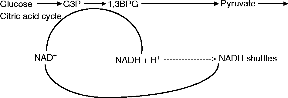
The reduced use of fatty acids makes way for the increased utilization of glucose (pyruvate) as the carbon source (acetyl-CoA) for oxidative metabolism. This shift in the dominant carbon source for oxidative metabolism has been documentedReference Brooks, Butterfield, Wolfe, Groves, Mazzeo and Sutton26–Reference Roberts, Butterfield, Cymerman, Reeves, Wolfel and Brooks28, and so too has the accompanying decline in blood [La]Reference Edwards1, Reference Beidleman, Muza, Rock, Fulco, Lyons and Hoyt4–Reference Pronk, Tiemessen, Hupperets, Kennedy, Powell and Hopkins16. These coinciding factors may explain why blood [La] for a given workload and/or VO2 decreases. As the use of glucose increases for a given workload (or the rate of glycolysis increases), pyruvate dehydrogenase (PDH) activity increases, allowing for the increased use of pyruvate to form acetyl-CoA for oxidative metabolism. With more pyruvate being oxidized by the citric acid cycle (CAC), less pyruvate accumulates within the cytosol, and therefore less lactate is produced.
The finding that peak blood [La] decreases linearly as ALT increases may be explained as the strength or extent to which pyruvate oxidation is increased. In other words, the higher the ALT an individual acclimatizes to, the greater the reduction in the use of fatty acids and the greater the increase in PDH (pyruvate dehydrogenase) activity, resulting in increased oxidation of pyruvate to acetyl-CoA.
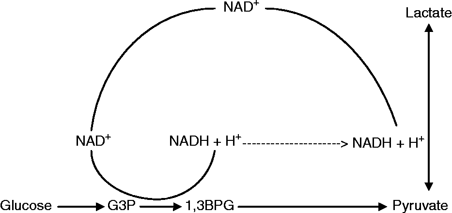
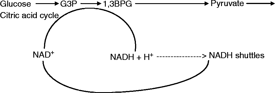
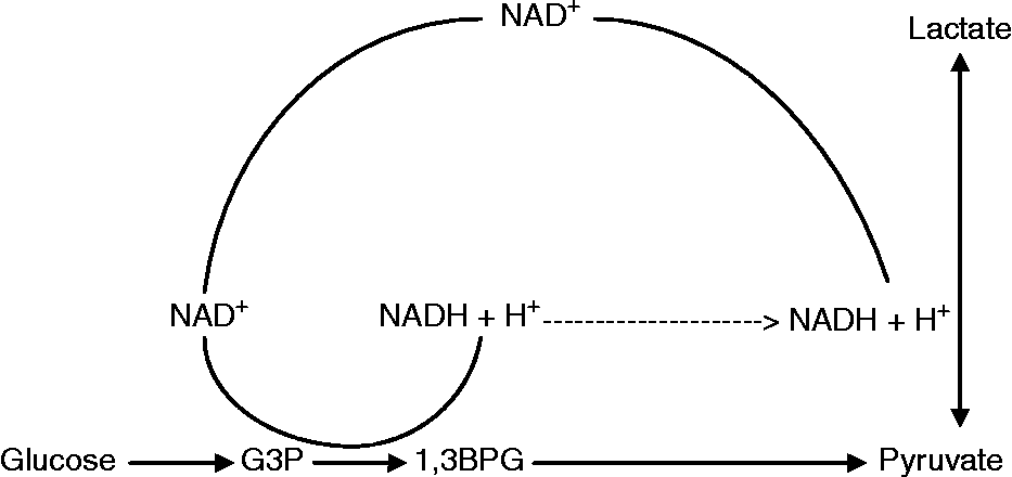
However, the increased use of pyruvate by the CAC leaves unoxidized NADH within the cytosol. In order to maintain cytosolic redox state, NADH must be oxidized back into NAD by the NADH shuttles (malate–aspartate or glyceraldehyde-3-phosphate). If the NADH shuttles do not oxidize NADH back into NAD, the glyceraldehyde-3-phosphate → 1,3bisphosphoglycerate (1,3BPG) reaction of glycolysis would become severely affected, endangering the rate of production of both glycolytic and CAC ATP. This shift in the maintenance of cytosolic redox state is depicted in Figs 3 and 4.
Cytosolic redox state regulation before acclimatization to high ALTs
Our proposed shift in cytosolic redox state regulation after acclimatization to high ALTs
In agreement with Lundby and Wagner, our proposed LP mechanism could be reversed by a significant loss of body mass similar to that observed in the studies of Lundby and van Hall. A significant loss in muscle mass would ultimately increase the metabolic rate (glycolysis) for the remaining muscles at any given workload. The increased rate of glycolysis would produce pyruvate faster than PDH would be able to flux it into the CAC (despite PDH being more active after ALT acclimatization than before). The down shift in the lactate–work rate relationship curve by increases in PDH activity would be counter balanced by the up shift in metabolic rate due to the significant loss of muscle mass.
One key aspect in a muscle mass scenario is that as the elevation at which exercise testing is conducted increases, the workload at which long-term steady-state exercise can be maintained decreases. At lower ALTs (Lundby and van Hall's studies were usually done at 5000 m+) or during very sub-maximal exercises, losses in body mass may not erode the presence of the LP, as the increased metabolic rate for remaining muscle mass may not be great enough to allow pyruvate to accumulate within the cytosol. The presence of the LP at different workloads/elevations in subjects who lose muscle mass would make for an interesting study.
The possible LP mechanism we have described illuminates the understanding that lactate production is not a function of oxygen debt, but rather a function of the competition between lactate dehydrogenase (LDH) and PDH over pyruvate and NADH accumulation within the cytosolReference Gladden38. Two separate studies done by Parolin et al. demonstrate how lactate production can be affected by changes in the number of PDH found in the active form. In the first study, subjects underwent two separate steady-state cycle ergometer tests at 55% of SL VO2max for 15 min while breathing either a low-oxygen gas mixture (11% O2) or SL ambient airReference Parolin, Spriet, Hultman, Hollidge-Horvat, Jones and Heigenhauser39. After 1 min of exercise, the active form of PDH (PDHa) within working skeletal muscles was found to be significantly less evident during acute hypoxia testing than during normoxia testing. However, after 15 min of exercise, PDHa within working skeletal muscles was found to be significantly more present during acute hypoxia than in normoxia. Despite the latter, blood [La] was still significantly higher after 15 min in hypoxia than in normoxia. Furthermore, blood [La] was higher in hypoxia than in normoxia, despite the rate of pyruvate oxidation by the CAC after 15 min of exercise being greater during hypoxia testing than during normoxia testing.
The second study used dichloroacetate (DCA) to activate PDH within skeletal muscles before exercise beganReference Parolin, Spriet, Hultman, Matsos, Hollidge-Horvat and Jones40. Subjects underwent two separate steady-state cycle ergometer tests at 55% SL VO2max while breathing a low-oxygen gas mixture (11% O2). One test involved infusion of a control solution (saline) and the other test involved infusion of DCA. Results showed that PDH was found significantly more in its active form for the DCA group at both 1 and 15 min of exercise. This led to an intramuscular [La] significantly lower for the DCA group after both 1 and 15 min of exercise. Furthermore, intramuscular [PCr] was significantly higher after 15 min of exercise in the DCA tests than in the control tests. This suggests that the increased oxidation of pyruvate at the onset of exercise may attenuate the production of lactate. In combination, these studies provide strong support for the LP mechanism proposed in this paper.
Our LP, however, also questions the use of lactate as a means to measure cytosolic NADH/NAD (redox state) in subjects acclimatized to high ALTs. Early measurements of cytosolic lactate and NADH/NAD, as well as mitochondrial NADH/NAD, were done at SLReference Williamson, Lund and Krebs41, Reference Sahlin, Katz and Henriksson42. Eventually, equilibrium-based equations were derived for calculating cytosolic NADH/NAD from intramuscular [La]. By our proposed mechanism, there is an upregulation of the NADH shuttles after acclimatization to high ALTs compared with SL. Therefore, while the calculations for cytosolic redox state have proved to be accurate and useful for studies carried out at SL, our proposed LP mechanism suggests that these calculations may not be suitable for subjects acclimatized to high ALTs.
Implications of the LP on models of muscular failure
The formation of lactate was long thought to be the cause of muscular failure as the source of exercise-induced metabolic acidosis, a belief that is still held by some researchers to this dayReference Böning, Beneke and Maassen43. However, a closer look at the biochemistry of lactate formation reveals otherwise. Comprehensive reviews by Robergs et al. Reference Robergs, Ghiasvand and Parker44, Reference Robergs and Parker45 have clearly shown that the formation of lactate actually causes a net uptake of hydrogen ions. However, this is not to say that lactate has no effect on intramuscular acidity. In a response to these reviews, Lindinger et al. Reference Lindinger, Kowalchuk and Heigenhauser46 pointed out that while lactate formation does not cause intramuscular acidosis per se, increased [La] is at least partially responsible for inducing acidosis because of lactate's nature as a strong acid anion.
Increased hydrogen ion concentration ([H+]), or decreased pH, was long thought to be the main cause of exercise-induced muscular failure. However, recent studies have indicated otherwise. Early studies examining the effect of increased [H+] either correlated exercise exhaustion with increased blood/intramuscular [H+] or studied the effect of varying pH solutions on the force production of single muscle fibres in situ. While early in situ techniques were limited to cold bath solutions ranging from 10 to 15°C, recent advancements in these techniques have allowed researchers to conduct studies at much higher, more physiologically accurate (28–32°C), temperatures. These advancements have led to the finding that while increased [H+] does have an effect on muscle force production at lower temperatures, its effect at temperatures closer to those in living mammals is minimalReference Pate, Bhimani, Franks-Skiba and Cooke47, Reference Westerblad, Bruton and Lannergren48. This has led to the conclusion that increased [H+] may have a small effect on muscular fatigue, but that it is not the main cause.
Currently, the most popular model of muscular fatigue revolves around an increased intramuscular concentration of PiReference Westerblad, Allen and Lannergren49, Reference Cooke50. Increased [Pi] is thought to either inhibit Ca2+ release from the sarcoplasmic reticulum, thus preventing the removal of troponin C from myosin, or decrease myosin ATPase activity, thereby leaving actin–myosin cross-bridges in the rigor state. It is also possible that both these processes occur simultaneously. The most notable criticism of this model of fatigue is that ‘increased Pi that reduces maximum cross-bridge force has been difficult to test in intact muscle cells, since it has proven difficult to increase myoplasmic Pi without imposing other metabolic changes as well’Reference Westerblad, Allen and Lannergren49.
Another model of muscular fatigue that is supported by our LP mechanism but has not gained much attention is a reduction of intramuscular [ATP]. Decreased [ATP] could affect force production either by dropping to levels insufficient to meet the demand of cross-bridge cycling or by affecting Na+–K+ pump activityReference Cairns and Lindinger51. The most notable criticisms of this mechanism are that (1) extreme depletion of the ATP reserves would kill the cell, and probably the organism; and (2) studies have shown that intracellular [ATP] is very tightly regulated and is not often shown to declineReference Cady, Jones, Lynn and Newham52, Reference Green, Sutton, Wolfel, Reeves, Butterfield and Brooks53. However, several studies have shown that intramuscular [ATP] does declineReference Karlsson and Saltin54–Reference Bogdanis, Nevill, Lakomy and Boobis57. It has also been significantly correlated with a decrease in muscular force productionReference Fitts and Holloszy58. This contradiction between different reports of [ATP] during exercise has led the authors to question the experimental conditions under which intramuscular [ATP] is measured. Measurements of [ATP] using muscle biopsies, in and of themselves, are limited by the fact that [ATP] is indeed very tightly maintained. In other words, if [ATP] is as tightly maintained as opponents of the decreased [ATP] model claim it is, then the transition time from exercise termination to the muscles being frozen should be enough time for a decreased [ATP] to be restored, limiting the ability to accurately correlate intramuscular [ATP] with muscular fatigue. While there is not enough direct evidence to fully support a decreased [ATP] model of muscular fatigue, current research methods are not sophisticated enough to completely negate it.
Failure after ALT acclimatization by our proposed LP mechanism occurs due to an imbalance between pyruvate oxidation by the CAC and NADH oxidation by the NADH shuttles. This imbalance leads to a corresponding imbalance in cytosolic redox state. Because pyruvate is increasingly oxidized by the CAC, it is not available to undergo the LDH reaction. NADH left in the cytosol must now be oxidized back into NAD by the NADH shuttles. Otherwise, the glyceraldehyde-3-phosphate → 1,3BPG reaction now becomes the rate-limiting reaction of glycolysis. A decreased glycolytic turnover rate would cause a decrease in the rate of production of both glycolytic ATP and pyruvate. This further hinders total rate of ATP synthesis by limiting the amount of pyruvate available for the CAC. If muscular contraction continues, this ultimately leads either to a significant decrease in the intracellular [ATP], or more likely, to inhibitory effects due to a sustained increase in [Pi].
Empirical examples of imbalanced cytosolic redox state leading to increased intracellular [Pi] or decreased intracellular [ATP] exist for several exercise intensities both before and after ALT acclimatization. As reported previously, all-out exercise at ALT lasting approximately 10 s yields power outputs and blood [La] that are nearly identical to those found at SLReference Grassi, Mognoni, Marzorati, Mattiotti, Marconi and Cerretelli11. This indicates that starting from rest, the maximal rates of creatine kinase and glycolysis are unaffected. Therefore, it is safe to assume that the mechanism for failure in all-out exercise lasting approximately 10 s is identical at SL and during acute/chronic ALT exposure. Exercise intensities eliciting failure in 10 s have an extremely high rate of ATP turnover. Because the limit to the amount of ATP that can be regenerated from the PCr stores is literally the amount of PCr available within the muscles, glycolysis is relied upon for sustaining the exercise. Failure under these conditions occurs when the rate of the LDH reaction is outmatched by the rate of the glyceraldehyde-3-phosphate → 1,3BPG reaction, causing NAD to be consumed faster than it can be replenished. Once again, depletion of NAD within the cytosol slows down glycolysis, leaving [ATP] vulnerable to depletion or preventing the inversely high [Pi] from being turned back into either PCr or ATP.
Aerobic exercise at ALT, on the other hand, does decrease maximal work output, and increasingly elicits the LP as exercise duration and elevation increase. It is well documented that arterial O2 saturation and VO2max decrease as ALT increasesReference West, Boyer, Graber, Hackett, Maret and Milledge2, Reference West3. Increasing ALT, therefore, hinders the ability of the ETC to oxidize NADH within the mitochondrial matrix back to NAD. The NADH shuttles require an NAD or FAD within the matrix of the mitochondria to function, and NAD/FAD availability within the matrix is dependent on the availability of oxygen to the ETC. With pyruvate being less available to undergo the LDH reaction due to its increased oxidation by the CAC, the NADH shuttles must compensate to maintain cytosolic redox state. Therefore, ultimately, the limiting factor to cytosolic NADH oxidation during aerobic exercise after ALT acclimatization becomes the rate at which NADH/FADH within the mitochondrial matrix can be oxidized by the ETC. When exercise reaches a work rate that causes the oxidation of pyruvate to exceed the oxidation of NADH by the NADH shuttles, the NAD availability within the cytosol begins to decrease, inhibiting the total rate of ATP synthesis.
The LP and the cell–cell lactate shuttle
Our proposed LP mechanism may provide new insight into Brooks' cell–cell lactate shuttle hypothesisReference Brooks, Fahey and Baldwin59. The original hypothesis suggested that La production by fast-twitch muscle fibres (types IIA and IIB) provides an alternative energy source for slow-twitch oxidative muscle fibres (type I). This increases the efficiency of glucose utilization by the working muscles as a whole. Instead of excess pyruvate building up inside fast-twitch fibres, it is converted to La whereby it can be absorbed and utilized by type I fibres. This allows slow-twitch oxidative muscle fibres to create ATP without drawing too much on glycogen and/or blood glucose. This in turn leaves more glucose in the blood to be absorbed and utilized by fast-twitch non-oxidative muscles. In essence, it creates an extremely efficient metabolic pathway where the maximum amount of energy available from a single glucose or glycogen molecule is utilized.
Our proposed LP mechanism suggests that maximization of energy production capabilities overrules homeostatic regulation. In evolutionary terms, if an organism was not able to produce the ATP (and therefore force) necessary to either escape a predator or catch prey, the organism did not survive and therefore did not pass on its genes. Further evidence of this principle is readily apparent through the fight-or-flight response. In essence, maximization of ATP production at ALT means decreasing fatty acid oxidation and increasing pyruvate oxidation through the CAC. In doing so, the organism is most efficiently adapted to withstand long periods of physical stress with low oxygen availability. Upon return to SL, however, where atmospheric pressure is high and SaO2/CaO2 are near maximal, long-term survival best suits the oxidation of fatty acids at light workloads to blunt the use of glucose. By sparing the use of glucose for mundane activities, an organism is better prepared for maximal exercise exertion during a potential fight-or-flight response. However, because the muscles resume oxidizing fatty acids, the capacity for oxidation of pyruvate by the CAC is diminished. This causes an accumulation of pyruvate within the cytosol, leading to increased LDH activity and higher blood [La].
Therefore, aerobic failure during exercise at SL (before acclimatization to high ALTs) occurs through the same mechanism as observed in all-out exercise lasting 10 s, just much more slowly. As reviewed earlier, Brooks et al. Reference Brooks, Wolfel, Groves, Bender, Butterfield and Cymerman25 showed that La uptake from the blood by working muscles is concentration dependent. When the concentration of La in the blood is high enough, working muscles both release and uptake La. When the rate of lactate flux out of working muscles exceeds its uptake by non-working tissues, blood [La] rises until the [La] in the blood is in near equilibrium with that of the muscles, causing lactate flux in and out of the muscles to be equal. This parallels a situation in which the muscles cannot remove La. If La cannot be removed, the increasing [La] will downregulate the LDH enzyme. This would in turn leave accumulated pyruvate and NADH in the cytosol, depleting NAD reserves, decreasing the rate of glycolysis, affecting ATP production rate, etc. Therefore, in order for working muscles to maintain a redox state, La must be removed from the blood by non-working tissues at the same rate working muscles are adding it.
In summary, when fatty acids are being oxidized by the mitochondria, the cell–cell lactate shuttle also serves to maintain redox state within the cytosol of working muscles. This dispersion of La throughout the body allows non-working tissues to help maintain whole-body redox state and allows working muscles to continue exercising.
Conclusion
Thirty years of research has shaped the phenomenon known as the LP into a broad and complex phenomenon. The list of conditions outlining the LP in this review is by no means conclusive in its interpretation of this phenomenon. Though several excellent studies have been done over the past 30 years, there are still several possibilities for future investigations. Changes in body mass during acclimatization to ALT, particularly in terms of lean versus fat mass, should be studied in greater depth in relation to the LP. This would help determine the effect of changes in body mass on the La–work rate relationship as described by Lundby and WagnerReference Lundby and Wagner36. Studying the presence of the LP during very light workloads and varying ALTs in subjects who have lost muscle mass would contribute to our understanding.
Fitness status during acclimatization to ALT and its effect on the LP need to be further investigated as well. Though fitness has several components and can be defined by several performance parameters, both anaerobically and aerobically fit subjects should be studied. Measures defining aerobic fitness at ALT might include time to exhaustion at a given workload as well as VO2max. Anaerobic measures might include 1 rep max as well as the established 10 s maximal power output on the stationary bicycle.
The LP has been investigated both during incremental and sub-maximal aerobic exercise protocols at a wide range of elevations, including extreme ALTs of 6000 m and higher. Data on all-out exercise to exhaustion lasting approximately 10 s have only been collected at ALTs up to 5050 m. The performance of this type of work needs to be duplicated at higher elevations (6000–8000 m). This will help determine whether the linear decline in peak blood [La] with increasing elevation occurs solely in incremental aerobic exercise protocols.
Finally, the mechanistic hypothesis we have proposed to explain the LP needs to be tested. We hypothesize that the LP occurs due to pyruvate being more readily oxidized by PDH. NADH in the cytosol must be oxidized back into NAD by the NADH shuttles to supply the glyceraldehyde-3-phosphate → 1,3BPG reaction of glycolysis. For a given workload or VO2, the combination of (1) acetyl-CoA being increasingly derived from pyruvate and (2) the rate of the NADH shuttles being increased in proportion to the increase in pyruvate oxidation ultimately overrides the oxidation of fatty acids to acetyl-CoA. This improves the efficiency of oxygen utilization at ALT, and allows muscles to work at a higher workload than could be sustained in acute hypoxia. Therefore, PDH should be found in its active form in greater abundance after acclimatization to ALT than before. The increased presence of PDH should be consistent from the onset of exercise and throughout exercise duration. Also, the presence of PDH in its active form should continually increase as the elevation of acclimatization increases. This should be consistent both during and at the onset of exercise. Reports indicate that the LP is reversible upon return to SL within a few weeks. Therefore, the reversal of this adaptation should be observed within the same time course.
The LP is an energy phenomenon that continues to perplex scientists to this day. Though it was originally brought to light by studies designed to measure the human responses to extreme ALTs, its consequences may be more far-reaching. Along with its impact on high-ALT medicine and physiology, it also has implications for research in biochemical metabolic regulation and muscle fatigue/failure. What started with a basic observation in blood lactate is now leading to an in-depth understanding of metabolic regulation and the physiological response to high-ALT exposure.