





Background
Non-seminomatous germ cell tumours (NSGCTs) are highly chemosensitive malignancies for which platinum-based chemotherapy remains central to treatment in the primary, recurrent and metastatic settings. Reference Feldman, Bosl, Sheinfeld and Motzer1,Reference Oldenburg, Berney and Bokemeyer2 Radiotherapy has historically had a limited role because of perceived radioresistance and excellent outcomes with systemic therapy. Reference Francolini, Trodella and Marvaso3
NSGCT spreads through retroperitoneal nodal chains and may extend into the suprahilar and retrocrural compartments. Retrocrural disease lies beyond the standard retroperitoneal lymph node dissection (RPLND) field and occurs in approximately 4·5–7·5% of patients in surgical series. Reference Venkatesh, Phillips and Krishnamurthy4
Recurrent or metastatic NSGCT is generally managed with systemic therapy and resection where appropriate, creating a therapeutic challenge when the disease is unresectable and further chemotherapy is contraindicated.
Stereotactic ablative radiotherapy (SABR) delivers a high biologically effective dose with a steep dose fall-off, allowing dose escalation while limiting exposure to adjacent normal tissues. SABR may therefore offer a metastasis-directed therapy option for selected patients when surgery is not feasible and further systemic therapy is contraindicated. Evidence supporting SABR in NSGCT remains limited to isolated case reports, while a growing body of evidence supports its use in oligometastatic and oligoprogressive disease in other tumour types.
Clinical Presentation
A 53-year-old man was diagnosed with NSGCT in 2002 and underwent left orchidectomy and RPLND, demonstrating differentiated teratoma, followed by bleomycin, etoposide and cisplatin (BEP) chemotherapy.
In December 2022, he developed a left supraclavicular fossa (SCF) mass with 105 ng/mL alpha foetoprotein (AFP). Biopsy demonstrated recurrent mature teratoma. Progression on etoposide/cisplatin prompted paclitaxel, ifosfamide and cisplatin (TIP), complicated by refractory seizures requiring intensive care in the context of pre-existing Lennox–Gastaut syndrome. Chemotherapy was discontinued. Palliative external beam radiotherapy (EBRT), 20 Gy in five fractions, produced no response. Left neck dissection in June 2024 demonstrated an 85-mm mature cystic teratoma.
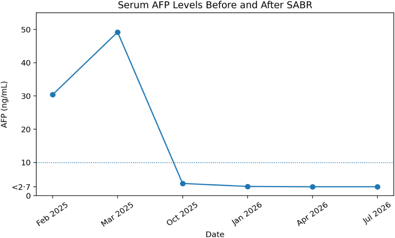
During surveillance, AFP increased from 30·4 to 49·2 ng/mL. Positron emission tomography–computed tomography (PET–CT) in March 2025 demonstrated a solitary intensely Fluorodeoxyglucose (FDG) avid 2·9 × 1·5 cm right retrocrural lymph node (SUVmax 15·9), with no other progression (Figure 1). He remained asymptomatic.
Pre-stereotactic radiation PETCT (A) and contrast-enhanced CT thorax abdomen and pelvis (CTTAP) (B) demonstrated a 2·9 × 1·5 cm intensely FDG avid retrocrural mass.

Figure 1. Long description
Panel A: A PETCT scan showing a cross-sectional view of a retrocrural mass. The image highlights areas of intense FDG uptake, indicated by bright spots. The scale on the left shows the intensity of the uptake, with a range from 0.00 to 10.00. Panel B: A contrast-enhanced CT scan of the thorax, abdomen, and pelvis, displaying a cross-sectional view of the same retrocrural mass. The mass is clearly visible and measured at 2.9 by 1.5 centimeters.
At multidisciplinary review, the node was considered unresectable because of proximity to major vascular and neural structures. Salvage chemotherapy was not advised, given the previous life-threatening neurological toxicity.
His disease was considered oligoprogressive, and SABR was proposed as a metastasis-directed treatment with curative intent, aiming to achieve durable local control while avoiding the substantial risks associated with further systemic therapy.
Treatment
A CT simulation was performed supine with stereotactic immobilisation using a customised vacuum cushion, indexed position, arms by the side and knees supported.
Four-dimensional CT assessed respiratory motion. An internal target volume (ITV) was generated from respiratory phases and expanded uniformly by 5 mm to create the planning target volume (PTV).
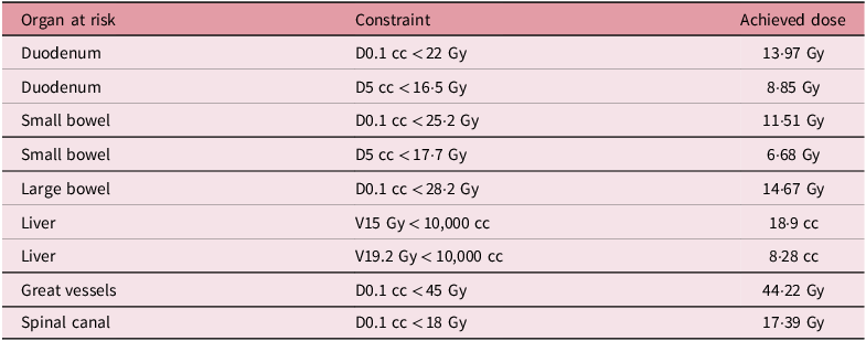
Given the proximity of tumour sites to the aorta, right kidney and bowel, 42 Gy in three fractions was prescribed using volumetric modulated arc therapy (VMAT) with inverse optimisation. Dose volume histogram analysis (Figure 2) demonstrated PTV D95 96%, D99 91·98% and Dmax 116·12%. All organs at risk constraints from the UK SABR Consortium guidance were met 5 ; (Table 1). There was no overlap with previous radiotherapy. Treatment was delivered on alternate days with daily cone beam CT guidance.
Dose volume constraints for surrounding organs at risk

Table 1. Long description
A table with three columns and eight rows. The columns are labeled Organ at risk, Constraint, and Achieved dose. The rows list different organs and their respective constraints and achieved doses. Row 1: Duodenum, D 0.1 cubic centimeters less than 22 Gray, 13.97 Gray. Row 2: Duodenum, D 5 cubic centimeters less than 16.5 Gray, 8.85 Gray. Row 3: Small bowel, D 0.1 cubic centimeters less than 25.2 Gray, 11.51 Gray. Row 4: Small bowel, D 5 cubic centimeters less than 17.7 Gray, 6.68 Gray. Row 5: Large bowel, D 0.1 cubic centimeters less than 28.2 Gray, 14.67 Gray. Row 6: Liver, V 15 Gray less than 10,000 cubic centimeters, 18.9 cubic centimeters. Row 7: Liver, V 19.2 Gray less than 10,000 cubic centimeters, 8.28 cubic centimeters. Row 8: Great vessels, D 0.1 cubic centimeters less than 45 Gray, 44.22 Gray. Row 9: Spinal canal, D 0.1 cubic centimeters less than 18 Gray, 17.39 Gray.
Follow-up and Outcomes
Treatment was completed without interruption or acute gastrointestinal toxicity. Grade 1 fatigue (CTCAE v5.0) resolved by 1 month.
Cross-sectional imaging at four months showed reduction of the retrocrural node from 2·9 × 1·3 cm to 1·2 × 0·9 cm (Figure 3). At 7 months it measured approximately 1 cm. Imaging at 12 and 15 months showed no residual disease.
Dose distribution from DMax 48·72 Gy to the prescribed dose of 42 Gy. (A) DMax to 50% achieved with SABR by volumetric modulated arc therapy, (B) low-dose spill to 50% to surrounding organs at risk, (C) PTV coverage from DMax to 95% prescribed dose and (D) DVH parameters. Yellow – GTV, red – PTV, light yellow – duodenum, green – kidneys, magenta – small bowel, gold – great vessels, brown – liver, green – stomach, orange – large bowel, purple – spinal canal.

Figure 2. Long description
Panel A: A cross-sectional image showing dose distribution achieved with SABR by volumetric modulated arc therapy. The image includes color-coded regions representing different dose levels, with yellow indicating GTV, red indicating PTV, and other colors representing surrounding organs. Panel B: A cross-sectional image illustrating low-dose spill to 50% to surrounding organs at risk. The image uses color coding to show the distribution of the dose, with specific colors representing different organs such as the duodenum, kidneys, small bowel, great vessels, liver, stomach, large bowel, and spinal canal. Panel C: A cross-sectional image showing PTV coverage from DMax to 95% prescribed dose. The image uses color coding to indicate the dose distribution and the coverage of the PTV. Panel D: A graph displaying DVH parameters. The graph shows dose-volume histograms for different organs and structures, with lines representing the dose distribution and volume coverage for each organ.
Contrast-enhanced CTTAP demonstrating a 2·9 × 1·3 cm retrocrural mass pre-SABR (A), a 1·2 × 0·9 cm mass 4 months post-SABR (B) and a stable 1 cm mass 7 months post-SABR (C).

AFP fell to 3·7 ng/mL at 4 months and 2·8 ng/mL at 7 months, remaining below 2·7 ng/mL at 12 and 15 months (Figure 4).
Serum alpha foetoprotein (AFP) levels before and after stereotactic ablative radiotherapy (SABR). AFP rose prior to treatment, peaking in March 2025, and declined following SABR (42 Gy in three fractions) delivered in April 2025, with sustained biochemical response through 15 months of follow-up. The dotted horizontal line represents the upper limit of normal AFP (10 ng/mL).

Discussion
Historically, radiotherapy has played a limited role in NSGCT because of effective platinum chemotherapy and perceived radioresistance. This perception largely derives from conventional fractionation, where dose escalation may be limited by normal tissue tolerance. SABR permits delivery of a substantially higher dose per fraction and tumour biologically effective dose while maintaining a steep dose gradient around the target. Reference Fowler6
SABR has challenged concepts of radioresistance, with high local control demonstrated in malignancies such as renal cell carcinoma, Reference Siva, Bressel and Sidhom7 suggesting that apparent resistance may partly reflect limitations of dose delivery, rather than intrinsic tumour biology.
Published experience of SABR in NSGCT is extremely limited, with this being the second reported case for nodal recurrences to our knowledge. Gulia et al. Reference Gulia, Anand and Punnakal8 reported local control 6 years after 45 Gy in six fractions for an inoperable chemorefractory retroperitoneal nodal relapse. Satyanarayan et al. Reference Satyanarayan, Mooney and Bhanvadia9 reported complete radiological and serological remission at 10 years following 54 Gy in three fractions to a chemorefractory pulmonary metastasis, with complete radiological and serological remission maintained at 10 years. These reports suggest that high-dose hypofractionated radiotherapy can achieve a meaningful control in selected germ cell tumours.
Our patient had an unresectable retrocrural metastasis and could not receive further chemotherapy because of severe neurological toxicity. SABR therefore provided a non-invasive local treatment option in a setting where conventional management was not feasible. The reduction and subsequent disappearance of the lesion, together with sustained AFP suppression at 15 months, provide concordant radiological and biochemical evidence of response. Treatment was also well tolerated, with no acute gastrointestinal toxicity, no treatment interruption and only a transient Grade 1 fatigue.
This case highlights a potential role for metastasis-directed treatment in oligoprogressive NSGCT, where the aim is to control a limited site of progression while avoiding or delaying a further systemic therapy. In carefully selected patients unable to tolerate chemotherapy, SABR may provide durable local control with low treatment-related morbidity.
Limitations include the single patient nature of this report and limited follow-up. Larger datasets are required to define patient selection and optimal dose fractionation, but this case adds to the limited evidence supporting SABR when standard NSGCT treatments are not feasible.
Author contributions
Niall O’Sullivan – conceptualisation, data collection, manuscript drafting.
Lorna Keenan – supervision, critical revision, final approval.
Financial support
No external funding was received for this study.
Competing interests
The authors declare no conflicts of interest.
Consent
Informed consent was obtained from the patient for publication of this case report and accompanying images.




 Open access

