Case report
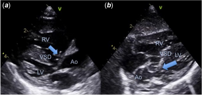
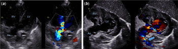
A 2-month-old female infant, diagnosed after birth with pulmonary atresia, ventricular septal defect, major aortopulmonary collateral arteries, and hypoplastic central pulmonary arteries (basal SpO2 80%), was admitted to the emergency department due to episodes of cyanosis and hypotonia while crying. Clinically, she had severe hypoxemia (SpO2 50%), poor peripheral perfusion, continuous murmur, and single second sound on cardiac auscultation. Her condition improved after fluid challenge and oxygen supplementation. The echocardiogram (Fig 1a and b) disclosed a small sub-aortic ventricular septal defect (4 mm) partially closed by tricuspid valve tissue, with exclusive right-to-left shunt (70 mmHg of interventricular gradient); no main pulmonary artery visualised; dilated and hypertrophic right ventricle; moderate tricuspid regurgitation (100 mmHg of gradient between the right ventricle and right atrium); interventricular septum bulging to the left side; accelerated flow through the left ventricular outflow tract (maximum velocity 2 m/second); and preserved systolic left ventricle function. On the third day of hospitalisation, she was transferred to the paediatric ICU due to hypoxemia and bradycardia while crying, which she recovered rapidly after a brief period of non-invasive positive pressure ventilation. A cardiac catheterisation was performed, revealing multiple major aortopulmonary collateral arteries (3–4 mm) for both lungs and hypoplastic pulmonary arteries (1.7 mm). On the following day, she suffered two cardiac arrests requiring short courses of cardiopulmonary resuscitation and the subsequent echocardiogram revealed a newly dome-like elongation of tissue dynamically moving alternatively into the right and left ventricles protruding through the ventricular septal defect into the left ventricular outflow tract leading to a flow acceleration with a maximum gradient of 94 mmHg and medium of 48 mmHg. Considering this, a direct aortopulmonary anastomosis (“Melbourne” shunt) was performed, as well as fixation of the septal leaflet of tricuspid tissue, which was passing through the ventricular septal defect and causing left ventricular outflow tract obstruction, to the right side of interventricular septum. The post-operative echocardiogram (Fig 2a and b) showed patency of the central aortopulmonary shunt with unrestrictive systo-diastolic flow (maximum/medium gradients 40/20 mmHg), hypertrophic but less dilated right ventricle, preserved left ventricle contractility, sub-aortic ventricular septal defect with right-to-left shunt (75 mmHg of interventricular gradient), but no tissue extension through the left ventricular outflow tract. No more events of bradycardia/cardiac arrest occurred, so the infant was discharged on acetylsalicylic acid, with SpO2 75–80% and no neurologic impairment. Future interventions are under discussion, depending on the growth of the pulmonary arteries.
Transthoracic echocardiogram of a 2-month-old infant with pulmonary atresia, ventricular septal defect and major aorto-pulmonary collateral arteries. ( a ) Parasternal long-axis view showing a dilated and hypertrophic right ventricle (RV), budging of the interventricular septum to the left side, small sub-aortic ventricular septal defect (VSD), tricuspid valve tissue protruding through the VSD into the left ventricle (LV) outflow tract (arrow); ( b ) subcostal long-axis view denoting dome-like elongation of tricuspid valve tissue beneath the aortic valve (Ao).

Postoperative transthoracic echocardiogram: Melbourne shunt and septal leaflet of TV fixation to the right side of interventricular septum. ( a ) Apical 5-chambers view showing turbulent right-to-left shunt through the sub-aortic ventricular septal defect and also turbulence in the left ventricle (LV) outflow tract and through the aortic valve (Ao).

Discussion
Pulmonary atresia with ventricular septal defect and major aortopulmonary collateral arteries is a rare CHD with a poor prognosis. Its management and treatment options heavily rely on the pulmonary blood flow pattern that enormously affects the surgical plan. Reference Fouilloux, Bonello and Kammache1
Dynamic obstruction of left ventricular outflow tract caused by aneurysmal tricuspid valve tissue protruding through the ventricular septal defect is an extremely atypical finding in the setting of this entity. We believed it resulted from suprasystemic right ventricle pressures created by the pulmonary atresia and restrictive right-to-left shunting through the small sub-aortic ventricular septal defect, pushing the tricuspid valve tissue to the left ventricular outflow tract. This dynamic aortic obstruction caused episodes of hypoxemia and bradycardia that ultimately precipitated several episodes of cardiac arrest. Fortunately, they were not associated with neurological impairment or other known complications. This finding is similar to the appearance of tricuspid valve tissue elongation and aneurysmatic tissue valve formation frequently seen in the context of a perimembranous ventricular septal defect that partially, or even totally, closes the defect.
This is, at our best knowledge, the first case of left ventricle outflow tract obstruction due to tricuspid valve tissue elongation in a patient with pulmonary atresia, ventricular septal defect, and major aortopulmonary collateral arteries described in the literature. In 1980, a group of Italian surgeons reported a similar case of a 2-year-old girl initially diagnosed with tetralogy of Fallot that progressed to acquire pulmonary atresia and restrictive ventricular septal defect during follow-up. At the age of 6 years old, she presented a severe sub-aortic stenosis that was found to be due to a dome-like elongation of the membranous septum and accessory tricuspid valve tissue protruding through the ventricular septal defect into the left ventricle outflow tract. Reference Di Benedetto, Crupi and Villani2
This case highlights a unique finding in the setting of pulmonary atresia, ventricular septal defect, and major aortopulmonary collateral arteries. Left ventricle outflow tract obstruction caused by tricuspid valve aneurysmatic tissue is a complication that has never been reported in the literature, although it can further interfere with the clinical management of this already troublesome and complex CHD.
Acknowledgements
None.
Financial support
This research received no specific grant from any funding agency, commercial or not-for-profit sectors.
Conflict of interest
None.
Ethical standards
The authors assert that all procedures contributing to this work comply with the ethical standards of the relevant national guidelines on human experimentation and with the Helsinki Declaration of 1975, as revised in 2008, and has been approved by the institutional committees.