Introduction
Swimmer's itch, also known as cercarial dermatitis, represents a common non-communicable waterborne cutaneous allergic disease (Kouřilová et al., Reference Kouřilová, Hogg, Kolářová and Mountford2004) which develops as a consequence of repeated infections by larval stages (cercariae) of schistosomatid flukes. Causative agents of the disease develop in water snails from which they escape and penetrate into the skin of mammals (including humans) or birds. Depending on the species, larval development takes place in snails of fresh water (Schistosoma, Bivitellobilharzia, Heterobilharzia, Schistosomatium, Bilharziella, Trichobilharzia, Dendritobilharzia, Gigantobilharzia) or salt/brackish water (Austrobilharzia, Ornithobilharzia, Gigantobilharzia) (Kolářová, Reference Kolářová2007). The genus Orientobilharzia, the life cycle of which is connected with mammals and freshwater snails, is probably invalid; phylogenetic analysis of rDNA of O. turkestanicum revealed that the species belongs to Schistosoma (Wang et al., Reference Wang, Ni, Zhai, Chen, Chen and Zhu2009). Larval stages and intermediate hosts of Jilinobilharzia, Macrobilharzia and Allobilharzia (Blair & Islam, Reference Blair and Islam1983; Kolářová et al., Reference Kolářová, Rudolfová, Hampl and Skírnisson2006) remain unknown.
Swimmer's itch is usually associated with swimming in recreational freshwater lakes, and mostly cercariae of avian schistosomes of the genus Trichobilharzia are reported as a source of infection. Bathing in the sea or brackish waters is rarely mentioned with respect to cercarial dermatitis, and only larval stages of Austrobilharzia spp. are known as the causative agent in South Africa, Australia and North America (Kolářová, Reference Kolářová2007). In Europe, furcocercariae morphologically related to Cercaria nassa (i.e. Gigantobilharzia larval stage) found in Nassa reticulata were probably the causative agent of dermatitis of humans collecting marine molluscs in Venice lagoon (Canestri-Trotti et al., Reference Canestri-Trotti, Fioravanti and Pampliglione2001).
Cercarial dermatitis of humans occurs all over the world (Horák et al., Reference Horák, Kolářová and Adema2002), the newest data come, for example, from Austria, Iceland, UK, The Netherlands, Iran, China, Chile and USA (Farahnak & Essalat, Reference Farahnak and Essalat2003; Hörweg et al., Reference Hörweg, Sattmann and Auer2006; Brant, Reference Brant2007; Schets et al., Reference Schets, Lodder, van Duynhoven and de Roda Husman2008, Reference Schets, Lodder and De Roda Husman2009; Valdovinos & Balboa, Reference Valdovinos and Balboa2008; Wang et al., Reference Wang, Chen, Zhano, Chen, Zhai, Li and Zhu2008; Fraser et al., Reference Fraser, Allan, Roworth, Smith and Holme2009; Skírnisson et al., Reference Skírnisson, Aldhoun and Kolářová2009). In moderate climates, cercarial dermatitis is most prevalent during warm summer months, when both the release of cercariae from snail hosts and the number of people that have contact with water reach peak levels (Chamot et al., Reference Chamot, Toscani and Rougemont1998). Under certain circumstances, the disease can be acquired during winter, e.g. after bathing in geothermally warmed lakes (Skírnisson & Kolářová, Reference Skírnisson and Kolářová2005). It is usually reported from lowland waters but the number of outbreaks from cold lake areas at higher latitudes is increasing at present (Silan et al., Reference Silan, Dubois and Halpenny2001; Larsen et al., Reference Larsen, Bresciani and Buchmann2004; Ferté et al., Reference Ferté, Depaquit, Cardo, Villena and Léger2005; Jouet et al., Reference Jouet, Ferté, Depaquit, Rudolfová, Latour, Zanella, Kaltenbach and Léger2008, Reference Jouet, Ferté, Hologne, Kaltenbach and Depaquit2009).
Although no details on the total number of afflicted persons are available, cercarial dermatitis has been discussed as an emerging disease (Chamot et al., Reference Chamot, Toscani and Rougemont1998). However, reflecting historical data and definition of emerging pathogens (Woolhouse & Dye, Reference Woolhouse and Dye2001), we should speak about a re-emerging disease, as suggested by Caumes et al. (Reference Caumes, Felder-Moinet, Couzigou, Darras-Joly, Latour and Léger2003). An emerging pathogen is defined as an infectious agent incidence of which is increasing after its first introduction into a new host population (Woolhouse & Dye, Reference Woolhouse and Dye2001), and this is not the case of schistosomes causing cercarial dermatitis at the present time.
Factors explaining the re-emergence of cercarial dermatitis are not fully known. High eutrophication of water reservoirs (Allgöwer & Effelsberg, Reference Allgöwer and Effelsberg1991; Valdovinos & Balboa, Reference Valdovinos and Balboa2008), colonization of ponds by susceptible snail species and nesting ducks, and long periods of sunshine in the summer are certainly important factors that have led to an increase in the number of outbreaks of cercarial dermatitis (de Gentile et al., Reference de Gentile, Picot, Bourdeau, Bardet, Kerjan, Piriou, Le Guennic, Bayssade-Dufour, Chabasse and Mott1996). Global warming, influencing behaviour of final hosts, reproduction of intermediate hosts and development of schistosomes in a particular ecosystem, seems to be the most important factor. It has been observed that some species/populations of European waterfowl, the most important definitive hosts of Trichobilharzia flukes, have ceased to migrate between nesting and wintering areas and have become resident in Central European lakes (Caumes et al., Reference Caumes, Felder-Moinet, Couzigou, Darras-Joly, Latour and Léger2003). These behavioural changes may facilitate the transmission of schistosomes, leading to a higher prevalence of larval stages in snails, a higher number of generations of intermediate hosts and schistosomes and, consequently, to a higher density of cercariae in water bodies (Mas-Coma et al., Reference Mas-Coma, Valero and Bargues2008, Reference Mas-Coma, Valero and Bargues2009).
The attack of human skin by schistosome cercariae is facilitated by a native ability of schistosome cercariae to respond to specific physical and chemical cues of the skin of both birds and humans; this similarity of attractive factors allows bird schistosome larvae to penetrate into the skin (Haas, Reference Haas2003) and cause cercarial dermatitis.
Human infections by avian schistosomes are usually associated with skin symptoms only, and it was assumed that parasites die soon after the penetration. However, the studies that focused on the course of Trichobilharzia infection in rodents revealed that these schistosomes can escape from the skin and migrate further through the mammalian body (Horák et al., Reference Horák, Mikeš, Rudolfová and Kolářová2008). Similar to the situation in experimental rodents, human infections by bird schistosomes may, under certain circumstances, be linked to more than just cercarial dermatitis (Bayssade-Dufour et al., Reference Bayssade-Dufour, Martins and Vuong2001).
Frequently, natural lakes as well as man-made water reservoirs (e.g. ponds, flooded sand-pits, water dams) with the occurrence of bird schistosomes are primarily mentioned in reports on outbreaks of cercarial dermatitis during recreational seasons. However, a risk of infection by the parasites can be predicted before the season: presence of both water snails and nesting waterfowl (but also the transitory presence of other birds) may indicate potential occurrence of schistosomes in a certain area. In addition to examination of water samples, a search for the parasites in snails and birds can be performed during the whole year and findings of schistosomes may further lead to application of selected control measures against cercarial dermatitis. However, in comparison with the situation in the Czech Republic, France, Iceland and the USA (Rudolfová et al., Reference Rudolfová, Littlewood, Sitko and Horák2007; Brant & Loker, Reference Brant and Loker2009a; Jouet et al., Reference Jouet, Ferté, Hologne, Kaltenbach and Depaquit2009; Skírnisson et al., Reference Skírnisson, Aldhoun and Kolářová2009) data on the occurrence of animal schistosomes causing cercarial dermatitis are accidental and incomplete at present, e.g. either larvae from snails or adults from vertebrate hosts are known in particular areas. The lack of data about the distribution of particular schistosome species is caused partly by difficulties with detection and identification of the parasites in intermediate as well as final hosts. Based on personal experience and information in the literature we, therefore, summarize in this paper methodical approaches enabling detection of bird schistosomes in their hosts as well as in the environment. As a consequence of such an examination, the areas representing risk of infection by bird schistosomes might be better identified and relevant protective measures in control of cercarial dermatitis applied.
Search for schistosome cercariae
Collection of snails
It is generally believed that bird schistosomes exhibit a degree of specificity towards their intermediate hosts, parasitizing closely related species of snails. In Europe, Trichobilharzia spp. larval development takes place mostly in snails of the family Lymnaeidae, whereas in North America physids, planorbids and lymnaeids are mostly used as intermediate hosts of the flukes (e.g. Brant & Loker, Reference Brant and Loker2009a, Reference Brant and Lokerb). Besides lymnaeid snails, European schistosome cercariae (not only Trichobilharzia spp.) were also found in Gyraulus parvus in Austria (Obwaller et al., Reference Obwaller, Sattmann, Konecny, Hörweg, Auer and Aspöck2001), Anisus vortex, Gyraulus albus and Segmentina nitida in the Czech Republic (Rudolfová, Reference Rudolfová2001), and Valvata (Tropidina) microstoma in Finland (Aldhoun et al., Reference Aldhoun, Faltýnková, Karvonen and Horák2009a). Bilhaziella polonica from Europe is frequently found in Planorbarius corneus; however, similar to Uzbekistan (Shakarbaev & Azimov, Reference Shakarbaev and Azimov2001), cercariae can also develop in Planorbis planorbis and A. septemgyratus (Rudolfová, Reference Rudolfová2001). Except for Schistosoma bovis found in Corsica, Sardinia, Sicily and Spain (Moné et al., Reference Moné, Mouahid and Morand1999), there are no data about the detection of mammalian schistosomes in Europe. Cercariae morphologically similar to Heterobilharzia americana, a species maturing in carnivores in North America, were found in Aplexa hypnorum in France (Gérard, Reference Gérard2004). According to the author's opinion, these cercariae belong to a different species of an unknown genus. However, the study was performed only with larval stages (cercariae), the morphology of which has a disputable/limited value for taxonomical purposes.
Water snails are usually collected in summer and early autumn but the parasites survive in hibernating snails (McMullen & Beaver, Reference McMullen and Beaver1945) in which they can be detected also during winter. The abundance of snails, their distribution and proportion of infected individuals depend on biotic (e.g. preference for different types of aquatic vegetation, immunological susceptibility and physiological suitability of particular snail species, distribution and behaviour of definitive hosts) and abiotic (e.g. water temperature, pH and chemical composition) factors (Webbe, Reference Webbe, Jordan and Webbe1982; Sturrock, 1993; Mas-Coma et al., Reference Mas-Coma, Valero and Bargues2008). The prepatent period within snails varies (about 3–5 weeks) and it appears that lower water temperature may prolong the life of snails and their parasites, and the total number of cercariae released from molluscs can be inversely proportional to temperature (Zbikowska, Reference Zbikowska2001).
Although outbreaks of swimmer's itch can be recorded repeatedly in a lake, the following examination of molluscs can bring negative results (Bei et al., Reference Bei, Salter, Lim and McKerrow2001). This can be explained partly by a low prevalence of bird schistosomes in snails. Trichobilharzia infection rates in European lymnaeids usually ranges between 0.3 and 5.2% (Loy & Haas, Reference Loy and Haas2001), and only for Radix auricularia may the prevalence exceed 5%. Therefore, examination of a low number of snails may produce false results (Loy & Haas, Reference Loy and Haas2001). In the past, a higher prevalence of infection was seldom recorded, e.g. 22 and 26% for R. auricularia in Bohemia and Germany, respectively (Kolářová et al., Reference Kolářová, Gottwaldová, Čechová and Ševcová1989; Müller & Kimmig, Reference Müller and Kimmig1994). Recently, the number of reports of a high prevalence in snails seems to have increased; in Iceland and Chile, 24.5% prevalence in R. peregra and 52.4% in Chilina dombeyana, respectively, have been detected (Valdovinos & Balboa, Reference Valdovinos and Balboa2008; Skírnisson et al., Reference Skírnisson, Aldhoun and Kolářová2009). These high values can result from disturbances of the ecological balance of lakes, including accelerated eutrophication which can lead to an increase in the abundance of susceptible snails in a particular water body (Kolářová et al., Reference Kolářová, Gottwaldová, Čechová and Ševcová1989; Leighton et al., Reference Leighton, Zervos and Webster2000; Valdovinos & Balboa, Reference Valdovinos and Balboa2008).
Detection of cercariae in snails
The exclusive use of methods focused on examination of snails shedding cercariae can produce false results on parasite prevalence, because it ignores developing larval stages which are not shed at the time of examination. The difference given by false results may reach 59.1% (Curtis & Hubbard, Reference Curtis and Hubbard1990) and, therefore, microscopical examination of snail organs (hepatopancreas) is more accurate.
Storage of naturally infected snails in the laboratory for a long period is one of the important tasks for further study of cercariae. It is optimal to keep snails at an appropriate temperature in water originating from the locality where they were collected. Some snails, such as Lymnaea stagnalis infected by Trichobilharzia sp. or P. corneus by B. polonica, can be kept in the dark and at low temperatures (e.g. in a refrigerator at 4°C) which induces retardation of both larval development and pathogenic processes. Then cercariae can be collected and analysed during the entire period of prolonged maintenance. In order to get the maximum number of cercariae, their release from snails exposed to room temperature for at least 1 h can be stimulated by a lamp, preferably at the appropriate time for a particular species, e.g. Trichobilharzia szidati cercariae are usually shed from snails in high quantities in the morning (Neuhaus, Reference Neuhaus1952). Long-lasting storage of naturally infected snails under laboratory conditions can result in the ‘disappearance’ of schistosome cercariae; this situation happens if the snails are simultaneously infected by other trematode species which may be dominant over schistosomes (Lim & Heyneman, Reference Lim and Heyneman1972).
Cercarial life span seems to be different for various schistosome species. Whereas Trichobilharzia cercariae can survive for 2 days at lower temperatures, larvae of B. polonica have a short life (a few hours). At present there are few data comparing the age of bird schistosome cercariae with their penetration ability and subsequent development in the skin and body of the host. Studies on S. mansoni showed that the ability of cercariae to penetrate the tails of mice remained constant throughout their life in an aqueous environment, but their capacity to establish themselves and reach maturity decreased as they aged (Lawson & Wilson, Reference Lawson and Wilson1983). It seems that the same situation may happen with infections by bird schistosomes and, therefore, it is strongly recommended that only freshly released cercariae are used.
Detection of cercariae in water samples
Detection of cercariae in water samples can be difficult due to the low parasite concentration. Various methods (filtration, use of phototactic response equipment and continuous flow centrifugation) which have been developed to detect human schistosomes (Théron, Reference Théron1986) may also be applied in the search for bird schistosomes. Graczyk & Shiff (Reference Graczyk and Shiff2000) proposed using a trap containing a matrix with unsaturated fatty acids (linoleic acid) which stimulate penetration of cercariae. Subsequently the immobilized and collected larvae can be visualized for counting. To detect pathogenic cercariae causing swimmer's itch, Linder et al. (Reference Linder, Thors and Jacks2001) proposed using a matrix with skin lipids on which the parasites deposit water-insoluble glyco-substances originating from penetration glands; these can subsequently be visualized specifically, e.g. with fluorochrome-labelled lectins or antibodies. However, the method cannot provide any valuable data on density of cercariae in a particular water body. On the other hand, high sensitivity and specificity of detection of schistosome cercariae in the aqueous environment can be achieved by molecular approaches. Polymerase chain reaction (PCR) assay based on a tandem repeated DNA sequence of T. ocellata ( = T. szidati) (Rudolfová et al., Reference Rudolfová, Hampl, Bayssade-Dufour, Lockyer, Littlewood and Horák2005) allows detection of a single cercaria of T. ocellata, T. franki and T. regenti in plankton (0.5 g) and snail tissues (0.25 g) (Hertel et al., Reference Hertel, Hamburger, Haberl and Haas2002).
Identification of cercariae
Morphological characteristics enable a relatively easy group identification of known European bird schistosome cercariae which are characterized as brevifurcate larvae with well-developed eyespots – they are known as ocellate furcocercariae. However, cercariae of different species and/or genera are very similar and thus exact taxonomical determination under the microscope is often impossible (Rudolfová et al., Reference Rudolfová, Hampl, Bayssade-Dufour, Lockyer, Littlewood and Horák2005). Dimensions of the parasites of different species do not represent a useful criterion for identification because intra-specific variability often exceeds inter-specific variability. Recent data of Podhorský et al. (Reference Podhorský, Hůzová, Mikeš and Horák2009) showed that chaetotaxy, a technique using silver nitrate for staining sensory papillae (Richard, Reference Richard1971), remains a promising way for discrimination of particular species, although some papillae do not stain sufficiently. Precise identification requires molecular techniques (e.g. Dvořák et al., Reference Dvořák, Vaňáčová, Hampl, Flegr and Horák2002; Aldhoun et al., Reference Aldhoun, Kolářová, Horák and Skírnisson2009b; Brant & Loker, Reference Brant and Loker2009b; Jouet et al., Reference Jouet, Ferté, Hologne, Kaltenbach and Depaquit2009) and relevant sequences of bird schistosomes are available in GenBank. Contrary to these modern approaches, a valid description of a new schistosome species also requires data on the morphology of adult flukes and their location in birds (see Experimental infections of birds, below).
Search for adult schistosomes in birds
Examination of birds, detection and fixation of adult flukes
Birds can be examined throughout the year. Valuable results can be obtained by examination of birds at necropsy collected during the hunting season, i.e. in the autumn and winter. In comparison with snails, the prevalence in birds seems to be quite high. A survey of schistosomes in mallards hunted in winter showed a 75% and 33% prevalence of Trichobilharzia sp. flukes in the French Lake of Annecy and the Lake Der-Chantecoq, respectively (Kolářová et al., Reference Kolářová, Skírnisson, Rudolfová, Jouet, Léger and Ferté2005). Skírnisson et al. (Reference Skírnisson, Aldhoun and Kolářová2009) reported a 75% prevalence of the parasites in mallards in Iceland.
The search for avian schistosomes in birds is commonly focused on the examination of bird faeces; this technique permits the detection of visceral schistosomes but fails to show the presence of nasal flukes. To identify infection by nasal schistosomes, fresh lavage of the nasal cavity can be examined for eggs or miracidia; the samples can be obtained by rinsing out the nasal cavity with saline or water (Horák et al., Reference Horák, Kolářová and Adema2002). In the case of faecal examination, the number of eggs is usually very low and, therefore, it is necessary to examine whole bird excrements (Appleton, Reference Appleton1986). To increase the probability of egg detection, sedimentation methods (e.g. formol–ether technique) or Kato–Katz technique (Appleton, Reference Appleton1986; Kassai, Reference Kassai1999) are recommended. Helminthological examination of birds at necropsy represents the method of choice; freshly killed birds as well as frozen ones can be examined. This approach offers more relevant information on helminth infections and their pathological consequences.
In birds, all stages of developing schistosomes damage the affected organs. Visceral species cause pathologies similar to those induced by S. mansoni in mammals (Horák et al., Reference Horák, Kolářová and Adema2002; van Bolhuis et al., Reference van Bolhuis, Rijks, Dorrestein, Rudolfová, van Dijk and Kuiken2004). Nasal T. regenti induces severe inflammation in various parts of the avian central nervous system (CNS) and the nasal cavity (Kolářová et al., Reference Kolářová, Horák and Čada2001).
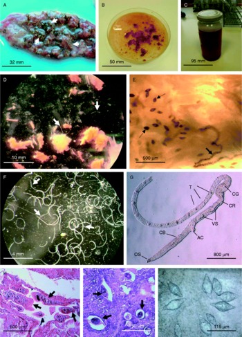
We recommend examining mainly the blood system and surrounding tissues of the preferred organs; visceral flukes and eggs are mostly found in the intestinal wall, mesentery veins, liver, lungs and the heart. In the case of severe infection, attention should be paid to the inflammatory lesions (fig. 1A) where schistosomes can be detected with high probability. Nasal worms can be recovered from the nasal tissues as well as various parts of the CNS (Horák et al., Reference Horák, Dvořák, Kolářová and Trefil1999; Hrádková & Horák, Reference Hrádková and Horák2002). Heavy and progressing infection can be characterized by ectopic location of parasites and eggs; e.g. adults of visceral Trichobilharzia can invade bile ducts, their eggs can be disseminated to the CNS. The evaluation of results can be complicated in birds simultaneously infected by different species of schistosomes.
Investigation of birds for schistosomes. A, Inflammatory lesions (arrows) caused by Trichobilharzia eggs in the large intestine of a mallard. B, Adult schistosomes are freed from the blood vessels by tearing the affected organ to small pieces which are, shaken further in saline (C). D, Thin and long fragments of thread-like schistosomes (arrows) released from the pieces of tissues. E, Part of the infected tissue with eggs (dotted arrows) and adult flukes (solid arrow) compressed between two microscope slides. F, Isolated fragments of bird schistosomes; arrows point at canalis gynaecophorus of males. G, The morphology of the anterior end of Trichobilharzia sp. adult male: oral sucker (OS), caecal bifurcation (CB), acetabulum (AC), vesicula seminalis externa and interna (VS), caeca reunion (CR), canalis gynaecophorus (CG) and testes (T). Nomarski interference contrast. H, Adult schistosomes (arrows) and (I) eggs (arrows) on histological mounts. Periodic acid–Schiff. J, Schistosome eggs in scrapings of intestinal mucosa compressed between two microscope slides.

Eggs of visceral schistosomes can be detected in scrapings of the intestinal mucosa; fresh pieces of the tissue should be compressed between two slides and immediately examined (fig. 1J). To obtain the maximum number of parasites, the whole organ needs to be sliced or torn with pinsetters down to small pieces (fig. 1B) in saline, because adult flukes located in the bloodstream should be freed from vessels. Fresh as well as thawed pieces can also be compressed between two slides and examined microscopically. Parasites and their eggs can be detected under low magnification; the detection of adult worms is facilitated by their dark intestine which is filled with digested blood (fig. 1E). Remaining pieces should be immersed in saline and shaken by hand in a jar with a lid (fig. 1C) or using a shaker. The suspension should be allowed to settle for at least 30 min, then the supernatant is poured off and the sediment again washed in saline. This procedure should be repeated at least three times.
Examination of the sediment under the stereomicroscope may reveal worms and eggs. Usually, only thin and long fragments of thread-like worms (fig. 1D and F) are isolated. Further morphological characterization of worms depends mainly on examination of the anterior body parts of both sexes; i.e. in males the most valuable part ends behind canalis gynaecophorus (fig. 1G), in females behind the receptaculum seminis. The observation can be performed microscopically with fresh material (preferably using Nomarski interference contrast, fig. 1G) or on fixed and stained worms. Isolated worms are usually fixed in 70% ethanol or 4% formaldehyde for morphological studies, but in 90% ethanol for DNA analysis; staining by, for example, borax-carmine or Gram Weigert and, finally, mounting in Canada balsam or solacryl.
Biopsies can be fixed in 70% ethanol or 4% formaldehyde and then prepared for further histological examination. Usually, staining by haematoxylin–eosin is performed but the detection of worms can be facilitated by staining with periodic acid–Schiff (fig. 1H and I).
Infections by schistosomes can also be diagnosed by indirect techniques, i.e. by serological detection of antibodies against the parasite antigens. Kouřilová & Kolářová (Reference Kouřilová and Kolářová2002) showed that antibodies against gut-associated antigens of adult flukes can be detected in the sera of ducklings harbouring T. regenti infection. Various serological techniques which have been used to assess specific antibody responses against bird schistosomes in sera of humans with cercarial dermatitis (Kolářová et al., Reference Kolářová, Sýkora and Bah1994) can also be applied for diagnosis of the infection in birds, but these methods are not species/genus specific. Nevertheless, recent data showed that detection of antibodies against the 34 kDa antigen of T. regenti cercariae might be promising in differential diagnosis of human cercarial dermatitis (Lichtenbergová et al., Reference Lichtenbergová, Kolbeková, Kouřilová, Kašný, Mikeš, Haas, Schramm, Horák, Kolářová and Mountford2008), and, hypothetically, this antigen might also be used for examination of birds.
Identification of adult schistosomes
Taxonomical determination is based on the evaluation of all available data, i.e. host species, location and morphological characters of the worms and their eggs. Unfortunately, morphological characterization is a complicated matter and, therefore, additional techniques can help to identify species. Cytogenetic study (karyology) has been used for 11 avian schistosome species (A. variglandis, B. polonica, Gigantobilharzia huronensis, Ornithobilharzia canaliculata and seven Trichobilharzia species), and some intergeneric/interspecific differences were found (Špakulová et al., Reference Špakulová, Horák and Müller1997, Reference Špakulová, Horák and Dvořák2001). Recently, the validity of species and phylogenetic relationships have been studied by sequencing rDNA and mtDNA (e.g. Webster et al., 2007; Brant & Loker, Reference Brant and Loker2009b). Based on comparison of T. szidati, T. regenti and T. franki, it has been concluded that ITS1 and ITS2 sequences can be useful for species identification (Dvořák et al., Reference Dvořák, Vaňáčová, Hampl, Flegr and Horák2002).
In the case where only schistosome eggs are found, exact determination should be based on morphological characteristics as well as DNA analysis of the material. When viable eggs (i.e. with living miracidia) are found, further experimental infections of laboratory-reared snails of relevant species can be realized (Horák et al., Reference Horák, Kolářová and Adema2002). In case of susceptible/suitable snails, cercariae of schistosomes are produced after several weeks and these can serve for experimental infection of appropriate birds (see below) in which the parasites mature. The requirement for suitable intermediate and final hosts represents a disadvantage of such infection experiments.
Experimental infections of birds
Also, experimental infections of ducks (Anas platyrhynchos f. dom., Cairina moschata f. dom.) may help with identification of bird schistosomes. The feet of 1-week-old ducklings can be exposed (usually for 1 h) to cercariae released from naturally infected snails (Meuleman et al., Reference Meuleman, Huyer and Mooij1984). After 2–3 weeks (the beginning of patent infection), the target organs/tissues of the infected ducklings (visceral or nasal) should be examined for the presence of adult worms and/or eggs (Kolářová et al., Reference Kolářová, Horák and Čada2001; Horák et al., Reference Horák, Kolářová and Adema2002; Chanová & Horák, Reference Chanová and Horák2007); eggs and miracidia can also be found in faeces and nasal lavage. In addition, in the prepatent phase of infection, migrating schistosomula can be detected in different species-specific organs/tissues, such as the spinal cord and the lungs (Horák et al., Reference Horák, Dvořák, Kolářová and Trefil1999; Horák & Kolářová, Reference Horák and Kolářová2000; Blažová & Horák, Reference Blažová and Horák2005). Obviously the results of examination may be negative, because the above species of waterfowl are not necessarily the best (i.e. suitable) final hosts.
Conclusion
The association between avian schistosomes and cercarial dermatitis has been known for many decades. The parasites can be collected and further determined in water samples, and from snails and birds, using different methods, including molecular techniques. Description of new species of schistosomes is a complicated matter which requires a multidisciplinary approach (morphological characterization, infection experiments, analysis of parasite behaviour, histopathological evaluation, molecular analyses, etc.). Methods used for the above-mentioned observations need to be standardized in order to allow comparison of data from different laboratories and areas. Successful isolation and characterization of bird schistosomes from snails, birds and water bodies may contribute to better understanding of parasite transmission and life cycles, and lead to a better control of the agent, including diagnosis, prevention and treatment of bird and human infections.
Acknowledgements
This study was partially supported by the Czech Science Foundation 206/09/0926; Czech Ministry of Education MSM 0021620828, MSM LC06009, MSM 0021620806, MSM 0021620812 and the Research Fund of the University of Iceland.