INTRODUCTION
The basic structure of the body wall of parasitic nematodes consists of the cuticle, an underlying syncytial or cellular layer, called the hypodermis, and the longitudinally oriented somatic musculature. The entire cuticle is shed at each of 4 moults and is replaced with a new cuticle at each stage of the life-cycle of all nematodes. Moulting proceeds in 3 major steps; (i) the old cuticle separates from the hypodermis (apolysis), (ii) components of the new cuticle are synthesized in the hypodermis and are secreted across the hypodermal membrane into the space between it and the old cuticle, and (iii) the old cuticle is ruptured and shed (ecdysis) (Lee, 2002). In parasitic nematodes, moulting is a crucial process essential for subsequent development and maturity, but very little is known about the underlying mechanisms regulating this complex biological process at the molecular level (Boag et al. 2001; Nisbet et al. 2004). Although a few studies have confirmed the role of transglutaminase and soluble inorganic pyrophosphatase in the moulting processes of parasitic nematodes (Lustigman et al. 1995; Chandrashekar and Mehta, 2000; Islam et al. 2005) and of a cathepsin Z-like cysteine protease in the free-living nematode, Caenorhabditis elegans (see Hashmi et al. 2003), no study has yet focused on changes in protein expression (profiles) associated with moulting and development processes of human and animal parasitic nematodes. Investigations into the moulting and development-specific protein expression profiles through establishing simple in vitro moulting assays of pre-parasitic or parasitic larvae have enormous implications for understanding the molecular mechanisms of moulting and development processes of parasitic roundworms.
The anion, fluoride, is known to play an important role in the reduction of dental caries and has been added to drinking water and toothpastes as an anticaries agent. Sodium fluoride (NaF) was also used as a drug against A. suum (see Soulsby, 1968). However, it was completely replaced with modern anthelmintics. Studies have shown that NaF can reduce the expression of proteins and virulence factors in microbes (Sutton et al. 1987; Thongboonkerd et al. 2002). In addition to its ability to inhibit microbial proteins, NaF is also known to affect proton-translocating pyrophosphatases from plants (Rea and Poole, 1993), trypanosomatids (Scott et al. 1998) and apicomplexan protozoa (Marchesini et al. 2000). In agreement with these observations, we also have found that NaF can inhibit a novel Family I-soluble inorganic pyrophosphatase in A. suum (AsPPase) (Islam et al. 2003). Furthermore, we have found that NaF is able to inhibit endogenous AsPPase activity in vitro and block the moulting process of lung-stage larvae of A. suum at micromolar and millimolar concentrations, respectively. We hypothesized that a complete moulting blockade in the lung-stage larvae induced by NaF in vitro is preceded by a partial and/or complete inhibition of the synthesis of a wide range of proteins potentially involved in the moulting and development processes of the parasites. Therefore, we conducted this proteomics study to gain a better insight into the proteins involved in these processes in aerobic third-stage larvae (L3) to anaerobic fourth-stage larvae (L4) of Ascaris parasites using NaF as a potent inhibitor. A. suum L3s obtained from an infected rabbit's lungs (designated AsLL3) were cultured in the presence of NaF to block moulting and its absence to further allow moulting to L4 (AsLL4). The soluble proteins in whole larval extracts were subjected to analysis by two-dimensional electrophoresis (2D–PAGE), a powerful and sensitive tool for detecting and separating proteins (O'Farrell et al. 1977; Barrett et al. 2000). In addition, the potential of NaF to inhibit antigenic proteins and endogenous AsPPase activity/expression in the developing larvae, as well as the ultrastructural damage caused by NaF, were studied.
MATERIALS AND METHODS
Parasites
Adults of A. suum were collected from the small intestine/duodenum of infected pigs at a slaughter house in Tsuchiura, Japan. Eggs of A. suum were obtained after the in vitro culture of female and male worms and embryonated at 25 °C as described previously (Tsuji et al. 2000a). The AsLL3s were collected from the lungs of New Zealand white rabbits 7 days after infection with 1·5×105 embryonated, infective eggs of A. suum (see Islam et al. 2003). The larvae recovered were washed several times in phosphate-buffered saline (PBS) (37 °C) containing 100 μg/ml each of penicillin and streptomycin. Then, 200 AsLL3s per ml of RPMI 1640 medium (GIBCO/BRL) supplemented with 10% foetal bovine serum (Sigma) and 50 μg/ml each of penicillin and streptomycin were cultured in a 70 ml Falcon® polystyrene tissue culture flask (3082 blue plug seal cap, Beckton Dickinson and Company, NJ, USA). The cultures were incubated for 10 days at 37 °C and 5% CO2 in the presence or absence of 2 mM NaF. Animal experiments were performed in accordance with the protocols approved by the National Institute of Animal Health (NIAH) Animal Care and Use Committee (Approval no. 589).
Preparation of protein samples
Ascaris larvae grown (for 10 days) in vitro were removed from the culture, washed in cool PBS (0 °C) and homogenized under liquid nitrogen (Tsuji and Fujisaki, 1994). The larval homogenate was dissolved in PBS containing a protease inhibitor cocktail (Cat. no. 1836153, Roche Diagnostics GmbH, Penzberg, Germany) and sonicated on ice. To measure enzyme activity, larval homogenates were prepared in 20 mM Tris–HCl buffer (pH 7·5) without the protease inhibitor. The homogenate was centrifuged at 26300 g for 30 min, and the supernatant fractions were collected and frozen at −80 °C until use. Protein concentrations were measured using Micro-BCA protein assay reagent (Pierce, Rockford, IL).
Two-dimensional gel electrophoresis
Protein samples were mixed with an equal volume of urea mixture containing 9 M urea, 4% Nonidet P-40, 0·8% ampholine (pH 3·5–10; Pharmacia, Sweden), and 2% 2-mercaptoethanol and then subjected to 2D–PAGE. Non-equilibrium pH gradient electrophoresis (NEPHGE) was performed (O'Farrell et al. 1977) in the first dimension using a rectangular gel electrophoresis apparatus (AE-6050A, ATTO, Tokyo, Japan). After electrophoresis at 400 V for 2 h, gels were incubated in the equilibration buffer (0·125 M Tris–HCl, pH 6·8, 10% glycerol, 2% sodium dodecyl-sulfate, 0·05% 2-mercaptoethanol and 0·0015% bromophenol blue) for 10 min with gentle shaking (Tsuji et al. 2000b). Electrophoresis in the second dimension was performed on 12·5% SDS-PAGE gels under reducing conditions. The proteins were stained using a silver staining kit (Dai-ichi Pure Chemicals, Tokyo, Japan) or transferred to 0·2 μm nitrocellulose membranes (Schleicher & Schuell, Dassel, Germany) for immunoblotting. Molecular weight and isoelectric point (pI) values of proteins were estimated using 2D standards (Bio-Rad, Hercules, CA). The major protein spots resolved on 2D gels prepared from NaF-treated, unmoulted (AsLL3) and moulted larvae (AsLL4) (control) were compared. Protein spots were examined visually on an illuminated screen. At least 10 independent runs of each category of sample were carried out for the optimization of the resolution of protein spots (Islam et al. 2004).
Production of immune sera
Antibodies to A. suum were generated in specific pathogen-free rabbits by repeated oral inoculation with embryonated, infective eggs (Tsuji et al. 2001). The rabbits were bled 7 days after the final inoculation, and the sera were stored at −20 °C until use. The corresponding pre-immune serum was used as a control (at the same dilution as the antiserum). Antibodies to recombinant AsPPase (rAsPPase) were produced in BALB/c mice as described previously (Islam et al. 2003). Briefly, 50 μg of rAsPPase emulsified with the adjuvant TiterMax Gold™ (CytRx, Atlanta Norcross, GE) were injected subcutaneously, followed by another injection 2 weeks later using the same adjuvant. The mice were bled 2 weeks after the second injection, and the sera were collected and stored at −20 °C until use.
Enzyme assay
The endogenous AsPPase enzyme activity in NaF-exposed unmoulted and moulted (control) A. suum lung-stage larval extracts prepared in 20 mM Tris–HCl, pH 7·5 were determined essentially as described previously (Islam et al. 2003). Briefly, the larval extracts were assayed in the standard reaction mixture containing 5 mM Mg2+, 100 mM Tris–HCl (pH 7·5) and 1 mM pyrophosphate (PPi) (Na4P2O7) in a total volume of 200 μl (together with the protein solution) at 55 °C. The assay was conducted by adding 10 μl of larval extracts into the standard reaction mixture, and the reaction was stopped by addition of 1 ml of 200 mM glycine–HCl, pH 3·0. Then, 125 μl of 1% ammonium molybdate (in 25 mM H2SO4) and 125 μl of 1% ascorbic acid (in 0·05% KHSO4) were added to the mixture and incubated for 30 min at 37 °C. The amount of ortho-phosphate (Pi) liberated from the hydrolysis of PPi during the course of reaction was measured in comparison with a standard Pi sample using a spectrophotometer (Model 600, Shimadzu, Tokyo, Japan) at an optical density of 700 nm. The specific activity of PPase enzyme was defined as μmol Pi released per min per mg of protein.
Two-dimensional immunoblot analysis
This analysis was performed as described by Tsuji et al. (2001). The proteins separated by 2D–PAGE were transferred to nitrocellulose membranes, blocked with 5% (w/v) skim milk for 30 min and washed in Tris–buffered saline containing 0·1% (v/v) Tween 20 (TBS-T). The membranes were incubated overnight with rabbit (1[ratio ]250) or mouse serum (1[ratio ]1000). After washing 4 times with TBS-T, the membranes were incubated for 1 h with alkaline phosphatase-conjugated goat anti-rabbit/anti-mouse IgG (ICN Biomedicals, Aurora, OH) at a dilution of 1[ratio ]2500. After washing, the proteins bound to the secondary antibody were detected using 5-bromo-4-chloro-3-indolylphosphate and nitroblue tetrazolium (NBT/BCIP, Promega, Madison, WI).
Immunofluorescence studies
Lung-stage L3s cultured in vitro under an exposure to NaF were washed 3 times in cool PBS (4 °C) containing 0·1% (v/v) Tween 20 (PBS-T) and fixed in cool acetone for 2 min. After washing in PBS-T, the fixed larvae were blocked in 10% (v/v) goat serum (Wako) for 1 h and incubated with primary anti-rAsPPase antibody (1[ratio ]50) overnight at 4 °C. Larvae grown and moulted in the absence of NaF served as the control. Fluorescein isothiocyanate (FITC)-conjugated goat anti-mouse IgG (Sigma) was used as a secondary antibody (1[ratio ]100). After labelling, the larvae were washed in PBS-T, and whole larvae were mounted on slides with 70% glycerol. The larvae were analysed and photographed using a fluorescence microscope (Leica IM500) employing appropriate filters.
Light- and transmission electron-microscopy
To evaluate the effects of NaF on the cuticle and internal organs of developing AsLL3, cultures were examined daily using a light microscope from days 0 to 10. Lung-stage L3s which did not moult and showed damage to the internal organs in the presence of 2 mM NaF were photographed using a digital camera coupled to a microscope (Leica IM500). After the culture had terminated on day 10, larvae were removed and washed in PBS-T and fixed for 1 h at 4 °C with 3% (v/v) glutaraldehyde in 0·1 M phosphate buffer (pH 7·4), washed in the same buffer, and processed for examination by transmission electron microscopy (TEM) (Islam et al. 2005).
Internal amino acid sequencing
One protein spot that showed sensitivity to inhibition by NaF (i.e. whose activity and/or expression was affected by NaF) and showed an increased expression during moulting from AsLL3 to AsLL4 was excised and analysed for internal amino acid residues, in order to determine its putative biological functions in the moulting and development processes of A. suum larvae by peptide mass spectrometric and bioinformatic approaches. The protein spots excised from 2D gels stained with Coomassie Brilliant Blue R250 (You et al. 2003) were pooled, washed with 2% citrate and subjected to lysyl endopeptidase digestion overnight (12 h) at 35 °C (Rosenfeld et al. 1992). After in-gel digestion, peptides were collected by reverse phase-high performance liquid chromatography (RP-HPLC), and the internal amino acid sequences were analysed using the Procise 494 cLC protein sequencing system (Applied Biosystems, Foster, CA) (Majima et al. 1993; Furuse et al. 1998). The DDBJ/FASTA protein sequence database was searched for molecules with significant homology.
RESULTS
Identification of NaF-sensitive proteins in lung-stage larvae
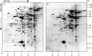
NaF-induced changes in protein expression were investigated, with a view to identifying proteins associated with moulting and development processes of lung-stage L3 to L4 using 2D electrophoresis. AsLL3s obtained from an infected rabbit's lungs were cultured in serum-supplemented RPMI medium in the presence or absence of NaF for 10 days. NaF exposures at a concentration of 2 mM blocked the moulting and development of AsLL3, whereas untreated AsLL3s developed well and moulted to AsLL4, further supporting previous observations (Islam et al. 2003). Also, 2D gel electrophoretic analysis of unmoulted AsLL3 vs. AsLL4 revealed that NaF exposure inhibited at least 16 proteins (spot nos. 1–16) of more than 200 proteins within the molecular mass range of 5 to 116 resolved on 2D gels (Fig. 1A,B). The expression of 6 major proteins (spot nos. 17–22) was reduced considerably following NaF exposure. A wide pH range was used to resolve soluble proteins with pI between 3·5 and 10·0. Each experiment was repeated at least 10 times to demonstrate the reproducibility of 2D gel results.

Fig. 1. Silver-stained 2D–PAGE gel (pI 3·5~10·0) of Ascaris suum lung-stage L3 (AsLL3). Larvae obtained from an infected rabbit's lung were cultured in vitro for 10 days in serum-supplemented RPMI 1640 medium in the presence of 2 mM NaF or its absence (control). Fifty micrograms of larval proteins were separated by 2D–PAGE and stained with a 2D silver staining kit. (A) Proteins expressed in moulted AsLL3 (i.e. AsLL4) grown in the absence of NaF in comparison with those inhibited by NaF in unmoulted AsLL3 (B) are indicated with a number.
Identification of stage-specific and moulting and development-associated proteins in lung-stage larvae
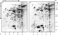
To further explore the stage-specific and moulting and development-associated expression profiles, 2D proteome maps from AsLL3 which had not cultured in vitro was included in this study for a comparison with those of AsLL4 (i.e. moulted larvae) and unmoulted AsLL3 (i.e. moulting had inhibited by NaF). Eight stage-specifically expressed proteins with molecular masses of 23 and 55 (spot nos. 23–30) were identified in AsLL4, of which spot nos. 23, 25 and 27 (corresponding spot nos. 10, 12 and 13, Fig. 1A) showed inhibition by NaF (Fig. 2A,B). More strikingly, spot nos. 31, 33, 34 and 35 which had a considerably decreased expression following NaF exposure, were expressed at high levels during the moulting from AsLL3 to AsLL4. The molecule represented by spot no. 32 showed a significantly reduced expression compared with that of AsLL3 (corresponding spot no. 43, Fig. 2B). By contrast, the 2D map of pre-cultured AsLL3 displayed 7 stage-specific proteins (spot nos. 36–42) (Fig. 2B), whereas spot no. 43 represented a reference spot for the corresponding spot no. 32 in AsLL4 (Fig. 2A). Together, these data differentiated: (i) proteins associated with sensitivity to inhibition by NaF (spot nos. 1–22); (ii) proteins stage-specifically expressed in AsLL4 (spot nos. 23–30) and in pre-cultured AsLL3 (spot nos. 36–42); (iii) proteins with considerably increased expression during moulting from AsLL3 to AsLL4 (spot nos. 31, 33–35); and finally (iv) proteins which are considered to play roles in the moulting and successful development of AsLL3 to AsLL4 (spot nos. 1–35).

Fig. 2. Silver-stained 2D–PAGE gel (pI 3·5~10·0) of Ascaris suum lung-stage L3 which had not been cultured. Fifty micrograms of larval proteins were separated by 2D–PAGE and stained with a 2D silver staining kit. (A) Proteins expressed in AsLL4 compared with those expressed in AsLL3 (B) which had not been cultured after removal from rabbit's lung are indicated with a number.
NaF exposure inhibited immunoreactive proteins in lung-stage larvae
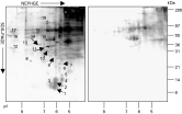
To identify the major immunoreactive proteins inhibited in larvae grown under exposure to NaF compared with those grown in an NaF-free control environment, proteins separated by 2D–PAGE were transferred to nitrocellulose membranes and probed with a hyperimmune serum raised in a rabbit. The results showed that 19 immunoreactive proteins, having a molecular mass of 10–97 kDa, were sensitive to inhibition by NaF and were resolved on 2D gels compared with controls (Fig. 3A,B). These data suggest that NaF acts as a potent inhibitor of a large number of immunoreactive protein antigens expressed in the developing AsLL3 in their mammalian host (pig).

Fig. 3. Immunoblot analysis of 2D–PAGE gel (pI 3·5~10·0) of Ascaris suum lung-stage L3 (AsLL3) major immunoreactive proteins. Two hundred micrograms of larval proteins were separated by 2D–PAGE, and the proteins transferred to a nitrocellulose membrane. The proteins bound to the rabbit anti-A. suum L3 serum (1[ratio ]250) were detected with alkaline phosphatase-conjugated goat anti-rabbit IgG (1[ratio ]2500) and NBT/BCIP. (A) Immunoreactive proteins expressed in AsLL4 grown in the absence of NaF compared with those in unmoulted AsLL3 inhibited by NaF (B) are represented with a number.
NaF exposure inhibited AsPPase endogenous enzyme activity in lung-stage larvae
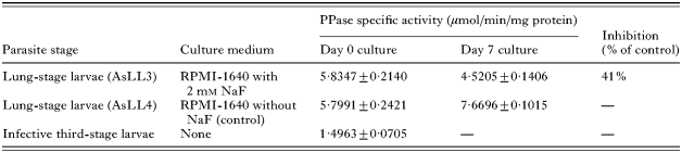
Endogenous AsPPase activity in NaF-exposed unmoulted AsLL3 compared with AsLL4 was measured to establish the possible role of NaF as a potent inhibitor of endogenous enzyme(s) of larval Ascaris. Data herein showed that NaF exposure at 2 mM concentrations inhibited endogenous AsPPase activity in unmoulted AsLL3 (4·5205±0·1406 μmol/min/mg protein) by 41% compared with AsLL4 (7·6696±0·1015 μmol/min/mg protein) (Table 1), suggesting that NaF is a potent inhibitor of a moulting-specific enzyme of Ascaris.
Table 1. Inhibition of endogenous inorganic pyrophosphatase (PPase) activity in Ascaris suum lung-stage larvae grown in the presence of NaF and its absence (Data presented are mean±S.D. from 5 independent experiments. Infective third-stage larvae were obtained from A. suum embryonated eggs by artificial hatching as described elsewhere.)

Inhibition of endogenous AsPPase in lung-stage larvae by NaF exposure
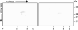
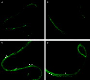
After showing an inhibitory effect of NaF on the moulting and AsPPase, we examined the inhibitory strength of NaF at 2 mM concentrations on expression of this protein/enzyme in the developing AsLL3 by immunochemical techniques. Two-dimensional immunoblot analysis revealed that endogenous AsPPase expression was considerably reduced in NaF-treated, unmoulted larvae compared with untreated control larvae (Fig. 4A,B). Similarly, immunofluorescent staining showed that the expression of endogenous AsPPase in the hypodermis of the unmoulted larvae was significantly decreased by NaF exposure compared with the control (Fig. 5A,B).

Fig. 4. Immunoblot analysis of 2D–PAGE gel (pI 3·5~10·0) of endogenous AsPPase in Ascaris suum lung-stage L3. One hundred micrograms of larval proteins were separated by 2D–PAGE and, the proteins transferred to a nitrocellulose membrane. Endogenous AsPPase reacted with mouse serum (1[ratio ]1000) raised against rAsPPase were detected with alkaline phosphatase-conjugated goat anti-mouse IgG (1[ratio ]2500) and NBT/BCIP. (A) Endogenous AsPPase in AsLL4. (B) Endogenous AsPPase was significantly inhibited by NaF in unmoulted AsLL3.

Fig. 5. Inhibition of expression of endogenous AsPPase enzyme in NaF-treated Ascaris suum lung-stage L3 detected by immunofluorescence. Immunofluorescent staining of whole mounted larvae was performed. (A) Moulted AsLL3 (i.e. AsLL4) were washed in cool PBS-Tween 20 (0·1%), fixed in acetone and incubated with anti-rAsPPase primary antibody (1[ratio ]50) served as the control. FITC-labelled goat anti-mouse IgG was used as a secondary antibody. (B) NaF-treated unmoulted AsLL3 were processed as in (A). Mouse anti-rAsPPase antibody bound to the endogenous AsPPase proteins expressed in the hypodermis of larvae were observed under a fluorescent microscope (Leica IM400) using appropriate filter sets and photographed (upper panel magnification×100, lower panel magnification×200).
Light and transmission electron microscopy of unmoulted lung-stage larvae
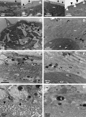
The effect of NaF (2 mM) on cuticular development and ecdysis and internal organs of AsLL3 was examined microscopically. Light microscopical examination revealed that there was no shedding of cuticular sheaths into the culture fluid and that none of the larvae had shown an initiation of moulting process until day 10 (when the culture was terminated). Although NaF exposure caused a considerable inhibition of larval development, larval viability was not affected (Fig. 6A–F). Larval mortality of <5% was recorded on day 10. Following NaF exposure, only a small proportion (~10%) of AsLL3 revealed visible damage to the oesophagus (e.g. narrowing and coiling of the organ) (Fig. 6D) and to the gut along its whole length (e.g. ‘rope-like’ shrinkage with an uneven surface and distinct vacuole formation) by light microscopy (Fig. 5E,F). AsLL3s in control culture were well developed and moulted to AsLL4 (over 70% population), leaving sheaths behind the culture fluid (Fig. 6A–C). TEM showed that no new cuticle was produced at the AsLL4 and there was no separation of old cuticle in AsLL3 after NaF exposure (Fig. 7A). Substantial degenerative changes were observed both in the contractile elements and cytoplasmic parts of the muscle cells and, on the gut wall, with formation of small vacuoles in the gut tissues (Fig. 7C,E,G). Distinct changes of destruction of microvilli leading to the formation of small vacuoles, wide distances between cells (arrow) and swelling of cells (arrowhead) were observed in NaF-treated, unmoulted larvae (Fig. 7G). By contrast, untreated control larvae (AsLL4) showed well-organized structures throughout the tissues (Fig. 7B,D,F,H).

Fig. 6. Light microscopy of Ascaris suum lung-stage larvae grown in the absence of NaF (A–C) (control) or its presence (D–F). (A–C) Larvae grown in the absence of NaF (control) which successfully moulted to AsLL4, arrow indicates cuticle shedding from tail end (A, ×50); B,C are larger views of (A) at ×200 and ×100, respectively. (D–F) AsLL3 did not moult to AsLL4 in the presence of 2 mM NaF. Under light microscopy, some larvae showed damage to the oesophagus (e.g. narrowing and coiling of the organ; arrow) (D, ×50) and to the gut along its whole length (e.g. ‘rope-like’ shrinkage with uneven surface and distinct vacuole formation; arrows) (E, ×100; F, ×200). Larvae were removed from the cultures and were photographed using a Leica IM500 microscope.

Fig. 7. Transmission electron micrographs of sections of Ascaris suum lung-stage larvae which did not moult in the presence of NaF. (A,C,E,G; left panel) Larvae grown in the presence of NaF did not moult to L4; (B,D,F,H; right panel) larvae in the absence of NaF, grown well and moulted to L4, were processed for transmission electron microscopy. (A) Sections highlight cuticular areas, arrows indicate epicuticle of AsLL3, no separation of old cuticle is seen. (B) Sections show cuticle of AsLL4, arrows indicate epicuticle of AsLL4 (left), separation between the AsLL4 epicuticle (arrows) and AsLL3 cuticle before ecdysis (arrowheads) is indicated (right). (C,E,G) Sections show degenerative changes in muscles and gut tissues, arrow indicates a wide distance between 2 microvilli, arrowhead indicates changes in the normal shape of microvilli. (D,F,H) Sections reveal normal shape of muscles and gut tissues including normal shape of microvilli. la, lateral alae; cu, cuticle; hp, hypodermis; nc, nerve chord; mu, muscle; mi, mitochondria; gu, gut; mv, microvilli; gl, gut lumen; gc, gut contents; dg, degenerative changes; v, vacuole; bm, membrane basalis; cy, cytoplasm; nc, nucleus; ncl, nucleolus; ed, electron-dense material; dm, dense material of intercellular gaps; sg, storage glycogen.
Analysis of amino acid sequences of selected protein
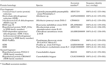
An NaF-sensitive and moulting-specific protein (spot no. 17; molecular mass of 33 kDa and pI of 8·8; Fig. 1A) was isolated from Coomassie-stained 2D gels and was analysed by peptide mass spectrometry to determine its partial amino acid sequence. After peptic digestion one of the fractions was analysed. The selected protein revealed 12 (1-GVIINTASIAAF-12) and 12 (1-GAIVGMTLPLAR-12) residue long amino acid sequences from the 2 major RP–HPLC purified fractions of the peptic digests, corresponding to 2 different locations of the whole protein sequence. The results of DDBJ/FASTA (Protein Query vs. Protein DB) homology searches indicated that the first fragment (1-GVIINTASIAAF-12) of the peptide sequence was 100% identical to 3–oxoacyl–(acyl carrier protein) reductase/short-chain dehydrogenase/reductase (SDR)/3–hydroxyacyl–CoA dehydrogenase (HAD) type II, of microbial origin (Table 2). While the second fragment of the peptide sequence was 100% identical to hypothetical proteins (belongs to the SDR enzyme family) of the model nematodes, Caenorhabditis elegans and C. briggsae (Table 2).

DISCUSSION
Since A. suum lung-stage larvae represent an excellent model with which moulting and development processes can be monitored in vitro (see Islam et al. 2003), we focused on investigating the protein expression profiles following NaF-induced moulting blockade in the larvae. We showed that NaF exposure can inhibit 22 proteins in unmoulted AsLL3 compared with AsLL4 which had moulted in the absence of NaF. This NaF-induced alteration in protein expression strongly suggests a crucial link between expression of a wide range of proteins and moulting and development processes of Ascaris larvae. To further explore the moulting and development-specific protein profiles, we conducted 2D gel electrophoretic analyses of AsLL3 that had not cultured (i.e. pre-cultured AsLL3) for a comparison with moulted larvae (i.e. AsLL4) and NaF-induced unmoulted larvae (i.e. AsLL3). The analyses revealed 8 stage-specific proteins expressed in AsLL4 and also 4 common proteins which showed sensitivity to inhibition following NaF exposure were over expressed during AsLL3 moulting process. The proteins specifically expressed in AsLL4 are likely to be involved in moulting and development processes of AsLL3 and, thus, may be regarded as being development-specific proteins. Again an elevated expression of NaF-sensitive proteins during larval moulting processes implies that these proteins may play roles in the moulting and developmental processes in Ascaris larvae. The pre-cultured AsLL3 also contained at least 7 stage-specific proteins. The increased expression of spot no. 43 in pre-cultured AsLL3 seems particularly interesting and warrants further investigation in 1st- and 2nd-stage larvae of A. suum. Together, these data strengthen the notion that a wide range of proteins may be expressed as a requirement of larval moulting and development.
In a biochemical investigation, we measured endogenous activity of AsPPase, a previously identified A. suum moulting and development-specific enzyme (see Islam et al. 2005), following exposure to NaF. The data showed that the elevated activity of endogenous enzyme during AsLL3 moulting was significantly inhibited (41%) by NaF exposure. Similarly, the expression of endogenous AsPPase in AsLL3 decreased following NaF exposure. These data show that NaF-induced inhibition of moulting and development processes of AsLL3–AsLL4 results in distinct changes in protein expression profiles. Also, suppression of a range of immunogenic proteins of AsLL3 by NaF suggests that this anion may be used to investigate pathogenicity of Ascaris roundworms during their invasive phase in their mammalian host. Ultrastructural studies indicated that NaF can potentially block the synthesis of components of the new cuticle by damaging the internal organs in addition to inhibiting a wide range of proteins associated with development in the AsLL3, justifying and validating the suitability of lung-stage A. suum-NaF as a model to investigate moulting-specific protein expression profiles of the parasitic roundworms. Together with the previous, successful application of the dsRNAi tool in lung-stage larvae of A. suum (see Islam et al. 2005), the present findings indicate that larval Ascaris may represent an exciting opportunity for studying not only the functional genomics but also the functional proteomics of Ascaris.
Analysis of the internal amino acid sequence of an NaF-sensitive and moulting-associated protein (spot no. 17) indicates that the protein is 100% identical in sequence to 3–hydrooxyacyl–CoA dehydrogenases/short-chain dehyrogenases/reductases which are members of the SDR superfamily of enzymes, which currently comprises ~3000 members (Kallberg et al. 2002). The 3–hydrooxyacyl–CoA dehydrogenase (HAD) is an essential enzyme which catalyses mitochondrial fatty acid β-oxidation (FAO) cycles (Yang et al. 2005), while short-chain dehyrogenases/reductases (SDR) are enzymes that represent different functions, including cell differentiation and signalling (Kallberg et al. 2002). A. suum undergoes a marked aerobic/anaerobic transition in metabolism during development. The egg, second-stage larva (L2) and L3 within the egg of A. suum are aerobic and rely mainly on the tricarboxylic acid (TCA) cycle for energy generation (Saz et al. 1968). In contrast, the L4 and adult worms are anaerobic and depend on fermentative metabolism (Ward and Fairbairn, 1970a; Behm, 2002). Although a range of mitochondrial enzymes has been reported to be involved in anaerobic energy metabolism in adult Ascaris (Köhler, 1985; Komuniecki and Vanover, 1987; Ma et al. 1993; Klingbeil et al. 1996; Kita and Takamiya, 2002; Rao et al. 2003), very little is known about HAD and SDR enzymes in aerobic/anaerobic metabolism in different life-cycle stages of Ascaris (Ward and Fairbairn, 1970a,b) and nothing is presently known about their roles in parasite moulting and development. Since mitochondrial enzymes are crucial for energy metabolism, developmental control and parasitic adaptation, the selected protein (spot no. 17), expressed during moulting and development processes of L4, might be a promising enzymatic target for chemotherapy against Ascaris. Further analysis by cloning and expression of the gene of the selected protein will be required to determine the functional significance of the recombinant protein.
In conclusion, A. suum lung-stage larvae-NaF may become a suitable tool for investigating moulting and development-specific protein expression profile in Ascaris parasites and may promote the discovery of potential chemotherapeutic targets.
This study was supported in part by a grant from the Zoonosis Control Program of the Ministry of Agriculture, Forestry and Fisheries and by a grant from the program for Promotion of Basic Research Activities for Innovative Biosciences (PROBRAIN). We are grateful to Toshio Fujisawa for preparing the photographs.