Introduction
Pneumolabyrinth describes a condition of acute inner ear dysfunction associated with the presence of air within the labyrinth. Several causes of pneumolabyrinth have been reported, including barotrauma, temporal bone trauma, dislocation or fracture of the stapes footplate, displaced stapes prosthesis, and cochleostomy with a cochlear implant. Air inside the inner ear is uncommon, and is rarely detected even following fractures of the otic capsule or transverse fractures of the temporal bone.Reference Gross, Ben-Yaakov, Goldfarb and Eliashar1, Reference Lyos, Marsh, Jenkins and Coker2
We present a patient with bilateral pneumolabyrinth secondary to blunt head trauma on the occiput after a fall, who had audiometrically confirmed, acute, bilateral, profound sensorineural hearing loss with severe vertigo. Nonetheless, his temporal bone computed tomography (CT) scan did not show any definite temporal bone fracture line or ossicular dislocation, or any other features apart from an air bubble inside the inner ear on each side.
Case report
A 49-year-old man was transferred to our otolaryngology department in order to evaluate his sudden, bilateral hearing loss and vertigo. He had fallen on his head from a 3-m high roof while fixing it. He did not lose consciousness. On arrival, the patient was shouting loudly and could not follow oral instructions, only written requests. There was no prior history of ear trauma, head trauma or ear surgery. The patient reported that he could not hear anything and complained of a constant, high pitched tinnitus starting immediately after the fall, together with the sudden onset of whirling vertigo.
On physical examination, no evidence of sensory or motor function deficit was found. Facial nerve function was intact. An otoscopic examination revealed that both tympanic membranes were intact, with mild hyperaemia around the malleus handle and with no evidence of haemotympanum or middle-ear fluid. No fracture lines were observed in the external ear canal. Tuning forks were not audible to the patient. He had spontaneous, left-beating, horizontal nystagmus. Fistula tests were negative.
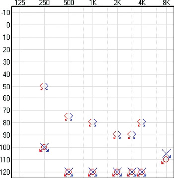
Pure tone audiometry revealed profound sensorineural hearing loss in both ears, with an air conduction threshold of over 110 dB (Figure 1). No auditory brainstem response was recordable in either ear.
Pure tone audiogram showing profound sensorineural hearing loss in both ears.

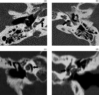
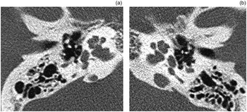
A high resolution CT scan of the temporal bones showed bilateral pneumolabyrinth, with air in the basal turn of the right cochlea as well as in the vestibule on the left side; however, it did not show a definite fracture line or any ossicular chain anomaly in the temporal bone. There was no fluid outside the oval window, in the middle ear or in the mastoid air cells, in either ear (Figure 2). On both sides, the stapes showed an intact shape and position, without any fracture or dislocation into the vestibule (Figure 3).
Axial (a and b) and coronal (c and d) high resolution computed tomography scans of both temporal bones. Parts (a) and (c) show a very low density air bubble inside the basal turn of the cochlea on the right side (arrow). Parts (b) and (d) show an air bubble in the vestibule on the left side (arrow).

Axial high resolution computed tomography views of the (a) right and (b) left temporal bone, showing that on both sides the stapes (asterisk) is in the correct position, without any fracture or dislocation.

The patient was admitted and treated conservatively, including absolute bed rest and prophylactic, intravenous antibiotic therapy. His vestibular symptoms disappeared gradually but there was no improvement in his hearing thresholds.
Two weeks after admission, a second temporal bone CT scan was performed. This revealed that, on both sides, the air which had been trapped in the vestibule and cochlea had been completely absorbed.
When reviewed two months after discharge, the patient reported a substantial improvement in vestibular symptoms; however, there still remained transient imbalance on rapid head movement. Unfortunately, there had been no improvement in hearing, and we recommended a cochlear implant; however, the patient never returned for further treatment.
Discussion
The temporal bones are pyramidal structures set within the thick bone of the skull base; consequently, they require great force to fracture. Temporal bone fractures can have associated intracranial injuries such as epidural or subdural haematoma, cerebral oedema and elevated intracranial pressure. Therefore, neurological manifestations are usually present.Reference Brodie, Cummings, Flint, Harker, Haughey, Robbins and Schuller3
A small number of studies have reported an association between pneumolabyrinth and temporal bone fracture. There have been even fewer cases of bilateral pneumolabyrinth reported after head trauma.Reference Lyos, Marsh, Jenkins and Coker2, Reference Woodson and Smith4 In those studies, temporal bone imaging revealed bilateral temporal bone fractures disrupting the otic capsule. Woodson and Smith reported that pneumolabyrinth is an excellent radiological marker for otic capsule fracture.Reference Woodson and Smith4 However, pneumolabyrinth without temporal bone fracture, as in the current case, is rarely present after head trauma.Reference Woo, Song, Kim and Bai5 Its bilateral occurrence is extremely rare. In this article, we describe a new case of bilateral pneumolabyrinth that was confirmed by temporal bone imaging, but for which there was no definite otic capsule fracture or neurological deficit apart from the VIIIth nerve.
The presence of air inside the inner ear is definite proof of a pathological connection between the inner ear and the air-filled mastoid or middle-ear cavities. Several causes of pneumolabyrinth have been proposed.Reference Woodson and Smith4, Reference Mafee, Valvassori, Kumar, Yannias and Marcus6 One is disruption of the otic capsule and membranous labyrinth by a temporal bone fracture. Another cause is rupture of the round window by an implosive force. Other causes include fracture of the stapes footplate (which can allow air to enter the vestibule from the oval window), perilymphatic fistulae (which might form a spontaneous connection between the inner and middle ear), and iatrogenic injuries such as stapedectomy and cochlear implantation.Reference Issacson, Laine and Williams7, Reference Hempel, Jäger, Baumann, Krause and Rasp8 Potential sites of gas entry include the oval and round window niche, microfissures between the posterior semicircular canal and the round window, and the fissula ante fenestram. In the current case, we suggest that pneumolabyrinth could have occurred as a result of a microfracture of the temporal bone or a slight displacement of the stapedial footplate, although temporal bone CT did not confirm the passage of air into the inner ear.
• Pneumolabyrinth (air within the labyrinth after head trauma) is very rare
• It is rarely detected, even after temporal bone and otic capsule fracture
• Temporal bone high resolution computed tomography is useful for otological complaints after head trauma
• A very rare case of bilateral pneumolabyrinth without temporal bone fracture is reported
There is no established algorithm for the diagnosis of pneumolabyrinth; early audiometry and high resolution CT are required.Reference Lyos, Marsh, Jenkins and Coker2
Similarly, because of the small number of cases of pneumolabyrinth reported in the literature, there is no consensus on management. The treatment options for pneumolabyrinth due to temporal bone trauma are observation or surgical exploration with closure of the connection between the inner ear and the air-filled tympanomastoid cavity. Current conservative management for pneumolabyrinth includes bed rest, head elevation, and avoidance of Valsalva's manoeuvre and straining. Surgical exploration may selectively require management of a displaced stapes, of progressive, fluctuating sensorineural hearing loss, or of persistent vertigo.Reference Gross, Ben-Yaakov, Goldfarb and Eliashar1 In our case, the observational option was chosen.