


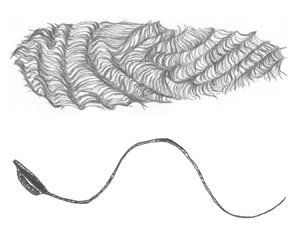

1. Introduction
The cellular scale is home to a wide variety of biological swimmers, both in the prokaryote and eukaryote domains (Yates Reference Yates1986). Examples that have long been studied comprise the spermatozoa of animals (including humans) (Gaffney et al. Reference Gaffney, Gadelha, Smith, Blake and Kirkman-Brown2011), algae and planktonic aquatic organisms (Pedley & Kessler Reference Pedley and Kessler1992; Guasto, Rusconi & Stocker Reference Guasto, Rusconi and Stocker2012; Goldstein Reference Goldstein2015), protozoa (Jahn & Votta Reference Jahn and Votta1972) and bacteria (Lauga Reference Lauga2016). Prokaryotic swimmers exploit the rotation of passive helical filaments to self-propel (Berg & Anderson Reference Berg and Anderson1973). In contrast, swimming eukaryotes employ a more complex propulsion machinery in which flexible flagella actuated internally by molecular motors deform periodically in a wave-like motion (Bray Reference Bray2000).
During recent decades, the biophysical and fluid mechanics communities have devoted considerable effort to quantify the interactions between cells and surrounding fluids (Lighthill Reference Lighthill1975, Reference Lighthill1976; Lauga Reference Lauga2020a). Most studies use mathematical, or computational, modelling to rationalise the dynamics of cells, from individual motion in bulk fluids to collective interactions in complex environments (Lauga & Powers Reference Lauga and Powers2009; Koch & Subramanian Reference Koch and Subramanian2011; Elgeti, Winkler & Gompper Reference Elgeti, Winkler and Gompper2015). These theoretical tools have in turn allowed the impact of fluid forces on shaping the geometry and kinematics of swimming cells to be explored, allowing in particular posing cellular swimming as an optimisation problem, with notable successes.
In the case of bacteria, in search of an optimal prokayotic flagellum, Purcell (Reference Purcell1997) determined using simple arguments the shape of the most efficient helical propeller. A later theoretical study of the polymorphic forms of the flagellum commonly found in most bacteria by Spagnolie & Lauga (Reference Spagnolie and Lauga2011) showed that the so-called ‘normal’ helical form is the most efficient by some significant margin, suggesting that fluid mechanical forces may have played a role in the evolution of the flagellum. In addition to the flagellum, overall swimming efficiency is also impacted by the shape of the cell body, as first shown and optimised by Fujita & Kawai (Reference Fujita and Kawai2001) for a monotrichous bacterium. Recent work considered the impact of other biological constraints on bacterial shape (Schuech et al. Reference Schuech, Hoehfurtner, Smith and Humphries2019).
In eukaryotic cells, optimal propulsion strategies result from an interplay between fluid dynamics, elasticity and active driving mechanisms. In a study of over 400 mammalian species, Tam & Hosoi (Reference Tam and Hosoi2011a) discovered that spermatozoa have an optimal tail-to-head length ratio and that their most efficient swimming mode involves symmetrical, non-sinusoidal travelling waves in the tail. Such travelling waves have been shown in many contexts to lead to optimal motion in an active flagellum (Pironneau & Katz Reference Pironneau and Katz1974, Reference Pironneau and Katz1975; Lauga Reference Lauga2020b). Lighthill (Reference Lighthill1975) found the two-dimensional hydrodynamically optimal travelling-wave profile to be sawtooth-like, but the singular kinks are regularised by elastic stresses (Spagnolie & Lauga Reference Spagnolie and Lauga2010) or by properly modelling the irreversible work of molecular motors within the active flagellum (Lauga & Eloy Reference Lauga and Eloy2013). In ciliated organisms, the beating pattern of individual cilia has also been shown to be optimal for propulsion (Osterman & Vilfan Reference Osterman and Vilfan2011; Eloy & Lauga Reference Eloy and Lauga2012). However, flagellar gaits may have local optima, suited e.g. to distinct feeding and swimming modes, as investigated by Tam & Hosoi (Reference Tam and Hosoi2011b) for biflagellated phytoplanktons. In larger organisms, multi-ciliated cells can increase their efficiency by choosing an optimal number of cilia (from hundreds to thousands) (Omori, Ito & Ishikawa Reference Omori, Ito and Ishikawa2020) and both optimal swimming and feeding have been shown to involve surface metachronal waves (Michelin & Lauga Reference Michelin and Lauga2010, Reference Michelin and Lauga2011).
Beyond biological systems, the question of optimality has received a lot of attention in the realm of biomimetic and engineered artificial swimmers. For example, swimming gaits and shapes maximising swimming efficiency have been determined for simple swimmers with only a few degrees of freedom (Tam & Hosoi Reference Tam and Hosoi2007; Alouges, DeSimone & Lefebvre Reference Alouges, DeSimone and Lefebvre2008; Golestanian & Ajdari Reference Golestanian and Ajdari2008; Alouges, DeSimone & Lefebvre Reference Alouges, DeSimone and Lefebvre2009; Nasouri, Vilfan & Golestanian Reference Nasouri, Vilfan and Golestanian2019), two-dimensional cell shapes (Avron, Gat & Kenneth Reference Avron, Gat and Kenneth2004; Montenegro-Johnson & Lauga Reference Montenegro-Johnson and Lauga2014) and idealised kinematics of treadmilling (Leshansky et al. Reference Leshansky, Kenneth, Gat and Avron2007) or surface slip flow driven swimmers (Vilfan Reference Vilfan2012). Other studies have sought shapes that would minimise absolute dissipation (Nasouri, Vilfan & Golestanian Reference Nasouri, Vilfan and Golestanian2021).
In this paper, we use data analysis to discover remarkable optimal properties of the geometry of cell shapes. We rely on our recently assembled BOSO-Micro database (Rodrigues, Lisicki & Lauga Reference Rodrigues, Lisicki and Lauga2021): a comprehensive collection of data from the experimental literature on cellular swimming that contains data on swimming speed and morphological characteristics of 382 unicellular organisms, including spermatozoa. Building on the data, we first demonstrate that the amplitude-to-wavelength aspect ratios of flagellated eukaryotes, including spermatozoa, are very close to the optimum predicted by hydrodynamic theory. Next, we demonstrate that the aspect ratios (length-to-width) of ciliate cell bodies are well predicted by the classical result for the shape of minimum drag force subject to a fixed-volume constraint.
2. Results
The BOSO-Micro database (Rodrigues et al. Reference Rodrigues, Lisicki and Lauga2021) provides us with novel means to examine swimming and morphological relationships across species. The open-source database contains swimming and morphological data published in scientific literature to date; we refer to the original paper for all details on data-gathering. Apart from swimming speed, many past investigations also contain data on the basic morphology of organisms, such as the size and shape of the cell body, or the length and number of flagella or cilia. Note that, in the database, the measurements and characterisation of organisms have been performed under different physiological conditions and in various environments, and therefore they do not represent a uniform sample. In cases where multiple values were reported for the morphology of swimming cells, we used average values.
2.1. Propulsion of flagellated eukaryotes
First, we focus on studies providing the details of actuation in flagellated eukaryotes and spermatozoa. We assume that the waveform of their flagellar oscillations can be captured by a characteristic amplitude  $h$ and wavelength
 $\lambda$, as sketched in figure 1(a). The database contains 23 different species of flagellated eukaryotes and 28 different species of spermatozoa for which we found measurements of
 $h$ and
 $\lambda$; note that these values were either directly reported in each paper, or were estimated by us using experimental images, see details in Rodrigues et al. (Reference Rodrigues, Lisicki and Lauga2021).
Aspect ratio of eukaryotic flagellar waves. (a) Sketch of flagellated eukaryotic cell with wave amplitude  $h$ and wavelength
 $\lambda$. (b) Histogram of aspect ratios
 $h/\lambda$ from experiments gathered in the BOSO-Micro database (Rodrigues et al. Reference Rodrigues, Lisicki and Lauga2021). (c) Mean value of
 $h$ vs
 $\lambda$ for all flagellated eukaryotes in our database. Spermatozoa are marked with circles, while the remaining flagellated eukaryotes are marked with squares. The solid line shows the least-square fit to the data and the shaded area is the 95 % confidence interval. The dashed line shows the prediction from the optimal theory from Lauga & Eloy (Reference Lauga and Eloy2013) for
 $Sp=4$ and the dotted line for
 $Sp=6$. Cells from the same class in the tree of life are plotted using the same colour scheme, with all classes represented here listed in the inset of (b).

For all our data, we plot in figure 1(b) the histogram of the amplitude-to-wavelength aspect ratio,  $h/\lambda$. This ratio does not exceed 0.6 in the data set, with most organisms showing the value below 0.25; hence the amplitude of a flagellar beat rarely exceeds a quarter of the wavelength. For further insight, we plot the amplitude
 $h$ against the wavelength
 $\lambda$ in figure 1(c). When multiple values are inferred for the same species, we plot in figure 1(c) the average value but include error bars to reflect the variability within the data reported for each species. Square markers represent flagellated eukaryotes, which are generally smaller in size as compared with spermatozoa, marked with circles.
It is clear from the data in figure 1(c) that there is a systematic increase of flagellar amplitude with wavelength. To rationalise and interpret these results, we performed a least-squares linear fit of the  $h/\lambda =\text {constant}$ relationship to the data. The solid line shows the result of fitting, with the 95 % confidence interval shaded in grey. For comparison with theoretical predictions, we use the work of Lauga & Eloy (Reference Lauga and Eloy2013) who computed numerically the optimal shape of an active flagellum. They considered an internally forced, elastic planar flagellum deforming periodically and moving at a constant speed through a viscous fluid and found the shape of the flagellum that maximises the swimming speed at a fixed energetic cost, representing irreversible work of molecular motors. They concluded that the optimal shape of the beating flagellum depends on a single dimensionless combination of parameters, termed the Sperm number
 $Sp$ (Lauga Reference Lauga2020a), and defined as the ratio of the wavelength
 $\lambda$ to the elasto-hydrodynamic penetration length
 $\ell$,
 \begin{equation} Sp = \frac{\lambda}{\ell}, \quad\text{with}\ \ell = \left(\frac{T B}{\zeta_\perp}\right)^{1/4}, \end{equation}
where  $T$ is the period of oscillatory actuation,
 $B$ the bending rigidity of the flagellum, and
 $\zeta _\perp$ the perpendicular resistance coefficient of the flagellum (i.e. the hydrodynamic force exerted on the flagellum per unit length for motion in the fluid perpendicular to its centreline). Thus the flagellum shape results from an interplay between the stiffness of the filament, viscous drag of the medium and internal forcing. Accordingly, decreasing the value of
 $Sp$ results in flatter, smoother kinks in the flagellar beating (Lauga & Eloy Reference Lauga and Eloy2013).
The typical values of the dimensionless  $Sp$ number for eukaryotes and spermatozoa have been reported to be within the range of 1–10 (Kumar et al. Reference Kumar, Walkama, Guasto and Ardekani2019). For sea-urchin Arbacia punctulata spermatozoa, Pelle et al. (Reference Pelle, Brokaw, Lesich and Lindemann2009) reported a bending rigidity of
 $B\approx 0.4 \unicode{x2013} 0.9\times 10^{-21}\,{\rm Nm}^2$, while Howard (Reference Howard2001) assumes it to be the upper limit of that range. In direct measurements, Okuno & Hiramoto (Reference Okuno and Hiramoto1979) found the stiffness of echinoderm sperm flagella to be in the range of
 $B\approx 0.3\unicode{x2013}1.5\times 10^{-21}\,{\rm Nm}^2$, while for Strongylocentrotus purpuratus, Okuno et al. (Reference Okuno, Asai, Ogawa and Brokaw1981) estimated
 $B\approx 0.8\times 10^{-21}\,{\rm Nm}^2$. Mechanical measurements for the flagella of wild-type Chlamydomonas yield
 $B = (0.84 \pm 0.28)\times 10^{-21}\,{\rm Nm}^2$ (Xu et al. Reference Xu, Wilson, Okamoto, Shao, Dutcher and Bayly2016), with similar values for mutant strains. In water, we have
 $\zeta _\perp \approx 2\times 10^{-3}\,{\rm Pa}\,{\rm s}$, and the typical periods
 $T$ vary across species from 0.02 to 0.05 s (Brennen & Winet Reference Brennen and Winet1977; Gray Reference Gray1955; Rodrigues et al. Reference Rodrigues, Lisicki and Lauga2021). From these numbers, and typical lengths of dozens of
 $\mathrm {\mu }$m (Rodrigues et al. Reference Rodrigues, Lisicki and Lauga2021), we therefore estimate the values of
 $Sp$ to be in the range of
 $Sp \approx 2\unicode{x2013}7$.
To compare the experimental measurements to predictions from theory, we use the work of Eloy & Lauga (Reference Eloy and Lauga2012) where, for a given value of  $Sp$, the shape of the optimal flagellum was computed; for each optimal shape, we can then extract the corresponding optimal aspect ratio,
 $h/\lambda$. In figure 1(c) we plot the aspect ratio as a linear curve
 $h/\lambda ={\rm constant}$ for
 $Sp=4$ (
 $h/\lambda =0.163$, dashed line) and
 $Sp=6$ (
 $h/\lambda =0.194$, dotted line). Both lines lie within the 95 % confidence interval of our dataset: the best fit for the data yields an aspect ratio
 $h/\lambda =0.188$, with the lower and upper bound of the 95 % confidence interval at
 $0.163$ and
 $0.213$, respectively (
 $R^2=0.562$).
2.2. Hydrodynamic drag force on ciliates
Ciliates constitute a large group of unicellular eukaryotic swimmers, characterised by the presence of hair-like cilia which cover their bodies in dense active carpets; a model organism, Paramecium, is sketched in figure 2(a). The internal structure of cilia is identical to eukaryotic flagella but they are typically shorter and used to propel cells with relatively much larger body sizes. This separation of scales has motivated mathematical models dating back to Blake (Reference Blake1971) to represent the action of cilia layers covering the cell bodies as a continuum boundary with prescribed actuation of the surrounding fluid. This surface forcing sets the fluid in motion, and leads to swimming. The balance of propulsion force and viscous drag force on the cell body determines the swimming speed  $U$ (Lauga Reference Lauga2020a). Because the latter results from the overall shape of the body, one possibility for cells to increase their swimming speed is to reduce the viscous drag opposing their propulsion by choosing the appropriate drag-minimising shape, which have been characterised theoretically (Pironneau Reference Pironneau1973, Reference Pironneau1974; Montenegro-Johnson & Lauga Reference Montenegro-Johnson and Lauga2014).
Aspect ratio of ciliates cell body. (a) Sketch of ciliate body showing the cell width  $W$ and length
 $B$. (b) Histograms of cell aspect ratios
 $W/B$ from experimental data gathered in the BOSO-Micro database (Rodrigues et al. Reference Rodrigues, Lisicki and Lauga2021). (c) Mean value of body length
 $B$ (in
 $\mathrm {\mu }$m) plotted as a function of width
 $W$ (in
 $\mathrm {\mu }$m) for all species in our database. The solid line shows the least-square fit to the data while the shaded area is the 95 % confidence interval. The dashed line shows the prediction from the theory of fixed-volume drag minimisation (
 $B/W=1.952$) (Pironneau Reference Pironneau1973) while the dotted line shows the drag minimum for a given surface area (
 $B/W=4.037$) (Montenegro-Johnson & Lauga Reference Montenegro-Johnson and Lauga2015). Cells from the same class in the tree of life are plotted using the same colour scheme, with all classes represented here listed in inset of (b).

To investigate if cells aligned with expectations from drag minimisation, we use our eukaryotic microswimmers database (Rodrigues et al. Reference Rodrigues, Lisicki and Lauga2021) to quantify the extent to which the cell bodies of ciliates minimise drag forces. In figure 2(b), we plot the histogram for the aspect ratio of the cell bodies,  $B/W$, of the 91 ciliates in our database. When multiple values were available for the same species, we use their average. Colours are used to code different classes of organisms. Clearly, the reported cells are typically prolate, and most of them have a length of approximately twice their width, although more slender cell bodies are also observed.
To further explore the correlation between the two length scales characterising the cell bodies of ciliates, we present in figure 2(c) a scatter plot of the data, which suggests a linear correlation between cell body and width. We plot the linear least-square fit to the data with a solid line, corresponding to the slope  $B/W=2.360$ (
 $R^2=0.503$), while grey shading marks the 95 % confidence interval, for aspect ratios in the range
 $(2.057, 2.664)$. Remarkably, these results are very close to the classical prediction of a minimal-drag shape in Stokes flow by Pironneau (Reference Pironneau1973), later computed by Bourot (Reference Bourot1974), who demonstrated numerically that the aspect ratio
 $B/W=1.952$ is hydrodynamically optimal for bodies with prescribed volumes (the corresponding optimal shape resembles a prolate spheroid with pointy ends). We mark this theoretical prediction with a dashed line in figure 2(c), and note that it lies just outside the confidence interval of our fit to data. In contrast, if instead one considers the shape that has minimum drag for a prescribed surface area (instead of a fixed volume), Montenegro-Johnson & Lauga (Reference Montenegro-Johnson and Lauga2014) found the optimal aspect ratio to be 4.037, marked in figure 2(c) with the dotted line; this lies well outside the data.
It is worth noting that the Stokes drag acting on an optimal fixed-volume prolate shape is only 4.5 % less than that experienced by a sphere of the same volume, suggesting that the energetic cost of diverting from the optimum is actually relatively low. It is therefore perhaps all the more remarkable that such a small energetic gain seems to be reflected in the empirical data. The small magnitude of the energetic improvement might in turn allow a larger cell shape variability and explain the diversity of observed micron-scale shape features.
3. Discussion
In this paper, we used morphological data gathered for swimming eukaryotic microorganisms in the BOSO-Micro database which assembled published data from the literature (Rodrigues et al. Reference Rodrigues, Lisicki and Lauga2021) to test two outstanding questions regarding hydrodynamic optimality of the geometric characteristics and shapes of the cells. First, we have shown that for 51 different species of flagellated unicellular eukaryotes and spermatozoa, which had the characteristics of their flagellar waves measured and reported in the literature, the ratio of the wave amplitude to the wavelength was close to the optimal hydrodynamic predictions for the shape of energetically optimal active filaments for Sperm numbers comparable to estimates for natural swimmers. Second, we have analysed a dataset of body widths and lengths of 91 species of ciliates and demonstrated that their cells have an aspect ratio close to the theoretical optimum for the minimal drag on a body of a fixed volume.
Our findings shed new light on the question of energetics of microscale swimming. There is a long-standing argument in biophysics that the energetic cost of locomotion is negligible when compared with metabolic constraints (Purcell Reference Purcell1977). Recent work focusing on 169 species of eukaryotes has estimated that the operating costs of flagella remain small, at a few per cent of the total energy budget, but flagellar construction costs can be quite significant (Schavemaker & Lynch Reference Schavemaker and Lynch2022). The results in the current work strongly indicate that, in multiple species of swimming cells that span almost three orders of magnitude in length, cellular shapes are consistent with energetic optimisation. This remarkable agreement suggests that hydrodynamic forces might have played a key role in shaping biological active swimmers.
Clearly, not all swimmers in figures 1 and 2 follow exactly the theoretical curves. Outliers in figure 1 that are the furthest from the best-fit line are Giardia lamblia, cricket, worm, midge and rabbit spermatozoa (below the trend line), and Poteriodendron, stick insect and fly spermatozoa (above the line). In figure 2 the outliers are Halteria grandinella, Mesodinium rubrum (below the line) and Spirostomum spp. and Trachelocerca spp. (above the line). Upon examining the shape of these specific organisms we could not identify any systematic distinguishing features. Other factors are therefore at play in determining the morphology of some species. In the case of bacteria, it is known that cell shapes are the result of a delicate trade-off between efficient swimming, the chemotaxis budget, and the costs related to cell construction (Schuech et al. Reference Schuech, Hoehfurtner, Smith and Humphries2019), which points to the complexity of the overall energetic landscape.
Furthermore, in the case of ciliated organisms, minimising the drag force might not be sufficient for maximising propulsion, since swimming is a balance between drag and thrust. And yet, the available experimental data strikingly show that drag minimisation appears to be important for ciliates, independently of the details of their propulsion efficiency. We conjecture that this might stem from the necessity of these cells to not only self-propel but also to respond to the external environmental flows to which they are inevitably exposed.
Funding
This project has received funding from the European Research Council (ERC) under the European Union's Horizon 2020 research and innovation programme (grant agreement 682754 to EL), from the National Science Centre of Poland (grant Sonata no. 2018/31/D/ST3/02408 to M.L.) and from Campus France (Eiffel Scholarship no. 812884G to M.F.V.R.).
Declaration of interests
The authors report no conflict of interest.
Data availability statement
The data that support the findings of this study are openly available in the OSF repository https://osf.io/4tyx6. They are also available and editable on GitHub: https://github.com/marcos-fvr/BOSO-micro.
Author contributions
M.L. and M.F.V.R. contributed equally to this work.



