

%20--%3e%3c!DOCTYPE%20svg%20PUBLIC%20'-//W3C//DTD%20SVG%201.0//EN'%20'http://www.w3.org/TR/2001/REC-SVG-20010904/DTD/svg10.dtd'%3e%3csvg%20version='1.0'%20id='Layer_1'%20xmlns='http://www.w3.org/2000/svg'%20xmlns:xlink='http://www.w3.org/1999/xlink'%20x='0px'%20y='0px'%20width='64px'%20height='64px'%20viewBox='5.5%20-3.5%2064%2064'%20enable-background='new%205.5%20-3.5%2064%2064'%20xml:space='preserve'%3e%3cg%3e%3ccircle%20fill='%23FFFFFF'%20fill-opacity='0'%20cx='37.785'%20cy='28.501'%20r='28.836'/%3e%3cpath%20fill='%23595959'%20d='M37.441-3.5c8.951,0,16.572,3.125,22.857,9.372c3.008,3.009,5.295,6.448,6.857,10.314%20c1.561,3.867,2.344,7.971,2.344,12.314c0,4.381-0.773,8.486-2.314,12.313c-1.543,3.828-3.82,7.21-6.828,10.143%20c-3.123,3.085-6.666,5.448-10.629,7.086c-3.961,1.638-8.057,2.457-12.285,2.457s-8.276-0.808-12.143-2.429%20c-3.866-1.618-7.333-3.961-10.4-7.027c-3.067-3.066-5.4-6.524-7-10.372S5.5,32.767,5.5,28.5c0-4.229,0.809-8.295,2.428-12.2%20c1.619-3.905,3.972-7.4,7.057-10.486C21.08-0.394,28.565-3.5,37.441-3.5z%20M37.557,2.272c-7.314,0-13.467,2.553-18.458,7.657%20c-2.515,2.553-4.448,5.419-5.8,8.6c-1.354,3.181-2.029,6.505-2.029,9.972c0,3.429,0.675,6.734,2.029,9.913%20c1.353,3.183,3.285,6.021,5.8,8.516c2.514,2.496,5.351,4.399,8.515,5.715c3.161,1.314,6.476,1.971,9.943,1.971%20c3.428,0,6.75-0.665,9.973-1.999c3.219-1.335,6.121-3.257,8.713-5.771c4.99-4.876,7.484-10.99,7.484-18.344%20c0-3.543-0.648-6.895-1.943-10.057c-1.293-3.162-3.18-5.98-5.654-8.458C50.984,4.844,44.795,2.272,37.557,2.272z%20M37.156,23.187%20l-4.287,2.229c-0.458-0.951-1.019-1.619-1.685-2c-0.667-0.38-1.286-0.571-1.858-0.571c-2.856,0-4.286,1.885-4.286,5.657%20c0,1.714,0.362,3.084,1.085,4.113c0.724,1.029,1.791,1.544,3.201,1.544c1.867,0,3.181-0.915,3.944-2.743l3.942,2%20c-0.838,1.563-2,2.791-3.486,3.686c-1.484,0.896-3.123,1.343-4.914,1.343c-2.857,0-5.163-0.875-6.915-2.629%20c-1.752-1.752-2.628-4.19-2.628-7.313c0-3.048,0.886-5.466,2.657-7.257c1.771-1.79,4.009-2.686,6.715-2.686%20C32.604,18.558,35.441,20.101,37.156,23.187z%20M55.613,23.187l-4.229,2.229c-0.457-0.951-1.02-1.619-1.686-2%20c-0.668-0.38-1.307-0.571-1.914-0.571c-2.857,0-4.287,1.885-4.287,5.657c0,1.714,0.363,3.084,1.086,4.113%20c0.723,1.029,1.789,1.544,3.201,1.544c1.865,0,3.18-0.915,3.941-2.743l4,2c-0.875,1.563-2.057,2.791-3.541,3.686%20c-1.486,0.896-3.105,1.343-4.857,1.343c-2.896,0-5.209-0.875-6.941-2.629c-1.736-1.752-2.602-4.19-2.602-7.313%20c0-3.048,0.885-5.466,2.658-7.257c1.77-1.79,4.008-2.686,6.713-2.686C51.117,18.558,53.938,20.101,55.613,23.187z'/%3e%3c/g%3e%3c/svg%3e)
%20--%3e%3c!DOCTYPE%20svg%20PUBLIC%20'-//W3C//DTD%20SVG%201.0//EN'%20'http://www.w3.org/TR/2001/REC-SVG-20010904/DTD/svg10.dtd'%3e%3csvg%20version='1.0'%20id='Layer_1'%20xmlns='http://www.w3.org/2000/svg'%20xmlns:xlink='http://www.w3.org/1999/xlink'%20x='0px'%20y='0px'%20width='64px'%20height='64px'%20viewBox='5.5%20-3.5%2064%2064'%20enable-background='new%205.5%20-3.5%2064%2064'%20xml:space='preserve'%3e%3cg%3e%3ccircle%20fill='%23FFFFFF'%20fill-opacity='0'%20cx='37.637'%20cy='28.806'%20r='28.276'/%3e%3cg%3e%3cpath%20fill='%23595959'%20d='M37.443-3.5c8.988,0,16.57,3.085,22.742,9.257C66.393,11.967,69.5,19.548,69.5,28.5c0,8.991-3.049,16.476-9.145,22.456%20C53.879,57.319,46.242,60.5,37.443,60.5c-8.649,0-16.153-3.144-22.514-9.43C8.644,44.784,5.5,37.262,5.5,28.5%20c0-8.761,3.144-16.342,9.429-22.742C21.101-0.415,28.604-3.5,37.443-3.5z%20M37.557,2.272c-7.276,0-13.428,2.553-18.457,7.657%20c-5.22,5.334-7.829,11.525-7.829,18.572c0,7.086,2.59,13.22,7.77,18.398c5.181,5.182,11.352,7.771,18.514,7.771%20c7.123,0,13.334-2.607,18.629-7.828c5.029-4.838,7.543-10.952,7.543-18.343c0-7.276-2.553-13.465-7.656-18.571%20C50.967,4.824,44.795,2.272,37.557,2.272z%20M46.129,20.557v13.085h-3.656v15.542h-9.944V33.643h-3.656V20.557%20c0-0.572,0.2-1.057,0.599-1.457c0.401-0.399,0.887-0.6,1.457-0.6h13.144c0.533,0,1.01,0.2,1.428,0.6%20C45.918,19.5,46.129,19.986,46.129,20.557z%20M33.042,12.329c0-3.008,1.485-4.514,4.458-4.514s4.457,1.504,4.457,4.514%20c0,2.971-1.486,4.457-4.457,4.457S33.042,15.3,33.042,12.329z'/%3e%3c/g%3e%3c/g%3e%3c/svg%3e)
%20--%3e%3c!DOCTYPE%20svg%20PUBLIC%20'-//W3C//DTD%20SVG%201.0//EN'%20'http://www.w3.org/TR/2001/REC-SVG-20010904/DTD/svg10.dtd'%3e%3csvg%20version='1.0'%20id='Layer_1'%20xmlns='http://www.w3.org/2000/svg'%20xmlns:xlink='http://www.w3.org/1999/xlink'%20x='0px'%20y='0px'%20width='64px'%20height='64px'%20viewBox='5.5%20-3.5%2064%2064'%20enable-background='new%205.5%20-3.5%2064%2064'%20xml:space='preserve'%3e%3cg%3e%3ccircle%20fill='%23FFFFFF'%20fill-opacity='0'%20cx='36.924'%20cy='28.403'%20r='28.895'/%3e%3cpath%20fill='%23595959'%20d='M37.442-3.5c8.99,0,16.571,3.085,22.743,9.256C66.393,11.928,69.5,19.509,69.5,28.5c0,8.992-3.048,16.476-9.145,22.458%20C53.88,57.32,46.241,60.5,37.442,60.5c-8.686,0-16.19-3.162-22.513-9.485C8.644,44.728,5.5,37.225,5.5,28.5%20c0-8.762,3.144-16.343,9.429-22.743C21.1-0.414,28.604-3.5,37.442-3.5z%20M12.7,19.872c-0.952,2.628-1.429,5.505-1.429,8.629%20c0,7.086,2.59,13.22,7.77,18.4c5.219,5.144,11.391,7.715,18.514,7.715c7.201,0,13.409-2.608,18.63-7.829%20c1.867-1.79,3.332-3.657,4.398-5.602l-12.056-5.371c-0.421,2.02-1.439,3.667-3.057,4.942c-1.622,1.276-3.535,2.011-5.744,2.2%20v4.915h-3.714v-4.915c-3.543-0.036-6.782-1.312-9.714-3.827l4.4-4.457c2.094,1.942,4.476,2.913,7.143,2.913%20c1.104,0,2.048-0.246,2.83-0.743c0.78-0.494,1.172-1.312,1.172-2.457c0-0.801-0.287-1.448-0.858-1.943l-3.085-1.315l-3.771-1.715%20l-5.086-2.229L12.7,19.872z%20M37.557,2.214c-7.276,0-13.428,2.571-18.457,7.714c-1.258,1.258-2.439,2.686-3.543,4.287L27.786,19.7%20c0.533-1.676,1.542-3.019,3.029-4.028c1.484-1.009,3.218-1.571,5.2-1.686V9.071h3.715v4.915c2.934,0.153,5.6,1.143,8,2.971%20l-4.172,4.286c-1.793-1.257-3.619-1.885-5.486-1.885c-0.991,0-1.876,0.191-2.656,0.571c-0.781,0.381-1.172,1.029-1.172,1.943%20c0,0.267,0.095,0.533,0.285,0.8l4.057,1.83l2.8,1.257l5.144,2.285l16.397,7.314c0.535-2.248,0.801-4.533,0.801-6.857%20c0-7.353-2.552-13.543-7.656-18.573C51.005,4.785,44.831,2.214,37.557,2.214z'/%3e%3c/g%3e%3c/svg%3e)
%20--%3e%3c!DOCTYPE%20svg%20PUBLIC%20'-//W3C//DTD%20SVG%201.0//EN'%20'http://www.w3.org/TR/2001/REC-SVG-20010904/DTD/svg10.dtd'%3e%3csvg%20version='1.0'%20id='Layer_1'%20xmlns='http://www.w3.org/2000/svg'%20xmlns:xlink='http://www.w3.org/1999/xlink'%20x='0px'%20y='0px'%20width='64px'%20height='64px'%20viewBox='5.5%20-3.5%2064%2064'%20enable-background='new%205.5%20-3.5%2064%2064'%20xml:space='preserve'%3e%3cg%3e%3ccircle%20fill='%23FFFFFF'%20fill-opacity='0'%20cx='36.944'%20cy='28.631'%20r='29.105'/%3e%3cg%3e%3cpath%20fill='%23595959'%20d='M37.443-3.5c8.951,0,16.531,3.105,22.742,9.315C66.393,11.987,69.5,19.548,69.5,28.5c0,8.954-3.049,16.457-9.145,22.514%20C53.918,57.338,46.279,60.5,37.443,60.5c-8.649,0-16.153-3.143-22.514-9.429C8.644,44.786,5.5,37.264,5.5,28.501%20c0-8.723,3.144-16.285,9.429-22.685C21.138-0.395,28.643-3.5,37.443-3.5z%20M37.557,2.272c-7.276,0-13.428,2.572-18.457,7.715%20c-5.22,5.296-7.829,11.467-7.829,18.513c0,7.125,2.59,13.257,7.77,18.4c5.181,5.182,11.352,7.771,18.514,7.771%20c7.123,0,13.334-2.609,18.629-7.828c5.029-4.876,7.543-10.99,7.543-18.343c0-7.313-2.553-13.485-7.656-18.513%20C51.004,4.842,44.832,2.272,37.557,2.272z%20M23.271,23.985c0.609-3.924,2.189-6.962,4.742-9.114%20c2.552-2.152,5.656-3.228,9.314-3.228c5.027,0,9.029,1.62,12,4.856c2.971,3.238,4.457,7.391,4.457,12.457%20c0,4.915-1.543,9-4.627,12.256c-3.088,3.256-7.086,4.886-12.002,4.886c-3.619,0-6.743-1.085-9.371-3.257%20c-2.629-2.172-4.209-5.257-4.743-9.257H31.1c0.19,3.886,2.533,5.829,7.029,5.829c2.246,0,4.057-0.972,5.428-2.914%20c1.373-1.942,2.059-4.534,2.059-7.771c0-3.391-0.629-5.971-1.885-7.743c-1.258-1.771-3.066-2.657-5.43-2.657%20c-4.268,0-6.667,1.885-7.2,5.656h2.343l-6.342,6.343l-6.343-6.343L23.271,23.985L23.271,23.985z'/%3e%3c/g%3e%3c/g%3e%3c/svg%3e)
Introduction
Amoebiasis, an infection caused by Entamoeba histolytica, is a neglected re-emerging disease, causing serious morbidity and mortality in humans [Reference Ximénez1, Reference Watanabe and Petri2]. This infection is manifested as either commensal or pathogenic forms of intestinal parasite [Reference Ximénez1]. Although E. histolytica, E. dispar and E. moshkovskii are morphologically identical, the pathogenicity of E. dispar and E. moshkovskii remains unclear [Reference Fotedar3, Reference Fotedar4].
According to reports from different parts of the world, most cases of amoebiasis are asymptomatic [5]. However, there are controversies regarding the pathogenesis of this disease. Some researchers believe that the species and strain of the parasite are involved in the pathogenesis, while some suggest that the severity of infection and host conditions can intensify the clinical symptoms [Reference MacFarlane and Singh6–Reference Ravdin and Guerrant8].
Traditionally, laboratory detection of Entamoeba species in human faeces was dependent on the microscopic examination of stool samples. However, this method cannot differentiate pathogenic E. histolytica, commensal E. dispar and ubiquitous E. moshkovskii. Also, researchers have recently identified a new species in humans, called E. bangladeshi, which is highly similar to other members of the Entamoeba complex [Reference Royer9]. Therefore, molecular methods are necessary for differentiating these amoebae [Reference Ayed10, Reference Anuar11].
Molecular investigations revealed that the prevalence of E. dispar is 10 times higher than that of E. histolytica worldwide [5]. So far, most molecular studies on Entamoeba species have used single polymerase chain reaction (PCR) assays to detect E. histolytica and E. dispar, while detection of E. moshkovskii has been disregarded [Reference Anuar12]. Therefore, nested multiplex PCR assay has been developed for the rapid detection and identification of these three Entamoeba species [Reference Khairnar and Parija13].
According to recent studies, gastrointestinal disorders (GIDs) are caused by E. moshkovskii, and humans may be proper hosts for this Entamoeba [Reference Fotedar4, Reference Ali14]. In addition, previous studies have indicated an association between E. dispar and clinical symptoms [Reference Herbinger15, Reference Ximénez16]. Therefore, we designed and implemented the present study to assess the prevalence of E. histolytica, E. dispar and E. moshkovskii in Sanandaj, capital of Kurdistan Province in west of Iran, using nested multiplex PCR and to investigate the relationship between these Entamoba species and clinical symptoms.
Methods
Study setting and sampling
This cross-sectional study was conducted from June 2015 to November 2016 on 1383 individuals, attended to 14 medical laboratories in Kurdistan Province, Iran. After collecting faecal samples from the medical laboratories and completing the questionnaires, the samples were directly transferred to the Research Laboratory of the Department of Parasitology and Mycology (School of Medicine, Kurdistan University of Medical Sciences, Sanandaj, Iran) for daily microscopic examinations.
Questionnaire
A structured questionnaire was used to collect information on the following causes of referral: (i) routine evaluation (i.e. check-up and receiving a health certificate); (ii) diagnosing agents of GIDs (i.e. diarrhoea, abdominal pain, weight loss, loss of appetite, vomiting, nausea, etc.); and (iii) detecting diseases other than GIDs (e.g. cancer, diabetes, autoimmune diseases, immunodeficiency disorders, etc.).
Microscopic examination
All faecal samples were examined to detect Entamoeba cysts or trophozoites, using direct wet mount examination and formalin–ether sedimentation technique under microscopic observation (Zeiss, Germany, 40× magnification). Following that, trichrome staining was performed for determining and confirming Entamoeba samples under high-power microscopic observation (Zeiss, 100× magnification).
All microscopy-positive isolates and those resembling the Entamoeba complex, in addition to 55 microscopy-positive isolates for Entamoeba coli, Endolimax nana and/or negative stool samples, were kept in 70% alcohol at 4 °C for DNA extraction and molecular analysis.
Molecular investigations were conducted in the Department of Parasitology and Mycology, School of Medicine, Shahid Beheshti University of Medical Sciences (Tehran, Iran). However, due to funding limitations, we were unable to perform PCR assays for all the samples.
Genomic DNA extraction
Almost 300 µl of faecal specimens were washed three times with triple-distilled water through centrifugation to remove any traces of alcohol. Then, genomic DNA was extracted directly from the samples, using FavorPrep® Stool DNA Isolation Mini Kit (YTA, FavorGen, Cat. No YT9032, Taiwan) with slight modifications. After adding a glass milk matrix and 1 ml of lysis buffer, the samples were frozen in liquid nitrogen and thawed at 90 °C in a water bath. The genomic DNA was then eluted in 50 µl of elution buffer and stored at −20 °C until PCR amplification.
DNA amplification by PCR
A nested multiplex PCR assay using species-specific primers was performed to amplify the region of 18S rRNA gene for the Entamoeba complex. The sensitivity and specificity of this method for the detection of the Entamoeba complex have been examined in the literature [Reference Khairnar and Parija13]. The first pair of primers, E-1 (5′-TAAGATGCACGAGAGCGAAA-3′) and E-2 (5′-GTACAAAGGGCAGGGACGTA-3′), was used to amplify about 900 bp of 18S rRNA gene. For the second round of nested multiplex PCR, the reaction conditions were optimised for amplifying species-specific product sizes (439, 553 and 174 bp for E. histolytica, E. moshkovskii and E. dispar, respectively).
The PCR assay was performed in a multiplex reaction mixture under similar conditions by combining three pairs of primers: EH-1 (5′-AAGCATTGTTTCTAGATCTGAG-3′) and EH-2 (5′-AAGAGGTCTAACCGAAATTAG-3′); Mos-1 (5′-GAAACCAAGAGTTTCACAAC-3′) and Mos-2 (5′CAATATAAGGCTTGGATGAT-3′); and ED-1 (5′-TCTAATTTCGATTAGAACTCT-3′) and ED-2 (5′-TCCCTACCTATTAGACATAGC-3′). The primer sequences were examined for specificity by conducting Basic Local Alignment Search Tool (BLAST) searches in the National Center for Biotechnology Information (NCBI). The primers were synthesised using the Macrogen® system (South Korea). For confirmation of the multiplex PCR, single-round PCR was also carried out using the described primers. The PCR assay was repeated four times in the samples (twice by multiplex-nested PCR and twice by single-nested PCR) under similar conditions.
The first PCR reaction was performed in a final volume of 25 µl, containing 12.5 µl of 2X PCR kit master mix (Ampliqon ApS, Literbuen 11, DK-2740 Skovlunde, Denmark), 15 ρM of each primer and 10 ng of extracted DNA. The second PCR reaction was performed in a final volume of 30 µl, containing 15 µl of 2X PCR master mix, 15 ρM of each primer and 10 ng of the first PCR product. The reaction conditions for the second PCR were optimised to combine the primers of E. histolytica (EH-1 and EH-2) with E. dispar (ED-1 and ED-2) and E. moshkovskii (Mos-1 and Mos-2) primers in a single reaction mixture under the same conditions.
For the first PCR assay, amplification was carried out in a thermocycler (Techne Ltd., Cambridge, UK) at 95 °C for 5 min; followed by 30 cycles at 94 °C for 30 s, at 58 °C for 30 s and at 72 °C for 30 s; and a final extension at 72 °C for 5 min. In addition, nested amplification included 35 cycles at 94 °C for 30 s, at 55 °C for 30 s and at 72 °C for 30 s under identical conditions for the initial denaturation and final extension.
Both positive and negative controls were included in each round of PCR to validate the results. Then, 3 µl of PCR products was electrophoresed on agarose gel 1.5%, stained with ethidium bromide and visualised under UV light. The positive control DNA was collected from axenically grown E. histolytica HM-1:IMSS, E. dispar SAW760 and E. moshkovskii Laredo strains. All positive control DNAs were provided by Dr Seiki Kobayashi (Department of Tropical Medicine and Parasitology, School of Medicine, Keio University, Tokyo, Japan) for A. Haghighi.
Sequencing of PCR products
The PCR-amplified products were subjected to direct sequencing, using a BigDye Terminator Cycle Sequencing Kit (PE Biosystems, Foster City, CA, USA) and a genetic analyser (3130 × 1; ABI Prism). The sequence chromatograms were observed using Chromas Version 1.0 (Technelysium Pty Ltd, Unit 406, 8 Cordelia St, South Brisbane QLD 4101, Australia). The nucleotide sequences were manually edited, and the sequence representatives for each identified species were submitted to the GenBank/EMBL/DDBJ database under accession numbers KY884295 and KY823418 to KY823428.
Statistical analysis
Data were entered in Microsoft Excel and analysed in STATA version 12.0 (StataCorp LP). The proportion percentage was measured to describe the characteristics of the participants, including the frequency of Entamoeba complex infection according to variables including age, sex, etc. The χ 2 test or Fisher's exact test was used to analyse the association between the Entamoeba complex and different subgroups. The odds ratios (OR) and 95% confidence intervals (CI) were also determined, based on the binary logistic regression analysis to identify the potential contribution of each variable to the acquisition of Entamoeba complex infection. P-value <0.05 was considered statistically significant.
Results
Microscopic analysis
Using microscopic methods, the Entamoeba complex cysts were detected in 0.58% (8/1383) of the isolates. Four (0.29%) isolates were also considered similar to the Entamoeba complex cysts (e.g. E. hartmanni).
PCR assay
Based on the nested multiplex PCR, all 12 microscopy-positive and Entamoeba complex-like isolates were considered positive for the Entamoeba complex. Additionally, among 55 microscopy-negative Entamoeba complex isolates, which were positive for other amoebae (e.g. E. coli and/or E. nana), two were detected as E. dispar and mixed of E. histolytica and E. dispar (Table 1).
Distribution of Entamoeba complex according to the multiplex PCR

a Only cysts form were seen under microscopy.
Prevalence and differential detection
Out of 1383 studied samples, 14 (1.01%) Entamoeba-positive isolates were identified. Two (14.28%) out of 14 samples were E. histolytica, eight (57.14%) were E. dispar, one (7.14%) was E. moshkovskii and three (21.43%) mixed E. histolytica and E. dispar (Table 1).
Relationship between clinical symptoms and Entamoba species
Table 2 presents the relationship between the clinical symptoms and Entamoba species. All infected patients with E. histolytica, E. moshkovskii, or had both E. histolytica and E. dispar showed GIDs, including abdominal pain, diarrhoea and chronic dysentery. It should be noted that one E. histolytica-positive patient and one mixed infected patient were immunocompromised. However, only three (21.43%) patients, infected with E. dispar, had abdominal symptoms and chronic diarrhoea.
Frequency of Entamoeba complex isolated from symptomatic and asymptomatic attended individuals
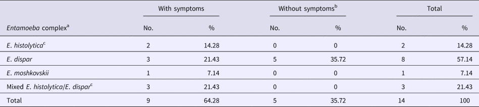
a Only cysts form were seen under microscopy.
b All Entamoeba complex isolates except five of E. dispar were associated with clinical symptoms.
c Two patients were found immunocompromised, one with E. histolytica and another with mixed E. histolytica/E. dispar.
Sequencing analysis of PCR products
Twelve out of 14 positive samples, including one E. moshkovskii, five E. histolytica and six E. dispar samples, were sequenced with species-specific primers in forward directions, using an ABI 3730XL sequencer (Macrogen® Corp., Seoul, South Korea). The BLAST analysis showed that sequences of six E. dispar amplicons under accession numbers KY823418 to KY823423 were 100% identical to the available GenBank sequences for E. dispar with the accession number KP722600.1. On the other hand, five E. histolytica sequences, with accession numbers KY823424 to KY823427 and KY884295, showed high homology (99–100%) to the GenBank sequences of E. histolytica under accession number KP233840.1. The only detected isolate of E. moshkovskii amplicon, under accession number KY823428, showed 100% homology to the sequences of E. moshkovskii under GenBank accession number KP722605.1.
Risk factors for Entamoeba complex infection
The results of single-variable logistic regression analysis for the evaluation of risk factors for Entamoeba complex infection and socio-demographic characteristics are presented in Table 3. According to Table 3, among the studied factors, none showed a significant relationship with Entamoeba complex infection.
Univariate analysis of risk factors associated with Entamoeba complex infection among individuals attended to the medical laboratories in Sanandaj County, Kurdistan, Northwest Iran(n = 1383)

N, number; OR, odds ratio; Reference, the subgroup is considered as baseline.
a Symptoms of gastrointestinal discomfort also occurred.
Discussion
Amoebiasis is one of the most common infections among humans worldwide [5]. The three studied species are morphologically similar, despite genetic and pathogenic differences [Reference Khairnar and Parija13]. E. histolytica is generally considered a pathogenic species, while other Entamoeba species are classified as non-virulent [Reference Anuar12, Reference Ali14]; therefore, distinguishing of these species is of great significance.
Microscopic methods, as well as molecular approaches, were used in this study for the detection of Entamoeba species and differentiation of E. histolytica, E. dispar and E. moshkovskii. In the medical laboratories of many countries, including our region, detection of Entamoeba is based on the microscopic identification of cysts or trophozoites. These methods are usually accompanied by misdiagnosis, and it is impossible to differentiate between the isolates of Entamoeba complex [Reference Tanyuksel and Petri17]. Therefore, molecular approaches have been developed to differentiate and detect Entamoeba species in faecal samples.
To the best of our knowledge, this study was the first to distinguish Entamobea species and to assess the prevalence of E. histolytica, E. dispar and E. moshkovskii in Kurdistan Province in west of Iran. Furthermore, we described the association of Entamoeba species with clinical symptoms among individuals, attended to 14 medical laboratories. The results of molecular studies showed that E. dispar, E. histolytica and E. moshkovskii infections are present in the study area. However, the prevalence of these amoebae and other parasites has dramatically decreased in recent years, similar to other regions of Iran.
According to the WHO/PAHO/UNESCO report and many conducted studies, the prevalence of E. dispar is greater than that of E. histoltyica and E. moshkovskii [5, Reference Anuar12]. Our findings also demonstrated that 78% (11/14) of the samples were attributed to E. dispar (eight samples with single infections and three samples containing both E. dispar and E. histolytica). The prevalence of E. dispar in the present study is close to most previous studies carried out in northern, central and southern Iran [Reference Hooshyar18], Khoramabad [Reference Kheirandish19], Gonabad [Reference Nejad20], Zahedan [Reference Haghighi21], Ahvaz and Hamidieh [Reference Rezaeian and Houshyar22] and Miandoab [Reference Fallah23], as well as studies from Malaysia [Reference Anuar12], Northern Ghana [Reference Verweij24], South Africa [Reference Samie25], Australia [Reference Fotedar3], Northwest Ethiopia [Reference Seong-Choon26] and the Netherlands [Reference Vreden27]. However, studies from Saqqez, Iran [Reference Mohammadzadeh28], south-west of Iran [Reference Pestehchian29], United Arab Emirates [Reference ElBakri30] and Gaza Strip [Reference Al-Hindi31] reported different results in areas where E. histolytica was more prevalent.
In 1997, WHO reported that most cases of E. histolytica may be in fact E. dispar, which is known to be non-pathogenic [5]. However, cases of E. histolytica infection have been reported in patients without symptoms. For instance, in studies from the Philippines [Reference Rivera, Tachibana and Kanbara32] and Japan [Reference Tachibana33], most positive cases of E. histolytica were considered asymptomatic. The prevalence of E. histolytica (single and mixed infections) in our population was 0.36% (5/1383). GIDs, including abdominal pain and chronic diarrhoea, were reported in all cases infected with E. histolytica (single and mixed infections). This finding is consistent with several reports from Pakistan [Reference Yakoob34] and South Africa [Reference Samie25], which showed that E. histolytica commonly produces clinical symptoms in patients.
It is commonly believed that E. dispar is a non-virulent species [Reference Ximénez1]. In this regard, Espinosa et al. reported that E. dispar is non-virulent under in vivo conditions [Reference Espinosa, Castañón and Martínez-Palomo35]. In addition, Oliveira et al. found that E. dispar was commensal and non-pathogenic to humans [Reference Oliveira36]. Dvorak et al. also suggested that E. dispar (SAW760 and SAW1734) strains are non-virulent [Reference Dvorak37]. These reports are in contrast with a study by Herbinger et al., which showed that most E. dispar isolates were associated with GIDs in returning travellers [Reference Herbinger15]. According to some studies, the Brazilian strain of E. dispar is pathogenic and can produce amoebic liver abscess under in vivo conditions [Reference Shibayama38]. Based on our findings, eight out of 11 patients with E. dispar had single infection, while three cases showed GIDs including abdominal pain.
It was initially hypothesised that E. moshkovskii is a non-virulent and free-living Entamoeba species [Reference Clark and Diamond39]. However, this is inconsistent with our findings, as gastroenteritis symptoms, such as abdominal pain and chronic diarrhoea, were observed in one patient infected with E. moshkovskii. Similarly, four studies from Australia, Tunisia, Malaysia and Bangladesh showed that humans can be true hosts for this species [Reference Fotedar3, Reference Ayed10, Reference Anuar11, Reference Ali14]. Also, some studies from Australia, Malaysia and India linked E. moshkovskii infection to GIDs [Reference Fotedar4, Reference Anuar12, Reference Parija and Khairnar40]. A study from Malaysia recommended that further investigations are necessary to determine the relationship between E. moshkovskii and GIDs and to identify the possible pathogenicity of this species [Reference Anuar12].
In the present study, considering the low number of positive cases, besides practical and financial limitations, other probable factors, such as bacterial, fungal and viral infections, or other non-infectious diseases associated with gastroenteritis symptoms were not examined and cannot be ruled out. Therefore, we cannot confirm the association between clinical symptoms and Entamoeba complex infection, and future investigations are necessary in this area.
In conclusion, this study reported the presence of E. histolytica, E. dispar and E. moshkovskii in Kurdistan Province, especially among patients with GIDs, although these species were not commonly detected. Based on the findings, the prevalence of E. dispar is greater than E. histoltyica and E. moshkovskii. Only a few cases of E. moshkovskii have been reported in Iran, and a single isolate of this amoeba was detected for the first time in our study. Overall, we found that E. dispar and E. moshkovskii might be associated with GID symptoms.
Supplementary material
The supplementary material for this article can be found at https://doi.org/10.1017/S0950268819000141.
Author ORCIDs
Ghasem Zamini, 0000-0002-7017-2647
Acknowledgements
We thank all technicians and personnel working in the medical laboratories, School of Medicine, Kurdistan University of Medical Sciences and also those who work at the Department of Parasitology and Mycology, School of Medicine, Shahid Beheshti University of Medical Sciences who contributed to the study.
Financial support
This work was a part of the Ph.D. thesis of Fares Bahrami under supervision of Professor Ali Haghighi and Dr Ghasem Zamini, which was supported financially by Kurdistan University of Medical Sciences (Grant No. 335) and Shahid Beheshti University of Medical Sciences (Grant No. 6598).
Conflict of interest
None.
Ethical approval
The authors assert that all procedures contributing to this work comply with the ethical standards of the Declaration of Helsinki, revised in 2008. The trial was reviewed and approved by the Ethics Committee of Shahid Beheshti University of Medical Sciences.


 Open access

