Introduction
Neospora caninum is an apicomplexan intracellular obligate protozoon, which causes neurological disorders in congenitally infected dogs and abortions in cattle (Dubey et al. Reference Dubey, Hattel, Lindsay and Topper1988, Reference Dubey, Lindsay, Anderson, Davis and Shen1992). In the pathogenesis of bovine neosporosis, cattle (an intermediate host) ingest oocysts shed by dogs (the definitive host of N. caninum) resulting in horizontal transmission of the parasite (Uggla et al. Reference Uggla, Stenlund, Holmdahl, Jakubek, Thebo, Kindahl and Bjorkman1998; Dijkstra et al. Reference Dijkstra, Barkema, Eysker, Hesselink and Wouda2002). N eospora caninum oocysts enter the tachyzoite phase in the gastrointestinal tract of cattle and the bradyzoite stage after invading neural and muscle tissues (Tunev et al. Reference Tunev, McAllister, Anderson-Sprecher and Weiss2002; Dubey, Reference Dubey2003). The dog is not only the definitive host but can also suffer severe disease as an intermediate host (Lindsay and Dubey, Reference Lindsay and Dubey1989). N eospora caninum bradyzoites can remain latent for years in cysts in neural and muscle tissue, but can re-enter the tachyzoite stage if host immunity is compromised (Tunev et al. Reference Tunev, McAllister, Anderson-Sprecher and Weiss2002; Dubey, Reference Dubey2003).
It remains controversial as to whether N. caninum can infect humans. Conflicting evidence has arisen from serological studies (Petersen et al. Reference Petersen, Lebech, Jensen, Lind, Rask, Bagger, Bjorkman and Uggla1999; Tranas et al. Reference Tranas, Heinzen, Weiss and McAllister1999; McCann et al. Reference McCann, Vyse, Salmon, Thomas, Williams, McGarry, Pebody and Trees2008). A study of 1029 blood donors in California found that 6·7% had antibodies against N. caninum (Tranas et al. Reference Tranas, Heinzen, Weiss and McAllister1999). Higher sero-prevalences for antibodies against N. caninum (26–38%) have been reported in some HIV positive human populations (Lobato et al. Reference Lobato, Silva, Mineo, Amaral, Segundo, Costa-Cruz, Ferreira, Borges and Mineo2006; Oshiro et al. Reference Oshiro, Motta-Castro, Freitas, Cunha, Dittrich, Meirelles and Andreotti2015). Conversely, two studies in the UK and Denmark found no serologic evidence of N. caninum infection but did not report data specifically on HIV positive or immunocompromised individuals (Petersen et al. Reference Petersen, Lebech, Jensen, Lind, Rask, Bagger, Bjorkman and Uggla1999; McCann et al. Reference McCann, Vyse, Salmon, Thomas, Williams, McGarry, Pebody and Trees2008). In vitro studies have shown that N. caninum can readily replicate in multiple human cell types, including primary monocyte-derived dendritic cells (Collantes-Fernandez et al. Reference Collantes-Fernandez, Arrighi, Alvarez-Garcia, Weidner, Regidor-Cerrillo, Boothroyd, Ortega-Mora and Barragan2012), brain microvascular endothelial cells (Elsheikha et al. Reference Elsheikha, McKinlay, Elsaied and Smith2013), trophoblastic (BeWo) cells, uterine cervical (HeLa) cells (Carvalho et al. Reference Carvalho, Alves, Cardoso, Mota, Barbosa, Ferro, Silva, Mineo, Mineo and Silva2010) and breast carcinoma (MCF-7) cells (Lv et al. Reference Lv, Li, Gong, Xing and Zhang2010). However, to our knowledge, no studies have established if N. caninum can replicate in human monocyte-derived macrophages and whether neosporosis is zoonotic remains inconclusive.
Macrophages have key roles in early responses to intracellular pathogens, including N. caninum. Macrophages were present in necrotic lesions in placentas of N. caninum-infected cows (Canton et al. Reference Canton, Katzer, Benavides-Silvan, Maley, Palarea-Albaladejo, Pang, Smith, Bartley, Rocchi, Innes and Chianini2013). Furthermore, macrophage responses were associated with survival in mice infected intraperitoneally with live N. caninum tachyzoites (NC-1 strain), whereas chemical depletion of macrophages resulted in earlier death in infected mice (Abe et al. Reference Abe, Tanaka, Ihara and Nishikawa2014). Macrophages have evolved effector functions to specifically combat intracellular microbes that also appear effective against N. caninum. Interferon-gamma (IFNγ) eliminated N. caninum tachyzoites in murine macrophages by inducing nitric oxide production (Tanaka et al. Reference Tanaka, Nagasawa, Fujisaki, Suzuki and Mikami2000). Mice with macrophages lacking the receptor for TNFα (TNFR2) had increased mortality and clinical signs when infected with N. caninum (strain NC-1) (Ritter et al. Reference Ritter, Kerlin, Sibert and Brake2002). Synthesis of other classical inflammatory cytokines (e.g. IL-1β and IL-8) were reported in bovine macrophages and umbilical endothelial vein cells infected with N. caninum, respectively (Taubert et al. Reference Taubert, Zahner and Hermosilla2006b; Flynn and Marshall, Reference Flynn and Marshall2011) but their functional significance remains elusive.
Host defence peptides, including cathelicidins, are cationic peptides with antimicrobial and immunomodulatory properties abundantly synthesized by leucocytes (Rajapakse et al. Reference Rajapakse, Uring-Lambert, Andarawewa, Rajapakse, Abou-Bacar, Marcellin and Candolfi2007; Yuk et al. Reference Yuk, Shin, Lee, Yang, Jin, Kim, Lee, Lee, Kim and Jo2009). Exogenous 1,25-dihydroxy vitamin D3, which is known to increase transcription of cathelicidin antimicrobial peptide (CAMP) (Gombart et al. Reference Gombart, Borregaard and Koeffler2005), reduced the growth of Toxoplasma gondii (intracellular protozoa closely related to Neospora) in murine intestinal epithelial cells (Rajapakse et al. Reference Rajapakse, Uring-Lambert, Andarawewa, Rajapakse, Abou-Bacar, Marcellin and Candolfi2007). It has also been shown that the upregulation of CAMP through stimulation with 1,25-dihydroxy vitamin D3 increased clearance of Mycobacterium tuberculosis in human macrophages through autophagy (Gombart et al. Reference Gombart, Borregaard and Koeffler2005; Yuk et al. Reference Yuk, Shin, Lee, Yang, Jin, Kim, Lee, Lee, Kim and Jo2009). However, the role of host defence peptides in N. caninum infection has apparently never been reported. Despite the importance of macrophages in controlling infection, N. caninum tachyzoites subvert cellular immune defenses to replicate in murine peritoneal macrophages (Dion et al. Reference Dion, Germon, Guiton, Ducournau and Dimier-Poisson2011). In this regard, specific subtypes of leucocytes (CD11c and CD11b) transported T. gondii tachyzoites to the brain of infected mice, suggesting this protozoon evades host leucocyte defences to propagate (Courret et al. Reference Courret, Darche, Sonigo, Milon, Buzoni-Gatel and Tardieux2006). Thus, the objective was to investigate interactions between monocyte-derived macrophages and N. caninum early in pathogen recognition and defence, namely the production of pro-inflammatory cytokines and host defence peptides.
Material and methods
Cell cultures
Human peripheral blood monocytic THP-1 cells (ATCC TIB-202) were chosen as a suitable in vitro model to study modulation of monocyte and macrophage functions (Daigneault et al. Reference Daigneault, Preston, Marriott, Whyte and Dockrell2010; Chanput et al. Reference Chanput, Mes and Wichers2014). THP-1 cells are good monocyte model for neosporosis because THP-1 cells and primary bovine monocytes appear to express similar cytokine profiles (TNFα, IL-1β, IL-8) in response to inflammatory stimuli, such as lipopolysaccharide (Daigneault et al. Reference Daigneault, Preston, Marriott, Whyte and Dockrell2010; Hussen et al. Reference Hussen, Duvel, Sandra, Smith, Sheldon, Zieger and Schuberth2013). These cells were maintained in RPMI Medium 1640 (Gibco) with 10% fetal bovine serum (FBS) (Benchmark Gemini), HEPES buffer solution (10 mm; Gibco), 2-mercaptoethanol (Gibco) and penicillin (100 U mL−1) and streptomycin (100 µg mL−1; HyClone Thermo, Fisher Scientific). To maintain N. caninum culture in vitro, African green monkey (Cercopithecus aethiops) kidney epithelial adherent cells (Vero; ATCC CCL-81) were used as host cells. These cells were maintained in Dulbecco's modified Eagle's medium (Gibco) with 10% FBS (Benchmark Gemini Bio-Products), sodium pyruvate (1 mm; Gibco) and penicillin (100 U mL−1) and streptomycin (100 µg mL−1; HyClone Thermo). Both THP-1 and Vero were grown in a humidified environment of 95% air and 5% CO2 at 37 °C.
Neospora caninum and derived soluble antigens
Tachyzoites of the N. caninum strain NC-1 (kindly provided by Dr J. R. Barta, Ontario Veterinary College, Guelph, ON, Canada) were maintained in vitro by a continuous passage in Vero cells and harvested when 80% of cells were infected, as described (Dubey et al. Reference Dubey, Hattel, Lindsay and Topper1988). The viability of harvested NC-1 tachyzoites was determined with 0·2% trypan blue staining (Gibco). For the experimental challenge, tachyzoites were harvested when >50% of Vero cells were infected, lysed, centrifuged (1350g; 15 min 4 °C), filtered (5 µm) and stained with trypan blue and enumerated with a hemocytometer. Tachyzoites used were immediately diluted in RPMI medium without FBS.
For the production of soluble N. caninum antigens, tachyzoites were harvested as described (Moore et al. Reference Moore, Echaide, Verna, Leunda, Cano, Pereyra, Zamorano, Odeon and Campero2011) and pellets subjected to 3 freeze/thaw cycles at −80 °C. Pellets were re-suspended in protease-free, sterile water with halt protease inhibitors (Thermo Scientific) and sonicated for 6 cycles of 15 s. Proteins were quantified with a BCA assay (Pierce BCA protein assay kit, Thermo Scientific). This preparation contains N. caninum antigens only and not any other immunologically active components (e.g. host cells or bacteria) because tachyzoites were exposed to freezing and thawing cycles, host cells were removed by centrifugation and bacterial contamination was absent. To confirm the specificity of the N. caninum antigen preparation, Western blotting was performed with specific antibodies against N. caninum. For this, the N. caninum antigen preparation was concentrated (Corning Spin-X UF 30 K MWCO concentrators). Each sample (0–100 µg protein) was mixed 1:1 with Laemmli sample buffer and electrophoresed through 12% SDS-PAGE gels. Proteins were subsequently transferred onto a PVDF membrane (BioRad) activated with methanol and stained with Ponceau S. Membranes were blocked with 5% skim milk powder dissolved in Tris buffered saline plus 0·1% Tween 20 solution (TBST) for 1 h and probed with anti-N. caninum antibody (5B6–24, VMRD; 1·25 µg mL−1) at 4 °C overnight. After incubation with a horseradish-peroxidase-conjugate secondary anti-mouse antibody (115–035–1461, Jackson ImmunoResearch; 1: 10 000) for 2 h at room temperature (RT), blots were developed using a Clarity Western ECL Detection System (BioRad).
Experimental design
Monocytic THP-1 cells were seeded at 1 × 10⁵ cells/well (24-well plates) (Greiner Bio-One) for gene expression and ELISAs; at 2 × 10⁵ cells/well (12-well plates) for Western blotting assays, and at 5 × 10⁴ cells/chamber (8-well chambers) or 1 × 10⁵ cells/chamber (4-well chambers) for confocal immunofluorescence microscopy. Monocytes were differentiated into phagocytic macrophage-like cells with phorbol 12-myristate 13-acetate (PMA; 50 ng mL−1) for 4 d, with PMA replenished once daily (Park et al. Reference Park, Jung, Yang, Yoo, Kim and Kim2007). Monocyte-derived macrophages were exposed to live N. caninum tachyzoites at various multiplicities of infection (MOIs 0, 1, 5 and 10) for 4 h, or incubated with soluble antigens of N. caninum (0, 5 or 10 µg mL−1) for 4 or 16 h. Multiplicities of infection (MOI) represented the ratio of N. caninum tachyzoites to challenged THP-1 cells and it is usually used to define the grade of a challenge with N. caninum and related Toxoplasma gondii (Clough et al. Reference Clough, Wright, Pereira, Hirst, Johnston, Henriques and Frickel2016; Wang et al. Reference Wang, Gong, Zhang, Wang, Tai, Wang, Wei, Yang, Yang, Li and Zhang2017). Treatment with PMA did not increase or decrease the number of THP-1 cells, which is consistent with previous studies (Daigneault et al. Reference Daigneault, Preston, Marriott, Whyte and Dockrell2010; Spano et al. Reference Spano, Barni and Sciola2013) (i.e. the numbers of THP-1 cells seeded per well and exposed to N. caninum challenge were the same).
To study signalling pathways involved in the innate response, THP-1 macrophages were pre-treated for 1 h with pharmacological inhibitors of ERK kinases MEK1 and MEK 2 [PD98059 (20 µ m); Cell Signalling], p38 MAPK [SB203580 (20 µ m); Tocris] or NF-κB [caffeic acid phenyl ester (10 µ m); Tocris]. These inhibitors were used at the concentrations recommended by the manufacturers. PD98059 is an effective and specific inhibitor of the MEK1/2 pathway. It functions by preventing the activation of MEK1/2 by upstream activators, such as Ras (Alessi et al. Reference Alessi, Cuenda, Cohen, Dudley and Saltiel1995). SB203580 is an effective inhibitor of p38 MAPK and has minimal effects on other protein kinases and phosphatases (Cuenda et al. Reference Cuenda, Rouse, Doza, Meier, Cohen, Gallagher, Young and Lee1995). Caffeic acid phenyl ester (CAPE) prevents NF-κB from binding DNA by blocking the translocation of the p65 subunit to the nucleus but does not affect other transcription factors (Natarajan et al. Reference Natarajan, Singh, Burke, Grunberger and Aggarwal1996). The control group was treated with dissolvent only (dimethyl sulfoxide, DMSO; Sigma-Aldrich).
To assess the infectivity of N. caninum tachyzoites when exposed to supernatant collected from infected macrophages, naïve N. caninum tachyzoites were pre-incubated with supernatants collected from macrophages previously challenged with N. caninum for 4 h or macrophages that were not challenged. Exposed N. caninum tachyzoites were washed and then incubated with naïve macrophages for 24 h to assess the tachyzoites’ ability to invade cells. Extracellular viable tachyzoites were counted using a hemocytometer. Intracellular tachyzoites were recovered by passing infected macrophages through a 25-gauge syringe and then enumerated.
It has previously been shown that THP-1 differentiated to a macrophage-like state with PMA phagocytose fewer latex beads than undifferentiated THP-1 monocytes (Daigneault et al. Reference Daigneault, Preston, Marriott, Whyte and Dockrell2010). To confirm that N. caninum tachyzoites were not being internalized through phagocytosis/endocytosis, THP-1 macrophages were pre-treated with the pharmacological inhibitor of endocytosis, D15 (2 m; Tocris) for 1 h, according to the manufacturer's instructions. Intracellular and extracellular tachyzoites were counted as described.
To study secreted factors released by macrophages infected with N. caninum, naïve THP-1 macrophages were incubated for 4 or 20 h with supernatants taken from THP-1 macrophages previously incubated with N. caninum (MOI 5; for 4 h). To obtain secreted cell products free of parasites, supernatants from infected THP-1 cells were centrifuged (1350g; 15 min 4 °C) and pellets discharged to eliminate N. caninum tachyzoites. Secreted cytokines IL-8, IL-1β, IL-10 and TNFα proteins in supernatants of macrophages were measured with human cytokine assay kits as described below (Quantikine® ELISA, R&D).
Intracellular identification of N. caninum in monocyte-derived macrophages
THP-1 macrophages infected with N. caninum were rinsed with phosphate buffered saline (PBS) solution and fixed in cold acetone for 10 min. Macrophages were then rinsed in cold PBS plus Tween 0·05% (pH 7·2) (PBS-Tw), then blocked in PBS-Tw, 1% bovine serum albumin, 10% donkey serum and 0·3 M glycine for 1 h at RT and rinsed with PBS-Tw. Cells were blotted with mouse anti-N. caninum mAb (5B6–24, VMRD; 1·25 µg mL−1) at 4 °C overnight. Macrophages were rinsed again with cold PBS-Tw, then blotted with Alexa Fluor® 594-conjugated AffiniPure Donkey Anti-Mouse IgG (H+L) (715–585–150, Jackson ImmunoResearch). Secondary antibodies were diluted 1:1000 in PBS-Tw plus 1% bovine serum albumin and incubated for 1 h at RT. Preparations were rinsed in cold PBS-Tw and nuclei counterstained with 4′, 6-diamidino-2-phenylindole (DAPI) (Thermofisher) and Alexa Fluor® 568 phalloidin (A12380, Thermo Scientific). Sections were rinsed with cold PBS-Tw and mounted with FluorSave reagent (Calbiochem, EMB Millipore). Slides were examined using a FluoView FV1000 confocal immunofluorescence microscope (Olympus). Uninfected macrophages were prepared in parallel as negative controls.
Gene transcription of cytokines and host defence peptides in monocyte-derived macrophages
Relative messenger gene (mRNA) transcription of human cathelicidin/LL-37 (cathelicidin antimicrobial peptide; CAMP) and cytokines (IL-8, IL-1β, TNFα, IFN-γ, IL-10) from macrophages was quantified by real-time qRT-PCR. Total RNA from THP-1 cells was isolated using TRIzol reagent (Invitrogen), as suggested by the manufacturer. Complementary DNA (cDNA) was prepared from 1 µg of total RNA using Moloney murine leukemia virus reverse transcriptase (iScript Reverse Transcription Supermix for RT-qPCR, BioRad). The quality and quantity of resulting RNA and cDNA were determined using a NanoVue Spectrophotometer (GE Healthcare). The absence of contaminating genomic DNA from RNA preparations was checked using a minus-reverse transcriptase control (i.e. a sample with all RT-PCR reagents except reverse transcriptase). Real-time qRT-PCR was performed using a CFX-96 real-time PCR system (BioRad). Each reaction mixture contained 100 ng of cDNA, 1X SsoAdvanced Universal SYBR Green Supermix (BioRad) and 0·5 µ m of each specific primer, in a final volume of 10 µL. Human primers for CAMP (PPH09430A), IL-8 (CXCL8; PPH00568A), IL-1β (PPH00171C), TNFα (PPH00341F), IFN-γ (PPH00380C), IL-10 (PPH00572C) and glyceraldehyde-3-phosphate dehydrogenase (GAPDH) (PPH00150F) (RT2 qPCR Primer Assay, Qiagen) were used. Primers used in this study (RT2 qPCR Primer Assay, Qiagen) were experimentally verified for specificity and efficiency. The RT-qPCR efficiency for each gene was calculated from the slope and determined by a linear regression model according to the equation: Efficiency = 10[−1/slope] – 1, as indicated in The MIQE Guidelines (Bustin et al. Reference Bustin, Benes, Garson, Hellemans, Huggett, Kubista, Mueller, Nolan, Pfaffl, Shipley, Vandesompele and Wittwer2009) (Table 1). R 2 values were also calculated using 10-fold serial dilutions of cDNA (Table 1). Reaction mixtures were incubated at 95 °C for 5 min, followed by denaturation for 5 s at 95 °C and combined annealing/extension for 10 s at 60 °C for a total of 40 cycles. Two housekeeping genes, GAPDH and β-actin, were initially tested and because neither had alterations under our treatments; we proceeded using the former gene. Negative controls for cDNA synthesis and PCR procedures were included in all cases. Values of target mRNA were corrected relative to the housekeeping gene coding GAPDH. Data were analysed using the 2−ΔΔCT method and results reported as mean fold change of the target transcription levels in infected groups (MOIs 5 and 10) vs. uninfected (MOI 0) control group.
Details of primers for mRNA relative quantification by real-time RT-PCR

a The RefSeq accession no. refers to the sequence used to design the RT2 qPCR Primer Assay.
Protein determination of cytokines in monocyte-derived macrophages
Protein levels of TNFα, IL-1β and phosphorylated CCAAT-enhancer binding protein-β (phospho-C/EBPβ) were determined by Western blotting in macrophages lysed in denaturing cell extraction buffer (DCEB; Invitrogen). Protein concentrations were determined with a BCA assay (Pierce BCA protein assay kit, Thermo Scientific). Each sample (15-μg protein) was mixed 1:1 with Laemmli sample buffer and separated on 10% SDS-PAGE gels. Subsequently, proteins were transferred onto a PVDF membrane (BioRad) activated with methanol. The membrane was blocked with 5% skim milk powder (or 5% bovine serum albumin for phosphorylated proteins) dissolved in Tris-buffered saline plus 0·1% Tween 20 solution (TBST) for 1 h. Membranes were probed with anti-TNFα (ab6671, Abcam; 1:500), anti-IL-1β (12242 Cell Signalling; 1:1000), anti-phospho-C/EBPβ (Thr235) (3084, Cell Signalling; 1:1000) or anti-GAPDH (CB1001, Calbiochem; 1:1000) antibodies at 4 °C overnight. After incubation with a horseradish-peroxidase-conjugate secondary anti-mouse or anti-rabbit antibody (115–035–146 or 111–035–144, Jackson ImmunoResearch; 1:10 000) for 2 h at RT, blots were developed using the Clarity Western ECL Detection System (BioRad). The image captures and densitometric analyses were performed with the ChemiDoc MP Imaging system and ImageLab 4·0·1 software (BioRad), respectively. Normalization was done with reference to the GAPDH lane protein. Results are reported as mean fold change of target expression in infected groups compared with an uninfected control group.
Levels of secreted IL-8, IL-1β, IL-10 and TNFα proteins in supernatants of THP-1 cells exposed to N. caninum were determined by human IL-8, IL-1β, IL-10 and TNFα cytokine assay kits (Quantikine® ELISA, R&D). All reagents including standard dilutions and samples were prepared and processed as recommended by the manufacturer. Briefly, sample, blank or standard and RD1–85 Assay Diluent (for IL-8) were loaded into each well of microplate strips coated with anti-IL-8, anti-IL-1β, anti-IL-10 or anti-TNFα antibody. Plates were sealed and incubated for 2 h at RT. RPMI Medium 1640 as described above (without FBS) was used as blank for both ELISAs. After binding of IL-8, IL-1β, IL-10 or TNFα peptide to an antibody wells were washed with wash buffer for 3 × 5 min2 each. Subsequently, IL-8, IL-1β, IL-10 or TNFα conjugate was added and incubated for 1 h at RT. Wells were washed again and substrate solution was added. Plates were read at 450/540 nm and the difference calculated to account for background absorption. Absorption for treatments/control was plotted on a standard curve and corresponding concentrations were obtained.
Statistical analyses
Graphs represent normally/Gaussian distributed (parametric) results represented as means and bars represent standard errors of the mean from a minimum of two independent experiments. Normality was assessed using the Shapiro-Wilk (Royston (1)) test. All comparisons were performed using the paired 2-tailed Student's t-test between the treated groups and the control group. P values < 0·05 were considered statistically significant. All statistical analysis was performed with Graph Pad Prism software (Graph Pad 7·0).
Results
Neospora caninum colonizes human monocyte-derived macrophages in vitro
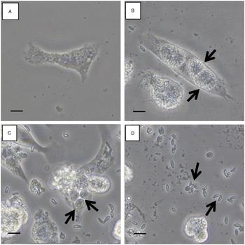
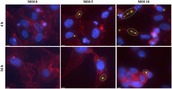
Limited studies have described the effects of N. caninum in bovine macrophages (Flynn and Marshall, Reference Flynn and Marshall2011). Moreover, it is unknown whether N. caninum can invade, colonize and persist in human phagocytic monocytes (THP-1), which are widely used for modelling host–intracellular pathogen interactions and monocyte and macrophage functions/response functions (Chanput et al. Reference Chanput, Mes and Wichers2014). To study early mononuclear responses to N. caninum, healthy THP-1 cells were differentiated into macrophages (Fig. 1A) and then, exposed to live tachyzoites for several days. Parasitophorous vacuoles inside THP-1 macrophages, characteristic of apicomplexan infection (Hemphill et al. Reference Hemphill, Gottstein and Kaufmann1996), were microscopically observed at different MOIs during the course of infection. For instance, N. caninum parasitophorous vacuoles were observed at 5 d post-infection (dpi) (MOI 1) (p.i.; Fig. 1B) as well as 1 dpi (MOI 5) (Fig. 1C). Each vacuole contained several tachyzoites (Fig. 1B, C). The cellular rupture was detectable as early as 2 dpi with high N. caninum challenge (MOI 5) (Fig. 1D). To confirm the presence of N. caninum inside cells, tachyzoites were immune-blotted with a monoclonal antibody against N. caninum surface antigens (NC) and counterstained with DAPI and phalloidin. Confocal microscopy studies identified N. caninum immune staining in the cytoplasm of THP-1 cells at 4 h and 16 h post incubation, thereby confirming N. caninum was internalized by macrophages (Fig. 2, Fig. S1). To confirm that N. caninum tachyzoites were invading macrophages as opposed to entering via endocytosis, THP-1 were treated with the endocytosis inhibitor, D15. No significant reduction in intracellular (or extracellular) N. caninum tachyzoites were observed in THP-1 treated with D15 compared with DMSO (Fig. 3A, B).
Neospora caninum was internalized in human monocyte-derived macrophages. (A) THP-1 not exposed to N. caninum (MOI 0). (B) Parasitophorous vacuoles (marked by arrows) in THP-1 macrophages after infection with N. caninum (MOI 1) visualized with optical microscopy at 5 d p.i. (C) Parasitophorous vacuoles (marked by arrows) in THP-1 macrophages after infection with N. caninum (MOI 5) visualized with optical microscopy at 1 d p.i. (D) Lysed macrophages and presence of extracellular N. caninum tachyzoites (marked by arrows) at 2 d p.i. after infection with N. caninum (MOI 5). Images are representative of one of three independent experiments. Scale bar = 12 µm.

Confocal immunolocalization of N. caninum internalized in monocyte-derived macrophages. Neospora caninum tachyzoites (circled) were identified inside macrophages after 4 and 16 h incubation with N. caninum MOI 0 (left), MOI 5 (center) or MOI 10 (right). N. caninum tachyzoites were stained with a mAb against a 65-kDa glycosylated N. caninum surface protein (yellow). Macrophages were counterstained with phalloidin, which binds actin (red) and the nucleus with DAPI (blue). Images are representative of one of three independent experiments. Scale bar = 15·78 µm.

Neospora caninum replicates in and invades monocyte-derived macrophages. (A-B) THP-1 macrophages (2 × 10⁵) were seeded (12-well plates) and were treated with an endocytosis inhibitor (D15) for 1 h before incubation with N. caninum tachyzoites (MOI 5 or MOI 10). (A) Intracellular and (B) extracellular tachyzoites were counted after 4 h by hemacytometer. Means + the s.e. are shown (n = 3 independent experiments run in duplicate). P < 0·05 (Student's t test), N.S. means not significant. (C-D) THP-1 macrophages (1 × 10⁵) were seeded (24-well plates) and were infected with N. caninum tachyzoites (either MOI 1 or MOI 5). Extracellular N. caninum tachyzoites were counted daily by hematocytometer (viability verified with trypan blue). (C) Viable extracellular N. caninum tachyzoites were at the highest concentration at 2 d p.i. with MOI 1 N. caninum after substantial lysis of macrophages (n = 3 independent experiments run in duplicate). *P < 0·05 (Student's t test). (D) The supernatant containing extracellular tachyzoites was transferred to uninfected naïve macrophages for 3 passages (generations) and viable extracellular tachyzoites were enumerated daily. Viable N. caninum tachyzoites (both MOI 1 and 5) were detected at 2 d p.i. in supernatants of exposed macrophages during all 3 passages and at higher concentrations compared to the initial inoculum. Means + the s.e. are shown (n = 2 independent experiments run in triplicate). N.S., not significant; *P < 0·05 (Student's t test).

Next, to determine if N. caninum tachyzoites were able to complete their life cycle in THP-1 macrophages, including replication and infection of surrounding cells, THP-1 cells were infected with various MOIs of N. caninum tachyzoites and followed for several days by enumerating live extracellular tachyzoites and changing the medium daily (Fig. 3C, D). The concentration of extracellular tachyzoites decreased 1 dpi, suggesting parasites were entering macrophages (Fig. 3C). However, the concentration of extracellular tachyzoites increased at 2 dpi (10- to 20-fold from the initial MOI; Fig. 3C), which was concurrent with microscopic confirmation of lysis of infected macrophages and the release of tachyzoites into the supernatant (Fig. 1D). Because tachyzoite counts 2 dpi were higher than our initial inoculum, we confirmed that N. caninum replicated in our in vitro model. To demonstrate that these N. caninum tachyzoites were able to infect naïve macrophages, the supernatant containing the released extracellular tachyzoites was transferred to uninfected naïve cells after lysis of infected macrophages (approximately 2 dpi). This process was repeated 3 times (Fig. 3D). The number of extracellular tachyzoites remained higher than our initial inoculum after macrophages lysis occurred, which confirmed that N. caninum tachyzoites retained infectivity in macrophages after multiple passages (Fig. 3D). Therefore, N. caninum was capable of infecting, replicating and persisting in human THP-1 monocytes, a classical cell line used for studying intracellular pathogens, as it does in bovine monocytes (Lindsay and Dubey, Reference Lindsay and Dubey1989).
Monocyte-derived macrophages respond to intracellular infection with N. caninum by producing pro-inflammatory cytokines and host defense peptide (cathelicidin)
When macrophages are exposed to inflammatory stimuli, they secrete a signature array of pro-inflammatory cytokines that include IFNγ, IL-8, TNFα and, IL-1β and sometimes, anti-inflammatory IL-10. These cytokines (IFNγ, IL-8, TNFα and, IL-1β) promote host defences by increasing vascular permeability and recruiting inflammatory cells to the site of infection. In terms of macrophage responses to N. caninum, live N. caninum tachyzoites (either MOIs 5 or 10) rapidly induced mRNA synthesis and secretion of neutrophil factor IL-8 (Fig. 4). This was accompanied by an increase in gene transcription (>100-fold increase) and release of TNFα (Fig. 5). Likewise, N. caninum tachyzoites induced gene transcription of IFNγ (Fig. 4) and gene transcription and release of IL-1β in macrophages (Fig. 5). In addition, a rapid increase in the rate of transcription of mRNA for anti-inflammatory IL-10 in macrophages infected with N. caninum was observed at 4 h (Fig. 4), but the secretion of IL-10 was not detectable (no data were shown). We also explored whether THP-1 macrophages infected with N. caninum produced inducible host defence peptides. Neospora caninum tachyzoites induced the gene synthesis of cathelicidin CAMP mRNA in macrophages (Fig. 4). Taken together, N. caninum tachyzoites rapidly invaded and induced the mRNA transcription of IFNγ, IL-8, TNFα, IL-1β, IL-10 and cathelicidins in THP-1 macrophages.
Effects of N. caninum infection on transcription and secretion of IL-8, and transcription of IFNγ, host defense peptide cathelicidin and IL-10. IL-8 mRNA transcription and secreted proteins and IFNγ, CAMP and IL-10 mRNA transcription were determined in N. caninum-infected macrophages. IL-10 secreted proteins were not detectable in the cell supernatant (data not shown). mRNA transcription levels were quantified by real-time qRT-PCR and means + the s.e. are shown (n = 3 independent experiments run in triplicate). *P < 0·05 (Student's t test). Secreted proteins were determined with ELISA and means + the s.e. are shown (n = 2 independent experiments run in triplicate). *P < 0·05 (Student's t test).

Effects of N. caninum infection on transcription, synthesis and secretion of TNFα and IL-1β. TNFα and IL-1β mRNA transcription, cell lysate and secreted proteins were determined in N. caninum-infected macrophages. mRNA transcription levels were quantified with real-time qRT-PCR. Means + the s.e. are shown (n = 3 independent experiments run in triplicate). *P < 0·05 (Student's t test). Protein fold-change was determined by Western blotting (n = 5 independent experiments) where bars (means + the s.e.) represent densitometry analysis for all the experiments. *P < 0·05 (Student's t test). Secreted proteins were determined with ELISA and represented as mean ± s.e.m. compared with control. Means + the s.e. are shown (n = 2 independent experiments run in triplicate). *P < 0·05 (Student's t test). N.S., not significant.

Soluble antigens of N. caninum partially contribute to the transcription of pro-inflammatory cytokines in monocyte-derived macrophages
To determine whether immune responses in macrophages are triggered by the invasion of N. caninum tachyzoites or soluble factors released by or contained in N. caninum, THP-1 cells were exposed to N. caninum antigens containing soluble cytoplasmic and membrane proteins. The presence of N. caninum in this antigenic preparation was confirmed by Western blot analysis using a monoclonal antibody against a 65-kDa N. caninum glycosylated surface antigen (Fig. 6A). Challenge of macrophages with up to 10 µg mL−1 of soluble N. caninum antigens for only 4 h induced gene synthesis of TNFα and IL-1β cytokines (Fig. 6B). IL-8 mRNA transcription was not affected in macrophages after 4 h stimulation with soluble N. caninum antigens (Fig. 6B). When macrophages were exposed to soluble N. caninum antigens for a longer interval (16 h), mRNA transcription of TNFα, IL-1β and IL-8 cytokines were significantly increased (Fig. 7). However, mRNA transcription of TNFα, IL-1β or IL-8 induced by soluble N. caninum antigens was approximately 10-times lower than transcription induced by live tachyzoites. mRNA transcription of IFNγ and CAMP in macrophages was not significantly altered after exposure to soluble N. caninum antigens after 4 or 16 h (Fig. 6B & 7). Therefore, soluble antigens of N. caninum contributed to innate TNFα, IL-8 and IL-1β responses in infected macrophages.
Effects of N. caninum antigens on the transcription of pro-inflammatory cytokines after 4 h. (A) N. caninum antigens were blotted with a monoclonal antibody against a 65-kDa N. caninum glycosylated surface protein (top). Western blot of N. caninum antigens (0, 25 or 100 µg mL−1) stained with Ponceau S (below, left) were conducted using a monoclonal antibody against a 65-kDa N. caninum glycosylated surface protein (below, right). (B) mRNA transcription levels for TNFα, IL-1β, IL-8, IFNγ, and CAMP were determined after exposure to N. caninum soluble antigens (0, 5 or 10 µg mL−1) for 4 h. mRNA transcription levels were quantified with real-time qRT-PCR. Means + the s.e. are shown (n = 3 independent experiments run in triplicate). *P < 0·05 (Student's t test).

Effect of N. caninum antigens on the transcription of pro-inflammatory cytokines after 16 h. Transcription levels for TNFα, IL-1β, IL-8, IFNγ, and CAMP were determined in monocyte-derived macrophages after exposure to N. caninum soluble antigens (0, 5 or 10 µg mL−1) for 16 h. Transcription levels were quantified with real-time qRT-PCR. Means + the s.e. are shown (n = 3 independent experiments run in triplicate). *P < 0·05 (Student's t test).

The MEK 1/2 signalling pathway is involved in the transcription of pro-inflammatory cytokines and cathelicidins in macrophages infected with N. caninum
Mitogen-activated protein kinase 1/2 (MEK 1/2) and p38 mitogen-activated protein kinase (p38 MAPK) are rapidly phosphorylated in THP-1 macrophages infected with T. gondii (Valere et al. Reference Valere, Garnotel, Villena, Guenounou, Pinon and Aubert2003). Moreover, it has recently been demonstrated that antigens secreted by N. caninum rapidly phosphorylate p38 MAPK in murine bone marrow-derived macrophages (Mota et al. Reference Mota, Oliveira, Davoli-Ferreira, Silva, Santiago, Nadipuram, Vashisht, Wohlschlegel, Bradley, Silva, Mineo and Mineo2016). To further investigate the importance of the MEK 1/2, p38 MAPK and NF-κB signalling pathways in the production of pro-inflammatory cytokines and host defence peptides in macrophages in response to N. caninum, THP-1 cells were pre-treated with pharmacologically specific inhibitors before challenge with live N. caninum tachyzoites for 4 h. Inhibition of MEK 1/2 with PD98059 significantly decreased TNFα and IL-1β mRNA transcription (Fig. 8). Inhibition of MEK 1/2 decreased IL-8 and CAMP mRNA transcription, but results were not significant (Fig. 8). Inhibition of p38 MAPK with SB203580 reduced TNFα mRNA transcription significantly, as well (Fig. 8). Conversely, inhibition of NF-κB with CAPE did not affect the transcription of these pro-inflammatory cytokines and cathelicidin (Fig. 8). Thus, MEK 1/2 and p38 MAPK signalling were important pathways in the innate response of macrophages infected with N. caninum.
Effects of inhibition of MEK 1/2, p38 MAPK and transcription factor NF-κB on transcription of pro-inflammatory cytokines. Monocyte-derived macrophages were treated with inhibitors of MEK 1/2 (PD98059), p38 MAPK (SB203580) or NF-κB (CAPE) or DMSO for the control for 1 h and then infected with N. caninum for 4 h. TNFα, IL-1β, IL-8 and CAMP. mRNA transcription levels was quantified with real-time qRT-PCR. Means + the s.e. are shown (n = 2 independent experiments run in triplicate). *P < 0·05 (Student's t test).

CCAAT-enhancer binding protein-β is phosphorylated in response to N. caninum infection in monocyte-derived macrophages
Since inhibition of NF-κB had no effect on the mRNA transcription of TNFα, IL-1β, IL-8 or CAMP in infected macrophages, we hypothesized that other transcription factors are active in the innate response to N. caninum. CCAAT-enhancer binding protein-β (C/EBPβ) represented a potential candidate, as it regulates transcription of cytokines and other mediators of innate immunity (Li et al. Reference Li, Gade, Xiao and Kalvakolanu2007). Moreover, IFNγ expression was induced in macrophages infected with N. caninum (Fig. 4), which is involved in the activation of C/EBPβ through the MEK1/2 pathway in murine RAW macrophages (Akira and Kishimoto, Reference Akira and Kishimoto1997; Hu et al. Reference Hu, Roy, Shapiro, Rodig, Reddy, Platanias, Schreiber and Kalvakolanu2001; Li et al. Reference Li, Gade, Xiao and Kalvakolanu2007). As expected, C/EBPβ in macrophages tended to be more phosphorylated in response to N. caninum infection, mostly at MOI 10 (Fig. 9). However, inhibition of MEK1/2 with PD98059 did not decrease levels of phosphorylated C/EBPβ in response to N. caninum infection (Fig. 9).
Phosphorylation of C/EBPβ in N. caninum infected macrophages. Western blotting for phosphorylated C/EBPβ was conducted in macrophages incubated with N. caninum MOI 0, 5 and 10 for 4 h pretreated with an inhibitor of MEK1/2 (PD98059). Bars represent densitometry analysis for the 19-kDa LIP protein bands. Figure represents one of two independent experiments.

Factors secreted by N. caninum infected monocyte-derived macrophages reduce invasion of N. caninum in naïve macrophages
Neospora caninum induced the mRNA transcription of cathelicidins and IFNγ and secretion of IL-8, TNFα and IL-1β in macrophages (Fig. 4 & 5). To determine whether products secreted from infected macrophages conferred host protection against N. caninum, the ability of N. caninum to infect naïve THP-1 macrophages when previously exposed to supernatant collected from infected macrophages was determined. There was a significant decrease in the concentration of intracellular N. caninum tachyzoites in naïve macrophages when N. caninum tachyzoites were previously incubated with supernatants from infected macrophages (Fig. 10A). However, there was a non-significant difference in the concentration of extracellular tachyzoites after treatment with supernatant from infected macrophages (Fig. 10B).
Infectivity of N. caninum tachyzoites in naïve macrophages after previous exposure to secretions from N. caninum infected macrophages. THP-1 macrophages (2 × 10⁵) were seeded (12-well plates) and infected with N. caninum tachyzoites (MOI 5) for 4 h. The supernatant was collected from both infected and uninfected macrophages and centrifuged to remove parasites. Naïve N. caninum tachyzoites were treated with those supernatants for 2 h, then washed. Tachyzoites (MOI 5) exposed or not to secreted factors from N. caninum-infected macrophages were incubated with naïve macrophages for 24 h. Concentrations of viable (A) intracellular and (B) extracellular tachyzoites were determined with trypan blue and a hemocytometer. Intracellular tachyzoites were recovered by scraping the plate, passing the supernatant containing the infected macrophages through a 25 G syringe, and removing cellular debris by centrifugation. Means + the s.e. are shown (n = 3 independent experiments run in duplicate). *P < 0·05 (Student's t test), N.S. means not significant.

To explore mechanisms by which these secreted products from N. caninum infected macrophages may further suppress N. caninum invasion, naïve THP-1 macrophages were stimulated with supernatants from macrophages previously infected with N. caninum. Naïve macrophages exposed to soluble factors released by infected macrophages and possibly N. caninum tachyzoites stimulated quick (4 h post incubation; Fig. 11A) and transitory (not significantly increased at 20 h post incubation; Fig. 11B) increases in the mRNA transcription of pro-inflammatory IL-8, TNFα and IL-1β in naïve macrophages. We inferred that innate factors secreted by infected macrophages reduced the capacity of N. caninum tachyzoites to invade or internalize into other cells and promoted rapid synthesis of pro-inflammatory cytokines in surrounding immune cells.
Macrophages infected by N. caninum released soluble factors that promoted the transcription of pro-inflammatory cytokines in naïve macrophages. Naïve THP-1 macrophages (2 × 10⁵) were seeded (12-well plates) and exposed to supernatants from THP-1 macrophages previously infected with N. caninum tachyzoites (MOI 5) for 4 h. The supernatant was collected from both N. caninum infected and uninfected macrophages and was centrifuged to remove parasites. Naïve macrophages were exposed to supernatants from N. caninum infected macrophages or uninfected macrophages for (A) 4 or (B) 20 h. mRNA transcription levels of TNFα, IL-1β and IL-8 was determined by qPCR. Means + the s.e. are shown (n = 3 independent experiments run in triplicate). *P < 0·05 (Student's t test).

Discussion
In this study, N. caninum was able to internalize and replicate in human macrophages, THP-1 cells. The internalization of N. caninum tachyzoites likely corresponds to the ‘active’ invasion of protozoa into host cells, because no significant difference in intracellular tachyzoites was observed in macrophages treated with a pharmacological inhibitor of endocytosis. Internalization of N. caninum was confirmed by staining with a monoclonal antibody against a glycosylated 65-kDa protein of N. caninum. Macrophages intracellularly infected by N. caninum showed high-levels of transcription of pro-inflammatory TNFα, IL-1β, IL-8, IFNγ cytokines, concomitant with mRNA transcription of host defence cathelicidins within the first hours after initiation of infection. In particular, macrophages infected with N. caninum showed significantly increased transcription of TNFα mRNA and secretion of TNFα proteins (ELISA), but no significant change in levels of intracellular TNFα (Western blotting). The observed expression of pro-inflammatory cytokines (TNFα, IL-1β, IL-8, IFNγ) suggests an M1 phenotype (Tarique et al. Reference Tarique, Logan, Thomas, Holt, Sly and Fantino2015). In this regard, murine models of neosporosis showed that macrophages are initially polarized to an M1 phenotype, but later express an M2 phenotype. This shift in macrophage polarization is thought to correspond to acute and chronic phases of infection, respectively (He et al. Reference He, Gong, Wei, Liu, Wang, Li, Yang and Zhang2017).
Transcription of anti-inflammatory IL-10, but not secretion, was observed in the first few hours of infection. Conversely, secretion of IL-10 was reported in murine macrophages (J774A.1) incubated with N. caninum for longer periods of time (7 days) (He et al. Reference He, Gong, Wei, Liu, Wang, Li, Yang and Zhang2017). Moreover, the non-synchronized relationship between IL-10 mRNA synthesis and peptide secretion implies differential mechanisms of expression and secretion in response to N. caninum stimulation. Likewise, defensin mRNA and peptide secretion acted independently in colonic cells exposed to ATP, indicating that stimuli may act as gene inducers and/or secretagogue agonists (Cobo et al. Reference Cobo, Kissoon-Singh, Moreau and Chadee2015). Furthermore, weak synchronization between RNA and protein synthesis has been reported in other biological systems. Two studies conducted in yeasts found that protein and mRNA levels were generally weakly correlated (Futcher et al. Reference Futcher, Latter, Monardo, McLaughlin and Garrels1999; Gygi et al. Reference Gygi, Rochon, Franza and Aebersold1999). Thus, much remains unknown about the role of IL-10 protein in regulating the production of pro-inflammatory cytokines in infections caused by N. caninum and closely-related T. gondii. In this regard, T. gondii was shown to be capable of regulating pro-inflammatory IL-12 and TNFα independently of IL-10 proteins by activating the STAT3 pathway (Butcher et al. Reference Butcher, Kim, Panopoulos, Watowich, Murray and Denkers2005).
We found that human (THP-1) macrophages infected with N. caninum expressed similar set of pro-inflammatory cytokines (TNFα, IL-1β, IL-8, IFNγ) to those observed in bovine cell culture and in vivo models of neosporosis (Carvalho et al. Reference Carvalho, Alves, Cardoso, Mota, Barbosa, Ferro, Silva, Mineo, Mineo and Silva2010; Lv et al. Reference Lv, Li, Gong, Xing and Zhang2010; Elsheikha et al. Reference Elsheikha, McKinlay, Elsaied and Smith2013). This expression of innate factors was only partially induced by soluble N. caninum antigens, consistent with other studies that described the entry of parasites into cells as a key event required to elicit a more prominent immune response (Hecker et al. Reference Hecker, Moore, Quattrocchi, Regidor-Cerrillo, Verna, Leunda, Morrell, Ortega-Mora, Zamorano, Venturini and Campero2013; Tosh et al. Reference Tosh, Mittereder, Bonne-Annee, Hieny, Nutman, Singer, Sher and Jankovic2016). N eospora caninum proteins were confirmed to be present in this sonicated antigenic preparation with Western blot analysis using a monoclonal antibody, which bound maximally to the 65-kDa N. caninum glycosylated surface antigen. Other unspecific bindings between the antibody and N. caninum proteins of other weights may be explained by cleavage of the 65-kDa surface antigens during sonication or minimal antibody affinity for other proteins of N. caninum. Secreted immune factors from macrophages infected with N. caninum reduced the ability of N. caninum to invade naïve macrophages and induced the secretion of pro-inflammatory cytokines in surrounding immune cells (and perhaps other macrophages at the site of infection). It is also possible that these secreted immune factors promote defensive mechanisms that allow host cells to eliminate intracellular tachyzoites, including the rapid release of reactive oxygen species (ROS) (da Silva et al. Reference da Silva, Ferreira Franca, Mota, de Macedo, Ramos, Santiago, Mineo and Mineo2017) and fusion of the parasitophorous vacuoles with the phagolysosome (Clough et al. Reference Clough, Wright, Pereira, Hirst, Johnston, Henriques and Frickel2016). In addition to secreted immune factors, supernatants from infected macrophages may have contained secretions from N. caninum tachyzoites (Pollo-Oliveira et al. Reference Pollo-Oliveira, Post, Acencio, Lemke, van den Toorn, Tragante, Heck, Altelaar and Yatsuda2013). Products secreted by N. caninum promoted the migration of murine monocytes in a CC-chemokine 5-dependent manner (Mineo et al. Reference Mineo, Oliveira, Silva, Oliveira, Abatepaulo, Ribeiro, Ferreira, Mineo and Silva2010), but their effects on other macrophage functions is unknown. The importance of TNFα and IFNγ in neosporosis was previously reported and may aid in resolving the infection. Synthesis of TNFα and IFNγ in macrophages exposed to N. caninum accounted for the production of these products by leucocytes that had infiltrated into the placentomes of N. caninum-infected cattle (Rosbottom et al. Reference Rosbottom, Gibney, Kaiser, Hartley, Smith, Robinson, Kipar and Williams2011; Canton et al. Reference Canton, Katzer, Benavides-Silvan, Maley, Palarea-Albaladejo, Pang, Smith, Bartley, Rocchi, Innes and Chianini2013). Moreover, TNFα production is associated with the elimination of N. caninum from bovine primary brain cells (Yamane et al. Reference Yamane, Kitani, Kokuho, Shibahara, Haritani, Hamaoka, Shimizu, Koiwai, Shimura and Yokomizo2000) and mice lacking the TNFα receptor had increased clinical signs of disease and mortality when infected with N. caninum (Ritter et al. Reference Ritter, Kerlin, Sibert and Brake2002). The effect of N. caninum in inducing TNFα in macrophages contrasted with previous reports in which T. gondii inhibited TNFα production in THP-1 cells by blocking the translocation of NF-κB to the nucleus (Butcher et al. Reference Butcher, Kim, Johnson and Denkers2001). In the present study, gene expression of TNFα in response to N. caninum was much higher in macrophages exposed to live tachyzoites compared with soluble antigens. Perhaps macrophages initially respond to the invasion of N. caninum tachyzoites by secreting TNFα. However, when infection ensues, N. caninum may modulate or even down-regulate the secretion of TNFα. The synthesis and roles of other cytokines, including IL-1β and IL-8 in macrophages in response to N. caninum, has not been extensively reported. We concluded that IL-1β was secreted in response to N. caninum infection, as described in bovine macrophages (Flynn and Marshall, Reference Flynn and Marshall2011). In addition, IL-8 was also secreted by macrophages in response to N. caninum infection, as described in bovine umbilical endothelial vein cells (Taubert et al. Reference Taubert, Krull, Zahner and Hermosilla2006a).
Our results demonstrated that intrinsic signalling through MEK 1/2 pathway was key for the production of innate factors by macrophages, particularly TNFα and IL-1β cytokines, in response to N. caninum. Interestingly, activation of the MEK 1/2 pathway is known to be induced by IFNγ (Li et al. Reference Li, Gade, Xiao and Kalvakolanu2007; Canton et al. Reference Canton, Katzer, Benavides-Silvan, Maley, Palarea-Albaladejo, Pang, Smith, Bartley, Rocchi, Innes and Chianini2013), a cytokine associated with clearance of intracellular protozoa, which was upregulated in infected macrophages in this study. Thus, the induction of IFNγ in N. caninum-infected macrophages may initiate monocytic defence responses by activating MEK 1/2 signalling pathways, thereby promoting the transcription of TNFα and IL-1β genes. Since the IFNγ-activated MEK 1/2 pathway phosphorylates transcription factor C/EBPβ (Hu et al. Reference Hu, Roy, Shapiro, Rodig, Reddy, Platanias, Schreiber and Kalvakolanu2001; Li et al. Reference Li, Gade, Xiao and Kalvakolanu2007), whether C/EBPβ was phosphorylated in response to infection was investigated. As expected, C/EBPβ was phosphorylated in response to infection with N. caninum. However, inhibition of MEK1/2 did not appear to reduce C/EBPβ phosphorylation in response to infection with N. caninum. There is evidence to suggest that there are additional pathways involved in IFNγ-dependent regulation of C/EBPβ phosphorylation (Li et al. Reference Li, Gade, Xiao and Kalvakolanu2007). Mixed-lineage kinase 3 (MLK3) in murine macrophages has been shown to participate in a MEK1/2-independent pathway in response to IFNγ resulting in the de-phosphorylation of C/EBPβ (Roy et al. Reference Roy, Shuman, Platanias, Shapiro, Reddy, Johnson and Kalvakolanu2005). Thus, it is possible that other factors, such as down-regulation of MLK3, resulted in increased phosphorylation of C/EBPβ, however, further study is required.
The p38 MAPK has been also involved in the activation of TNFα, although this was not extended to the other studied cytokines. The lack of p38 MAPK induction for CAMP, IL-1β, and IL-8 may correspond to distinctive signalling pathways or specific differentiation stages in macrophages. In this regard, it was recently reported that secreted N. caninum antigens downregulated IL-12 production in murine bone marrow-derived macrophages by phosphorylating p38 MAPK (Mota et al. Reference Mota, Oliveira, Davoli-Ferreira, Silva, Santiago, Nadipuram, Vashisht, Wohlschlegel, Bradley, Silva, Mineo and Mineo2016). NF-κB pathways did not appear to contribute to the production of innate factors early in monocytic infection with N. caninum. Although we cannot rule out differential cell permeability to inhibitors as an explanation for these responses, it is known that TNFα expression can occur through alternative pathways, including MEK1/2 through Egr-1 (Shi et al. Reference Shi, Kishore, McMullen and Nagy2002). Thus, we demonstrated the MEK 1/2 and p38 MAPK pathways are regulatory mechanisms involved in macrophage responses to neosporosis.
Intracellular pathogens are known to induce the production of host defence peptides in macrophages. For instance, macrophage expression of murine cathelicidins was increased after infection by Salmonella typhimurium and impaired survival of intracellular bacteria (Rosenberger et al. Reference Rosenberger, Gallo and Finlay2004). We described herein, apparently for the first time, the transcription of cathelicidin in macrophages infected by N. caninum. This novel mechanism of defence against N. caninum has been reported to play a role in the elimination of multiple intracellular pathogens. Elimination of the closely related T. gondii was impaired in murine macrophages lacking the purinergenic P2X7 receptor (Rajapakse et al. Reference Rajapakse, Uring-Lambert, Andarawewa, Rajapakse, Abou-Bacar, Marcellin and Candolfi2007), which is activated by cathelicidins (Tomasinsig et al. Reference Tomasinsig, Pizzirani, Skerlavaj, Pellegatti, Gulinelli, Tossi, Di Virgilio and Zanetti2008). Similarly, vitamin D3, a potent inducer of cathelicidin synthesis (Gombart et al. Reference Gombart, Borregaard and Koeffler2005), reduced the number of intracellular T. gondii in murine intestinal epithelial cells (Rajapakse et al. Reference Rajapakse, Uring-Lambert, Andarawewa, Rajapakse, Abou-Bacar, Marcellin and Candolfi2007). Stimulation of endogenous cathelicidin with vitamin D3 also promoted the autophagic elimination of the intracellular bacterium Mycobacterium tuberculosis in THP-1 and human primary monocytes (Rajapakse et al. Reference Rajapakse, Uring-Lambert, Andarawewa, Rajapakse, Abou-Bacar, Marcellin and Candolfi2007; Yuk et al. Reference Yuk, Shin, Lee, Yang, Jin, Kim, Lee, Lee, Kim and Jo2009). Moreover, autophagic elimination of M. tuberculosis was mediated through MEK 1/2 and C/EBPβ pathways (Yuk et al. Reference Yuk, Shin, Lee, Yang, Jin, Kim, Lee, Lee, Kim and Jo2009). Thus, the synthesis of cathelicidins and activation of the MEK 1/2 and C/EBPβ signalling pathways may participate in innate responses of macrophages against N. caninum infection.
Our findings complement studies demonstrating that N. caninum is able to invade multiple human cell culture models (Carvalho et al. Reference Carvalho, Alves, Cardoso, Mota, Barbosa, Ferro, Silva, Mineo, Mineo and Silva2010; Lv et al. Reference Lv, Li, Gong, Xing and Zhang2010; Collantes-Fernandez et al. Reference Collantes-Fernandez, Arrighi, Alvarez-Garcia, Weidner, Regidor-Cerrillo, Boothroyd, Ortega-Mora and Barragan2012; Elsheikha et al. Reference Elsheikha, McKinlay, Elsaied and Smith2013). Indeed, there are practical and biological advantages to using human cell lines to study neosporosis. Better immunoassays and reagents are available for human cell lines, compared with bovine and canine cell lines. Current evidence on the zoonotic potential of N. caninum is inconclusive (Petersen et al. Reference Petersen, Lebech, Jensen, Lind, Rask, Bagger, Bjorkman and Uggla1999; Tranas et al. Reference Tranas, Heinzen, Weiss and McAllister1999; Lobato et al. Reference Lobato, Silva, Mineo, Amaral, Segundo, Costa-Cruz, Ferreira, Borges and Mineo2006; McCann et al. Reference McCann, Vyse, Salmon, Thomas, Williams, McGarry, Pebody and Trees2008; Oshiro et al. Reference Oshiro, Motta-Castro, Freitas, Cunha, Dittrich, Meirelles and Andreotti2015). Studies in human cell lines provide insight into the potential for transmission of N. caninum between infected cattle or dogs and humans or alternatively help elucidate natural mechanisms of host resistance. However, this model is not without limitations. Despite expressing a similar cytokine profile to bovine cell culture and in vivo models (Daigneault et al. Reference Daigneault, Preston, Marriott, Whyte and Dockrell2010; Hussen et al. Reference Hussen, Duvel, Sandra, Smith, Sheldon, Zieger and Schuberth2013), further experiments in the natural hosts (dogs, cattle) of N. caninum are required corroborate findings from human cell culture models. Thus, human THP-1 monocytic cells are a useful tool for in vitro studies of innate defenses to neosporosis.
To conclude, phagocytic monocytes produced pro-inflammatory cytokines (TNFα, IL-1β, IL-8, IFNγ) and cathelicidins in response to N. caninum infection. This innate immune response may enable macrophages and surrounding immune cells to limit the propagation of N. caninum. This was demonstrated by the ability of soluble products secreted from N. caninum-infected macrophages to both: (1) reduce the number of intracellular tachyzoites in naïve human monocyte-derived macrophages; and (2) stimulate transcription of TNFα, IL-1β, and IL-8 in naïve macrophages. Intrinsic mechanisms in infected macrophages seemed to be mediated by MEK 1/2. The protective innate monocytic responses described herein contribute to our knowledge of neosporosis, in particular, roles of monocytes/macrophages in infection.
Supplementary material
The supplementary material for this article can be found at https://doi.org/10.1017/S0031182017002104
Acknowledgements
We thank Dr John Kastelic (University of Calgary) for editing this manuscript.
Financial support
This work was funded by the Margaret Gunn Endowment for Animal Research (University of Calgary), NSERC Discovery Grant (RGPAS-2017-507827) and the National Agency for Science and Technology Promotion (PICT 2013–0393), Argentina. This work was supported by the Live Cell Imaging Facility, funded by the Snyder Institute at the University of Calgary. EB was supported by the Markin USRP in Health and Wellness studentship (University of Calgary) and the Alberta Innovates Summer Research Studentship.