Introduction
The species described in this study is from the Guaporé river, Mato Grosso, Brazil, in which this fish is popularly known as ‘jatuarana’, being taxonomically identified as Brycon amazonicus by Lima (Reference Lima, Reis, Kullander and Ferraris2003). It is a native species of the Amazon Basin (Lima, Reference Lima, Reis, Kullander and Ferraris2003), widely cultivated in Brazil and until some years ago it was studied as Brycon cephalus (Gomes and Urbinati, Reference Gomes, Urbinati, Baldisserotto and Gomes2005).
According to Oliveira et al. (Reference Oliveira, Conte, Cyrino, Cyrino, Urbinati, Fracalossi and Castagnolli2004), research groups are intensifying studies aimed at improving culture of species in the Brycon genus, not only for fish farming but also to repopulate regions in which they are endangered due to ciliary deforestation, dam construction and river pollution.
One of the biggest problems in the culture of this species is related to high mortality at the larval stage when cannibalistic behaviour has been indicated as the main cause of death (Leonardo et al., Reference Leonardo, Hoshiba, Senhorini and Urbinati2008). However, other reasons are also related to low survival in the larviculture, such as lack of knowledge about their ontogeny, inadequate nutrition composition of fish foods, bad environmental conditions and unknown digestive capacity (Cuenca-Soria et al., Reference Cuenca-Soria, Álvarez-González, Ortíz-Galindo, Tovar-Ramírez, Guerrero-Zárate, Aguilar-Hernández, Perera-García, Hernández-Gómez and Gisbert2013; Nakauth et al., Reference Nakauth, Villacorta-Correa, Figueiredo, Bernardino and França2016; Alveal et al., Reference Alveal, Silva, Lohrmann and Viana2019). Upon hatching, fish larvae nutrition occurs through absorption from the yolk sac and, generally, their organic systems are simple and incomplete. Throughout this time, the larvae will acquire transitory structures that are adaptations related to the individual development stage, for example the intestinal bag that develops first and is subsequently replaced by the stomach (Nakauth et al., Reference Nakauth, Villacorta-Correa, Figueiredo, Bernardino and França2016; Silva et al., Reference Silva, Valentin, Paes, Faustino, Giannecchini, Viadanna, Nascimento, Fernandes and Nakaghi2016; Marques et al., Reference Marques, Faustino, Bertolucci, Do Carmo Faria Paes, Valentin and Nakaghi2017).
According to Luz (Reference Luz2007), as mortality in larviculture is quite high, this phase is the most delicate and important for the pisciculture, therefore an appropriate food protocol during this period is essential. In this context, this study analyzed the ontogeny of the digestive tract of B. amazonicum, from hatching up to the juvenile stage, aiming to provide basic knowledge for nutritional studies in this species and therefore increase its survival during larviculture.
Materials and methods
This research was approved by the Ethics Committee on Animal Use and conducted according to the Ethical Principles in Animal Experimentation adopted by the Brazilian College of Experimentation.
Water parameters
In incubators and nurseries, the mean (± standard deviation (SD)) values of water monitored daily during the sampling period were: dissolved oxygen 6.49 ± 1.57 mg l–1; alkalinity 87.82 ± 10.13 mg l–1; ammonia 0.64 ± 0.51 mg l–1; pH 7.65 ± 0.84; and temperature 30.07 ± 1.95°C. The temperature, pH and dissolved oxygen, were assessed by YSI 550A probe (www.ysi.com). Alkalinity and ammonia were determined by the methods of Goltermann et al. (Reference Goltermann, Clymo and Ohnstad1978).
Reproduction and sampling
Induced reproduction of B. amazonicus adults was conducted using a pituitary hormone trademark (acetone dried), with females receiving an initial dose of 0.5 mg/kg of live weight (LW), followed by 5.0 mg/kg of LW 10 h later when males received their single dose of 1.0 mg/kg LW. After extrusion of the gametes through abdominal massage, the oocytes were placed in a beaker and gently mixed with the semen. Water was added for activation of gametes and hydration of eggs.
The spawnings obtained were kept in 200 l conical fibreglass incubators, feeding on prey-fish larvae (Leporinus piau, Fowler 1941) from 29 h post-hatching (hPH) until 63 hPH, when the larvae were transferred to 1.5 × 1.0 × 0.75 m nurseries (useful volume of 0.75 m3), with a 2.0 mm diameter mesh, floating in a tank (600 m2) supplied with reservoir water (water flow 10 litres per minute), in which they remained until they were 25 days old. In each nursery there were 3000 larvae (four larvae per litre), feeding on fish food (40% crude protein) supplied four times a day, evenly distributed on the water slide.
Ten larvae were collected at hatching, every hour up to 11 hPH, and every 2, 3, 4, 6, 8 and 12 h, up to 35, 59, 83, 107, 123 and 147 hph (seventh day), respectively and then daily collections from the 8th to the 25th day.
Morphometry and histology
The collected larvae were anaesthetised with benzocaine hydrochloride (2 g diluted in 150 ml of 96% ethanol for each 20 l of water) and kept immersed until death occurred. Then, they were observed and measured related to the standard length (SL) under a stereomicroscope (Leica MZ 8) in millimetres (mm). Afterward, the samples were preserved in buffered formalin solution (4%), washed in 0.1 M phosphate buffer, pH 7.4, and dehydrated in a series of increasing ethanol concentrations, cleared in xylene and embedded in paraffin. Samples were serially sectioned (5 μm) and stained with haematoxylin–eosin. The sections were analyzed and photographed under a Leica DM 2500 microscope using the Leica Application Suite (LAS) software.
Results
Larvae between hatching and 29 hPH – 3.45 ± 0.38 to 5.78 ± 0.48 mm of SL
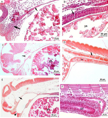
Newly hatched larvae (13 h post-fertilization) presented their mouth and anus closed, and the digestive tract was a straight tube dorsal to the yolk sac, lined with a simple epithelium (Fig. 1A, B, D).
Photomicrography of the digestive tract of B. amazonicus between hatching and 9 hPH. Hatching: (A) Buccopharyngeal region primordium (larger arrow), digestive tube in development (smaller arrow). (B) Posterior region of the digestive tube in development (arrow). 4 hPH: (C) Opening of the buccopharyngeal cavity (arrow). (D) Posterior region of the digestive tube in development (arrow). 7 hPH: (E) Buccopharyngeal cavity (arrow) with lips adhered (arrowhead). 9 hPH: (F) Formation of the urinary bladder and digestive tube. DT, digestive tube; U, urinary bladder; ys, yolk sac.

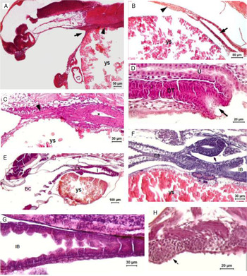
At 4 h later, the initial formation of the buccopharyngeal cavity was observed (Fig. 1C), but a membrane kept the lips closed (Fig. 1E). After 9 h post-hatching (hPH), a lumen in the tube could be observed (Fig. 1F, 2B and C). The mouth was open at 11 hPH (Fig. 2A) and the anus at 13 hPH (Fig. 2D).
Photomicrography of the digestive tract of B. amazonicus between 11 and 25 hPH. 11 hPH: (A) Buccopharyngeal cavity with the rest of the digestive tube still closed (arrow). (B) Lumen in the medium region of the digestive tube (arrow) and anterior region closed (arrowhead). 13 h PE: (C) Lumen (asterisk) in the anterior region of the digestive tube and tube near buccopharyngeal region still closed (arrowhead). (D) Anus (arrow), digestive tube and urogenital opening. 25 h PE: (E) Panoramic view of the anterior region. (F) Swim bladder in development (arrow) and intestinal bag. (G) Middle region of the digestive tube. (H) Taste buds on the buccopharyngeal cavity (arrow). BC, buccopharyngeal cavity; IB, intestine bag; DT, digestive tube; ES, oesophagus; U, urogenital opening; ys, yolk sac.

From 25 hPH, the digestive tube was completely open and divided into the buccopharyngeal cavity (presenting taste buds – Fig. 2H), oesophagus, and intestine (Fig. 2E), with the latter being more dilated at the proximal region, forming an intestinal bag lined by a simple cylindrical epithelium (Fig. 2F, G).
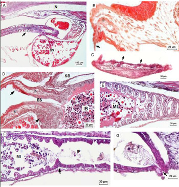
To avoid cannibalism, exogenous feeding with forage-fish larvae began at 29 hHP (Fig. 3A) although there were still endogenous reserves.
Photomicrography of the digestive tract of B. amazonicus at 29 and 35 hPH. 29 hPH: (A) Ingestion of forage larvae (arrow). 35 hPH: (B) Tooth (arrow) in the premaxilla. (C) Tooth alveoli in the mandible (arrows). (D) Pharynx (arrow), oesophagus, skeletal muscle (arrowhead), lamina propria, intestinal bag and swimming bladder. (E) Midgut with intestinal folds. (F) Septum (arrow) between middle and posterior intestine. (G) Anus (arrow). IB, intestinal bag; ES, esophagus; lp, lamina propria; MI, midgut; N, notocord; PI, posterior intestine; SB, swimming bladder; ys, yolk sac.

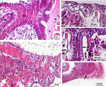
Photomicrography of the digestive tract of B. amazonicus at 59 hPH. (A) Pharynx with mucus cells (arrow), oesophagus and intestinal bag (black bar = longitudinal muscle, white bar = circular muscle). (B) Distal portion of the midgut. (C) Septum (arrow) between middle and posterior intestine. (D) Medium portion of the midgut. (E) Anus (arrow). ES, esophagus; IB, intestinal bag; MI, midgut; Ph, pharynx; PI, posterior intestine.

Larvae between 35 and 83 hPH – 5.90 ± 0.42 to 6.64 ± 0.40 mm of SL
At 35 hPH, the digestive tract was classified into cephalic intestine (buccopharyngeal cavity), anterior intestine (oesophagus), midgut (middle intestine) and posterior intestine (Fig. 3D, E) with the latter separated by a septum (Fig. 3F). In this time, food could be observed into the intestinal bag (Fig. 3D– F).
In the buccopharyngeal cavity, incisor teeth development was observed in the upper and in the lower jaw (Fig. 3B, C) at 35 hPH, but pharyngeal teeth were observed only from 83 hPH (Fig. 5H).
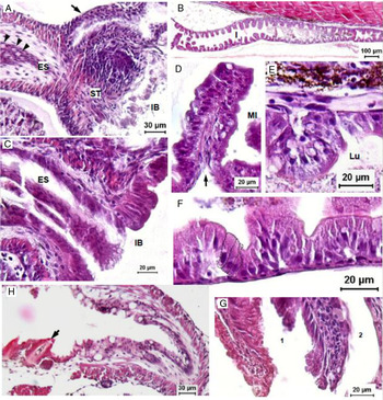
Photomicrography of the digestive tract of B. amazonicus at 83 hPH. (A) Oesophagus with mucous cells (arrowhead), stomach in development, intestinal bag and swim bladder duct (arrow). (B) Panoramic view of the intestine. (C) Oesophagus and intestinal bag. (D) Medial portion of the midgut and a fold (arrow) right after the intestinal bag. (E) Distal portion of the midgut. (F) Posterior intestine (dotted line: cylindrical cells with striated border). (G) Anus (1) and urogenital opening (2). (H) Pharyngeal teeth (arrow). ES, oesophagus; I, intestine; IB, intestinal bag; MI, midgut; ST, stomach.

From this period, a stratified squamous epithelium was observed protecting places exposed to friction such as the mouth, pharynx, oesophagus, and anus (Figs 3D, G, 4E, and 5G). In the pharynx and oesophagus cells were observed producing mucus and a thick skeletal muscle layer externally to the lamina propria was seen (Figs 4A and 5A).
A transition from squamous stratified to simple cylindric epithelium marked the border between the oesophagus and the intestinal bag (Figs 3D and 5C) with the midgut presenting cells with large vesicles in the cytoplasm (Figs 3E and 4B, D). At 83 hPH, these vesicles were observed in the distal portion of midgut, in a granular aspect (Fig. 5E), and were not observed in the posterior intestine (Figs 3F and 4C).
At the end of this period (83 hPH), vesicles that had been previously observed in the epithelial cells of the midgut had almost disappeared (Fig. 5B, D). In the distal portion of the midgut and in the posterior intestine, the epithelium presented cylindrical cells with a striated border and a nucleus in the basal position (Fig. 5E, F).
Larvae between 107 and 171 hPH – 10.25 ± 1.16 to 11.7 ± 0.92 mm of SL
Few morphological alterations were observed between 83 and 135 hPH.
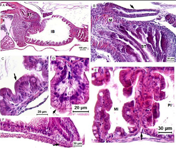
At 147 hPH, the stomach had a slightly curved shape with many mucosal folds (Fig. 6A). A thick muscular layer was observed externally, and a simple cylindrical epithelium was observed internally (Fig. 6B).
Photomicrography of the digestive tract of B. amazonicus at 147 hPH. (A) Panoramic view of the anterior portion of the digestive tract, with stomach in differentiation and intestinal bag. (B) Stomach, muscle layer and swim bladder duct (arrow). (C) Proximal portion of the midgut (intestinal bag) (arrow: absorption epithelium). (D) Posterior intestine (arrow: absorption epithelium). (E) Septum (arrow) between middle and posterior intestine (PI). (F) Rectum and anus (arrow). IB, intestinal bag; M, muscle; MI, midgut; PI, posterior intestine; ST, stomach.

In the intestinal bag and midgut, a simple epithelium of enterocytes brush border and goblet cells was observed (Fig. 6C, E). In the posterior intestine, the villus presented cells with small vesicles (Fig. 6D), while the rectal segment was differentiated from the rest of the intestine by the absence of mucosal folds and few vesicles cells in the epithelium (Fig. 6F). However, the number of these cells increased significantly from 171 hPH (Fig. 7F).
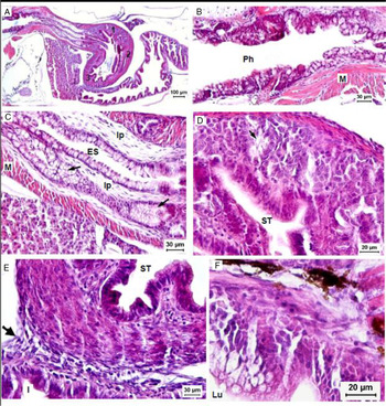
Photomicrography of the digestive tract of B. amazonicus at 171 hPH. (A) Panoramic view of the anterior portion of the digestive tract with stomach divided into a cardiac region (1), fundic (2) and pyloric (3). (B) Pharynx. (C) Oesophagus and glands (arrows). (D) stomach and gastric glands (arrow). (E) Pyloric sphincter (arrow) between stomach and intestine. (F) posterior intestine. ES, oesophagus; I, intestine; lp, lamina propria; Lu, lumen; M, muscle; Ph, pharynx; ST, stomach.

From 171 hPH, three regions of the stomach – cardiac, fundic and pyloric – were observed (Figs 7A and 8A). In the cardiac region, the first gastric glands were observed (Fig. 7D) and a thin muscle layer that became thicker in the fundic and pyloric regions (Fig. 7A, E).
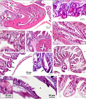
Photomicrography of the digestive tract of B. amazonicus at 243 hPH. (A) Panoramic view of the oesophagus, stomach, with regions: cardiac (1), fundic (2) and pyloric (3), and midgut. (B) Teeth alveoli (asterisk) and taste buds (dotted line) on the pharynx. (C) Oesophagus with internal circular muscle layer and external longitudinal muscle layer. (D) Stomach glands (arrow). (E) Lumen of the pyloric portion of the stomach, intestine and pyloric cecum. (F, G) Pyloric cecum (arrows). (H) Panoramic view of the midgut. (I) Distal portion of the midgut. (J) rectum and anus and mucous cells. A, anus; c, pyloric cecum; cm, circular muscle layer; ES, oesophagus; I, intestine; lm, longitudinal muscle layer; MI, midgut; ST, stomach.

The number of mucous cells in the pharynx and oesophagus epithelium increased significantly compared with the previous periods. The mucosal layer of the pharynx became thicker (Fig. 7B) and the oesophagus presented mucous glands in the lamina propria (Fig. 7C).
Larvae between 243 and 579 hPH – 13.3 ± 1.8 to 48.94 ± 10.97 mm of SL
At 243 hPH, teeth alveoli and taste buds were observed in the pharynx (Fig. 8B). In the oesophagus, a simple epithelium with mucous cells and pores at the apex were observed (Fig. 8C).
In the first half of the stomach, many glands were observed, and the second half presented a thick muscular layer (Fig. 8A, E). The gastric epithelium in the cardiac region presented a simple epithelium with cylindrical cells (Fig. 8D).
The pyloric caeca were also observed in the proximal region of the midgut (Fig. 8E, F), with a simple epithelium of enterocytes brush border and few presented vesicles cells (Fig. 8G). The quantity of these vesicles cells gradually increased towards the end of the midgut (Fig. 8H). However, at the distal portion of the midgut, all cells at the apex of the intestinal folds had small vesicles, whereas in the cells of the pits no vesicles were found (Fig. 8I). In the rectum and anus, mucus cells in the epithelium were observed (Fig. 8J).
From 412 hPH, the digestive tract was completely developed and mature.
Discussion
Most newly hatched fish larvae commonly exhibit a rudimentary digestive tract that undergoes several transformations until adulthood and, therefore, the larvae are less efficient in obtaining nutrients (Gisbert et al., Reference Gisbert, Moreira, Castro-Ruiz, Öztürk, Fernández, Gilles, Nuñez, Duponchelle, Tello, Renno, Garciá-Dávila and Darias2014; Nakauth et al., Reference Nakauth, Villacorta-Correa, Figueiredo, Bernardino and França2016; Silva et al., Reference Silva, Valentin, Paes, Faustino, Giannecchini, Viadanna, Nascimento, Fernandes and Nakaghi2016; Alveal et al., Reference Alveal, Silva, Lohrmann and Viana2019).
When the differentiation of the digestive tract begins, the rectilinear shape of the tube, without defined limits between segments and the absence of mucosa folds, facilitates the entrance of relatively large prey (Maciel et al., Reference Maciel, Lanna, Maciel Junior, Donzele, Neves and Menin2010; Nakauth et al., Reference Nakauth, Villacorta-Correa, Figueiredo, Bernardino and França2016). From 29 hPH, forage-fish larvae were offered and there was an overlap of endogenous and exogenous feeding, and a rapid absorption of the yolk sac was observed, indicating that, although the larvae received exogenous food, the assimilation of nutrients was not efficient.
The presence of taste buds and the development of teeth were observed during the beginning of exogenous feeding in B. amazonicus, which were also observed in B. moorei (Vanderwalle et al., Reference Vanderwalle, Germeau, Besancenet, Parmentier and Baras2005), B. orthotaenia (Gomes et al., Reference Gomes, Sato, Rizzo and Bazzoli2013), B. orbignyanus (Maciel et al., Reference Maciel, Lanna, Maciel Junior, Donzele, Neves and Menin2010) and B. gouldingi (Faustino et al., Reference Faustino, Makino, Neumann and Nakaghi2018a). The incisors teeth are used for hold and pre-digestive preparation of plant food or other kinds of food material, while the pharyngeal teeth assist in the seizure and maceration of soft body organisms (Rodrigues et al., Reference Rodrigues, Navarro and Menin2006; Faustino et al., Reference Faustino, Makino, Neumann, Satiko and Nakaghi2018b). Taste buds are chemoreceptors, with the purpose of capturing the smell and taste of saturated substances in water (Matsuoka, Reference Matsuoka2001). Therefore, larvae can select their preferred food from the food items offered and the taste buds in fish larvae can be related to the difficulty in acceptance of artificial food at weaning.
Characteristics such as dilatation in the proximal region of the midgut and the presence of mucosal folds at the beginning of development (25 hPH) allowed better food retention with increased absorption and digestion efficiency (Maciel et al., Reference Maciel, Lanna, Maciel Junior, Donzele, Neves and Menin2010; Faustino et al., Reference Faustino, Makino, Neumann, Satiko and Nakaghi2018b).
The presence of large supranuclear vacuoles occupying the cytoplasm of cells in the midgut after initiation of exogenous feeding is associated with the absorption and accumulation of lipids, which after luminal digestion are absorbed as fatty acids by enterocytes, in which are resynthesized and then directed to other parts of the body (Silva et al., Reference Silva, Valentin, Paes, Faustino, Giannecchini, Viadanna, Nascimento, Fernandes and Nakaghi2016; Marques et al., Reference Marques, Faustino, Bertolucci, Do Carmo Faria Paes, Valentin and Nakaghi2017). Peña et al. (Reference Peña, Dumas, Villalejo-Fuerte and Ortíz-Galindo2003) related the presence of supranuclear vacuoles of the midgut to the storage of lipids, which after luminal digestion are absorbed as fatty acids by enterocytes, in which they are resynthesized and directed to other sites in the organism.
The granular aspect of the cytoplasm of epithelial cells of the distal portion of the midgut observed in B. amazonicus at 83 hPH indicates the occurrence of protein pinocytosis as an alternative for digestion proteins in teleost larvae, as the digestive system is still not well developed (Gisbert et al., Reference Gisbert, Piedrahita and Conklin2004; Maciel et al., Reference Maciel, Lanna, Maciel Junior, Donzele, Neves and Menin2010). According to Sarasquete et al. (Reference Sarasquete, Polo and Yúfera1995), pinocytosis is an alternative for digestion of proteins in teleost larvae, as the digestive system is still not well developed.
The presence of mucous cells in the pharynx and oesophagus of B. amazonicus suggests the participation of these cells in swallowing the prey and protection of the epithelium from the friction of food passage (Faustino et al., Reference Faustino, Makino, Neumann, Satiko and Nakaghi2018b). The development of the skeletal muscles that surround the oesophagus probably also contributes to the rapid conduction of the prey through this segment.
In the stomach, the differentiation of the thicker muscular layer indicates mechanical digestion. And the similar aspect of mucosa folds in stomach and oesophagus indicates that chemical digestion is not yet functional (Galvão et al., Reference Galvão, Fenerich-Verani, Yamanaka and Oliveira1997). After the onset of stomach function in B. amazonicus, the size of the intestinal bag was gradually reduced, indicating that the function of food retention was transferred to the stomach.
According to Galvão et al. (Reference Galvão, Fenerich-Verani, Yamanaka and Oliveira1997), before the development of gastric glands, fish depend on their ability to select appropriate foods, mechanical digestion and pancreatic and intestinal enzymes. After the differentiation of the stomach in B. amazonicus, mucus cells were observed in the epithelium and among enterocytes of the midgut. These cells have the function of protect the epithelial cells from the hydrochloric acid produced by the gastric glands (Ghosh and Chakrabarti, Reference Ghosh and Chakrabarti2015) and also neutralize the acidity from the stomach, preparing the bolus for the action of pancreatic and intestinal enzymes (Ribeiro and Fanta, Reference Ribeiro and Fanta2000).
Mucus cells were also present in the epithelium of pyloric caeca, suggesting that these structures are important not only to increase the area of nutrient absorption but also to neutralize the pH of the stomach as there are no pyloric caeca in non-gastric fish (Zavala-Camin, Reference Zavala-Camin1996). This suggestion is reinforced by the fact that differentiation of pyloric caeca occurs only after the differentiation of the gastric glands, as seen in B. amazonicus and also in other teleost species (Drewe et al., Reference Drewe, Horn, Dickson and Gawlicka2004; Elbal et al., Reference Elbal, García Hernández, Lozano and Agulleiro2004).
Elbal et al. (Reference Elbal, García Hernández, Lozano and Agulleiro2004) reported that, in addition to the development and functionality of gastric glands, differentiation of the pyloric caeca mucosa establishes maturation of the digestive tract, defining the mature and definitive form.
In conclusion, the most critical period of the larviculture is the beginning of exogenous feeding and the second period is transition between live and artificial food. In the present study, three important phases of digestive tract development of B. amazonicus can be highlighted:
(i) beginning of exogenous feed (29 hPH, larvae with approximately 5.78 ± 0.48 mm of SL), with the exogenous food not efficiently digested, resulting in rapid consumption of the yolk sac;
(ii) improvement of the digestive capacity (25 to 35 hPH, larvae between approximately 5.50 ± 0.46 and 5.90 ± 0.42 mm of SL), with the dilatation of the midgut (intestinal bag) allowing food storage, and also mucosa characteristics, indicating the beginning of nutrient absorption, however, the digestive tract is still immature;
(iii) the recommended period for transition between live and artificial food (243 to 412 hPH, larvae between approximately 13.3 ± 1.8 and 25.79 ± 3.01 mm of SL) – phase between the start of absorption/assimilation of nutrients and the maturity of the digestive tract, with the complete differentiation and functionality of the gastric glands and pyloric caeca.
Financial support
The authors would like to thank the National Council for Scientific and Technological Development – CNPq and São Paulo Research Foundation – FAPESP for their financial support.
Conflicts of interest
None.
Ethical standards
This research was approved by the Ethics Committee on Animal Use and conducted according to the Ethical Principles in Animal Experimentation adopted by the Brazilian College of Experimentation.