Introduction
Clarias dussumieri (Valenciennes, 1840) is a sought-after freshwater catfish because of its admirable taste and rich nutritional value. This species predominantly inhabits the shallow rivers and wetlands of southwestern India (Gopalakrishnan and Ponniah, Reference Gopalakrishnan and Ponniah2000), making its conservation crucial. Known to grow up to 3 kg (Padmakumar et al., Reference Padmakumar, Bindu, Basheer and Gopalakrishnan2010), this indigenous species of the Western Ghats has a great potential for aquaculture. Its adaptive ability to breathe atmospheric air through an arborescent organ coupled with its hardy nature allows it to thrive at high stocking densities. This makes the fish suitable for various aquaculture systems, including biofloc and wastewater aquaculture, and facilitates live storage and transportation (Tharakan and Joy, Reference Tharakan and Joy1996). Despite its commercial and ecological importance, C. dussumieri populations have declined drastically, leading to its classification as ‘Near-Threatened’ on the IUCN Red List (Abraham, Reference Abraham2011).
Embryonic and larval development assessments are crucial for understanding the reproductive biology, habitat preferences, and aquaculture practices of teleost fishes, especially under changing environmental conditions (Behmene et al., Reference Behmene, Bachir Bouiadjra, Attou and Homrani2022; Mahalder et al., Reference Mahalder, Haque, Siddique, Hasan, Alam, Talukdar, Shohan, Ahasan, Hasan and Ahammad2023). Moreover, understanding the abiotic factors, such as temperature, on embryogenesis helps refine aquaculture practices, reduce abnormalities, and improve larval survival rates, ensuring the sustainability of the cultured species (Rey et al., Reference Rey, Nguyen, Singchat, Punthum, Kraichak, Panochit, Maneeaphai, Phuonnim, Sengtrakool, Sriphairoj, Hatachote, Muangmai, Chaiyes, Duengkae, Matsuda, Prasanpan and Srikulnath2025). Such knowledge is particularly vital for cultured fish to enhance aquaculture practices and fish production by tailoring conditions to suit the needs of each larval stage, thereby optimizing growth and survival rates (Puvaneswari et al., Reference Puvaneswari, Marimuthu, Karuppasamy and Haniffa2009).
As the early life stages of fish undergo significant cellular and morphological changes, the larvae rely completely on external feeding after yolk sac absorption. Recent advancements in nutritional formulations and rearing conditions have significantly enhanced larval growth and survival in Clariid aquaculture, establishing a link between performance and both feed composition and environmental management (Mir et al., Reference Mir, Srivastava, Bhat, Dar, Sushila, Varghese, Muralidhar and Jain2019; Hildebrand et al., Reference Hildebrand, Hentrich and Palm2024). Abnormal embryonic development under suboptimal conditions is associated with increased mortality rates in catfish species (Rey et al., Reference Rey, Nguyen, Singchat, Punthum, Kraichak, Panochit, Maneeaphai, Phuonnim, Sengtrakool, Sriphairoj, Hatachote, Muangmai, Chaiyes, Duengkae, Matsuda, Prasanpan and Srikulnath2025). A comprehensive understanding of developmental stages is essential for diagnosing risks across various habitats and aquaculture methods (Mahalder et al., Reference Mahalder, Haque, Siddique, Hasan, Alam, Talukdar, Shohan, Ahasan, Hasan and Ahammad2023; Hildebrand et al., Reference Hildebrand, Hentrich and Palm2024). Although developmental studies on other clariids are available (Behmene et al., Reference Behmene, Bachir Bouiadjra, Attou and Homrani2022; Rey et al., Reference Rey, Nguyen, Singchat, Punthum, Kraichak, Panochit, Maneeaphai, Phuonnim, Sengtrakool, Sriphairoj, Hatachote, Muangmai, Chaiyes, Duengkae, Matsuda, Prasanpan and Srikulnath2025), there is a notable lack of comprehensive descriptions of the embryonic and larval development of C. dussumieri. To address this gap, the present study aimed to characterize the embryonic and larval development of C. dussumieri to support conservation efforts and enhance its aquaculture potential.
Materials and methods
Broodstock selection and induced reproduction
Adult C. dussumieri were collected from Periyar river, near Thodupuzha, Kerala, transported to the hatchery and maintained in the cages at ICAR–NBFGR, Kochi. One month prior to breeding, broodstock were transferred to round fibre-reinforced plastic tanks (3 m diameter × 1 m height) half-filled with water and fitted with sponge filters and air-stones for aeration. Tanks were maintained under a natural 12L:12D photoperiod with steady ambient daylight at 100–300 lux measured using a lux metre (Sigma® – 101). A green shade net was placed over the tanks to diffuse light, reduce disturbance, and prevent fish from jumping out. PVC pipes (10 cm diameter, 100 cm length) were provided as hideouts. Induced breeding was conducted during the southwest monsoon (June–August), the natural breeding season (Padmakumar et al., Reference Padmakumar, Bindu, Basheer and Gopalakrishnan2010). Five pairs of mature males and females (150–250 g) were selected at a 1:1 ratio, and sexual identification was based on genital papillae and the release of eggs under gentle pressure. Males showed red-coloured pointed urogenital papillae (Padmakumar et al., Reference Padmakumar, Bindu, Sreerekha, Joseph, Krishnan, Manu and Basheer2017) (Figure 1A,B). Artificial breeding was done using WOVA-FH (Biostadt®, India), containing 20 µg salmon gonadotropin-releasing hormone analogue and 10 mg domperidone per millilitre, administered at 1 mL/kg body weight of fish intraperitoneally. After 12 ± 1.50 h, fish were stripped for eggs and sperm. Fertilization of C. dussumieri eggs used the wet method common in clariid catfish breeding (Padmakumar et al., Reference Padmakumar, Bindu, Sreerekha, Joseph, Krishnan, Manu and Basheer2017). Stripped eggs were mixed with sperm diluted in physiological saline (0.9% NaCl), which enhances viability of the milt (Olaniyi and Omitogun, Reference Olaniyi and Omitogun2014a). After mixing for two-three minutes, excess saline was drained. Fertilized eggs were rinsed with fresh water and transferred to incubation tubs (Olaniyi and Omitogun, Reference Olaniyi and Omitogun2014b). About 15–20 eggs were placed in a Petri dish for observation. Unfertilized eggs, identified by their opaque-white appearance, were removed before incubation. The fertilized eggs were incubated in an indigenously designed jar hatching system that maintained consistent water circulation and aeration to ensure optimal oxygenation and egg suspension. The incubation temperature was maintained at 26–27°C using aquarium heaters (Eheim thermocontrol 300, Germany) with thermostatic control, and hatching occurred within 24–27 h. While clariids tolerate temperatures between 20–35°C, optimal hatching occurs at 26–30°C (Prokešová et al., Reference Prokešová, Drozd, Kouřil, Stejskal and Matoušek2015), an incubation temperature of 26.5 ± 0.5°C was selected based on preliminary observations showing high fertilization success and robust hatching rates. Egg incubation proceeded under the same 12L:12D photoperiod in ambient daylight (100–300 lux) without direct artificial illumination. The fertilization rate, hatchability, and survival percentage were calculated using standard formulas (Okomoda et al., Reference Okomoda, Koh and Shahreza2018; Rurangwa et al., Reference Rurangwa, Roelants, Huyskens, Ebrahimi, Kime and Ollevier1998; Olaniyi and Omitogun, Reference Olaniyi and Omitogun2014b).
Sexual dimorphism in Clarias dussumieri (A) Female (B) Male.

 $$Fertilization\;rate\;\left( \% \right) = {{Number\;of\;fertilized\;eggs} \over {Total\;number\;of\;sampled\;eggs}} \times 100$$
 $$Hatchability\;\left( \% \right) = {{Number\;of\;hatchlings} \over {Total\;number\;of\;fertilized\;eggs}} \times 100$$
 $$Survival\;rate\;\left( \% \right) = {{Number\;of\;larvae\;survived} \over {Total\;number\;of\;larvae\;hatched}} \times 100$$
Larval survival rates were calculated by counting the number of live larvae at 14 days post-hatching (Ngo et al., Reference Ngo, Tran, Nguyen and Pham2024). Survival was defined as the proportion of larvae alive at counting, including those with deformities (< 1%), which was noted separately (Esa et al., Reference Esa, Dadile, Syukri, Christianus and Diyaware2023; Ngo et al., Reference Ngo, Tran, Nguyen and Pham2024)
Post-induced breeding management
Water quality parameters, including dissolved oxygen (mg L⁻1) and pH in the breeding tank, hatching jar, and larval rearing tank, were measured using the API® Freshwater Master Test Kit (Mars Inc., USA). Temperature (°C) was recorded with an immersion thermometer (Easy-Read®). Incubation jars and larval rearing tanks were kept in the same enclosed room under a 12L:12D photoperiod with ambient daylight illumination (100–300 lux) and no artificial lighting. Larvae were stocked in 50 L rearing tubs filled with aerated water to a depth of 10 cm, at a density of 10 larvae L⁻1. Mortality was monitored daily in the early morning, and dead larvae were promptly removed. From third day, the larvae were fed newly hatched Artemia nauplii until the 8th day, followed by Moina sp. until the 16th day. Frozen bloodworms were introduced on the 15th day and were continued for 10 days. On the 25th day, C. dussumieri juveniles were transitioned to an inert diet of 0.2 mm powdered feed (Marubeni Nisshin™), shaped into balls and fed for 20 days, followed by 0.8 mm commercial fish feed (Nutrila Growel™, India) with 42% crude protein. As the fish grew, feed size was gradually increased. The fish reached maturity within eight months.
Embryogenesis and larval measurement
Embryonic stage evaluation began when fertilized eggs were placed in the incubators. Eggs were examined using a stereo zoom microscope (Leica S8APO) with digital a camera (Leica DFC 280), and images were processed using the Leica Application Suite (LAS EZ v3.80). Development was monitored at 5–10 min intervals during cleavage stages, every 30 min during the morula to late blastula stages, and hourly from early gastrula until hatching. Stage transitions were determined when 50% of embryos showed characteristics of the next stage (Kupren et al., Reference Kupren, Nowosad and Kucharczyk2023). Ten eggs per stage were examined on cavity slides with distilled water. Larval samples were collected every 6 h for the first 72 h post-hatching, daily from 4–21 days, and at three-day intervals until day 30. Ten larvae per sample were preserved in 5% formalin for microscopic examination. Prior to fixation in formalin, the larvae were euthanized using an overdose (300 mg L⁻1) of tricaine methanesulfonate (MS 222) until complete cessation of movement was observed (Toomey et al., Reference Toomey, Dellicour, Kapusta, Żarski, Buhrke, Milla, Fontaine and Lecocq2021). This sampling regime ensured the capture of critical developmental changes throughout the early life stages.
Morphometric analysis of larvae
The larval development time and biometric measurements, including total length (TL), length up to notochord, length up to vent, height of yolk sac, length of yolk sac and maximum height of the body (Figure 2) were recorded to the nearest minute and millimetre, respectively, using Image analysis software (LAS EZ v3.80). Morphological changes in the larvae were documented daily using, images captured with a stereo zoom microscope (Leica S8APO), and specimens were labelled accordingly. During the larval rearing period, the relationship between total length and other morphometric parameters was analysed using an allometric power-law regression model (Y = aX b). The allometric exponent b was used to characterize the growth patterns (with b > 1 indicating positive allometry, b = 1 isometry, and b < 1 negative allometry). The objective was to examine growth patterns, verify proportional development, and evaluate gape limitations in larval feeding to facilitate the selection of appropriate feed size at various developmental stages (Rahmatullah et al., Reference Rahmatullah, Miah, Saha and Rahman2001; Babu et al., Reference Babu, Anirudhan, Madathumpady, Kurva, Dube, Kodi, Mhaddolkar, Pal, Ignatius and Achamveetil2024). The TL of the larvae was determined by measuring the distance from the tip of the snout to the tip of the tail, and the eye diameter (ED) was recorded in millimetres. Mouth opening (MO) was determined by measuring the maximum distance between the lower and upper jaws assuming that the mouth opens at an angle of 90° during prey capture (Nakagawa et al., Reference Nakagawa, Hashimoto, Murata and Miyashita2011). These measurements were performed with the specimen mounted on a glass slide and gently covered with a cover slip to enhance the optical clarity without causing damage (Balamurugan et al., Reference Balamurugan, Kathiresan and Meenakumari2018). Scatter plots were created, and correlation coefficients were calculated (Babu et al., Reference Babu, Anirudhan, Madathumpady, Kurva, Dube, Kodi, Mhaddolkar, Pal, Ignatius and Achamveetil2024). To provide a more comprehensive understanding of larval development, they were classified into various flexion stages (yolk sac larvae, pre-flexion, early flexion, late flexion, early post-flexion, late post-flexion, and juvenile), based on the development of caudal fin (Nowosad et al., Reference Nowosad, Kupren, Biegaj and Kucharczyk2021). The transition to the juvenile stage was determined based on established morphological criteria including complete fin ray segmentation, establishment of adult-like pigmentation patterns, and fully developed functional jaws, features commonly recognized as markers of metamorphosis completion in clariid catfishes (Morioka et al., Reference Morioka, Vongvichith, Phommachan and Ueno2013). Adult fin ray meristics (D, A, P1, P2 and C) were compiled and compared with stage wise counts.
Biometric parameters of larvae.

Statistical analysis
All data related to the timing of embryonic development (expressed in hours and minutes) and larval morphometry are presented as mean ± Standard Error of the Mean (SEM). Morphometric relationships between total length (TL) and traits such as mouth opening (MO), and eye diameter (ED) were analysed using least squares regression. The linear and power function models were tested. The coefficient of determination (R2) and slope (b) values were extracted to describe the strength and type of the growth relationships. All values were calculated using Microsoft Excel 2019.
Results
Induced breeding response
Ovulation occurred within 12 ± 1.50 h at 26.5 ± 0.50°C. The unfertilized eggs of C. dussumieri were brownish-orange, spherical, demersal, and slightly adhesive, measuring 1.5 ± 0.02 mm in diameter, with a thin perivitelline membrane. Water temperatures were maintained at 26–27°C in breeding tanks and hatching jars, and 27–29°C in larval tanks. Dissolved oxygen levels were 5–6 mg L⁻1 in breeding tanks, 4–6 mg L⁻1 in hatching jars, while pH ranged from 6.8–8.5. Induced spawning achieved 90.67 ± 6.03% fertilization, 87.33 ± 3.06% hatchability, and 90 ± 1.73% survival rate.
Embryonic development
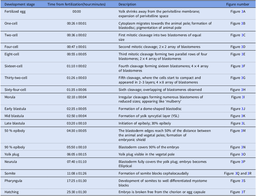
The following stages of embryonic development were observed in C. dussumieri after spawning and fertilization until hatching: zygote, cleavage, morula, blastula, gastrula, segmentation, pharyngula and hatching (Table 1).
Embryonic developmental stages in Clarias dussumieri at 27 ± 1°C

Time shown in the table for each developmental stage is the average value of five observations (Mean ± SEM). Time is expressed as h:min ± h:min.
Zygote stage
In fertilized eggs, a notable increase in the perivitelline space was observed, along with the migration of the cytoplasm towards the animal pole (AP). The nucleus and organelles became concentrated at the animal pole, while the yolk sac (YS) occupied the vegetative pole (VP). The average egg diameter expanded to approximately 1.7 ± 0.10 mm (Figure 3A). The one-cell stage is characterized by the formation of a protoplasmic bulge at the AP, referred to as the blastodisc (BDC) or germinal disc stage (Figure 3B).
(A) Fertilized egg. Perivitelline membrane (PVS) (B) 1-cell stage. Blastodisc (BDC), Animal pole (AP), Vegetal pole (VP) (C) 2-cell stage. Segmentation line (SL) (D) 4-cell stage. (E) 8-cell stage. (F) 16-cell stage. (G) 32-cell stage. (H) 64-cell stage. (I) Morula stage. (J) Early blastula stage. (K) Mid blastula stage. (L) 30% epiboly. Enveloping layer (EVL), Deep layer (DL), Yolk syncytial layer (YSL), Blastoderm (BD) (M) 50% epiboly. Embryonic shield (ES) (N) 90% epiboly. (O) Yolk plug (YP). (P) Neurula. Tail bud (TB), Neural keel (NK). (Q) 18-somite stage. Caudal region (CR), Notochord (N), Somites (S), Head region (HR) (R) 25-somite stage. Kupffer’s vesicle (KV), Sclerotome (SL) (S) Pharyngula. Optic placode (OPP), Otic placode (OTP), Yolk sac (YS), Spinal cord (SC) (T) Hatching. Eye (E), Oil droplets (OD).

Cleavage stage
Fertilized eggs of C. dussumieri exhibited meroblastic cleavage as it occurred only at the AP. The cleavage stage was characterized by a sequence of mitotic divisions in the active cytoplasm, culminating in 64 blastomeres. The first mitotic division was vertical, cleaving the blastodisc into two blastomeres of identical size. The second cleavage was vertical and perpendicular to the first, resulting in four blastomeres. The third division was vertical and parallel to the first, generating eight blastomeres with a 4 × 2 arrangement and the fourth was vertical and parallel to the second division, producing 16 blastomeres in a 4 × 4 arrangement. The fifth division was vertical and parallel to the first, producing 32 blastomeres in a 4 × 8 formation. The sixth cleavage was horizontal, resulting in two cell layers and 64 blastomeres. The blastomeres were reduced in size after each cleavage (Figures 3C–3H).
Morula stage
This stage was characterized by successive division of blastomeres to form small blastomeres that lost boundaries and appeared compacted over the AP (Figure 3I).
Blastula stage
The blastula stage was categorized into three stages: early blastula stage in which the blastoderm (BD) was approximately 256 cells, appeared dome-shaped over the yolk cell, and the cleavage pattern was irregular, but consistent. The mid blastula stage is characterized by the development of outer enveloping layer (EVL) and a peripheral yolk syncytial layer (YSL). The YSL is a syncytial layer located at the junction of the yolk sac (YS) and blastoderm (BD), and can be seen as a darker zone in live embryos under a light microscope (Rodrigues-Galdino et al., Reference Rodrigues-Galdino, Maiolino, Forgati, Donatti, Mikos, Carneiro and Rios2010). Late blastula stage displayed the initiation of epiboly, wherein the BD spread over the YS region due to blastomeres intercalation and the margins of BD covered 30% of the YS (Figures 3J–3L).
Gastrula stage
This stage began after 4 h and 30 min during embryonic development, when epiboly movements were notable. The morphogenetic movements, such as convergence and cell migration initiated at the BD border, when the embryo was approximately 50% through the epiboly stage. This commenced the formation of the germinal ring and embryonic shield, which concluded with the production of two embryonic layers known as the epiblast and hypoblast (Figure 3M and 3N). The BD progressively covered the entire yolk cell, as epiboly progressed (Figure 3O). In the neurula stage, the BD completely covered the yolk plug (YP), closing the blastopore, the neural groove became visible, and the embryo became elliptical in shape (Figure 3P).
Segmentation
The segmentation stage is characterized by the formation of somites (S), notochord (N) and rudiments of various organs and tissues. Somites were formed as a result of mesodermal segmentation. As the first three somites develop, the optic vesicles become more prominent. At this stage, the neural groove, which appeared during the neurula stage, closed to form the neural tube, and elongation of the embryo, along the head-tail axis was observed (Figure 3Q and 3R).
Pharyngula
Blood circulation is initiated during this phase. During the pharyngula period, distinct structures such as embryonic fins and gaps associated with the mouth and operculum became noticeable. The embryo’s body becomes elongated as the tail bud TB becomes free from the YS and presents approximately 30–35 myotomes. The pericardial cavity was easily visible and the first heartbeat was observed during this phase (Fig. 3S). Additional anatomical structures such as Kupffer’s vesicle (KV), optic placode (OPP), otic placode (OTP) eye and caudal region (CR) became evident during this stage.
Hatching stage
Hatching occurred between 25:30 ± 01:30 h after fertilization at 26–27°C and the larval stage began. At this stage, the enclosed embryo exhibited vigorous muscular twitches, using its head region (HR) to tear through the chorion and release itself (Figure 3T). Oil droplets (OD) were also observed as energetic reserves within the YS, supporting larval survival immediately after hatching.
Larval development
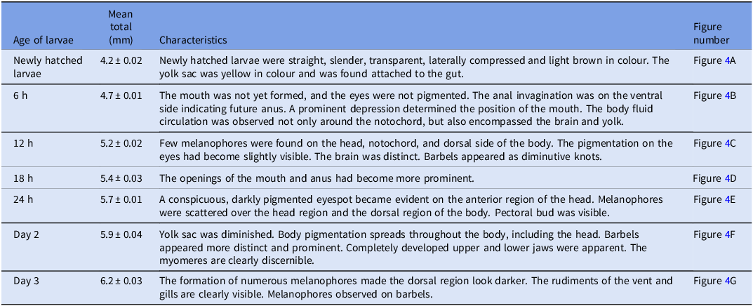
The larval development of C. dussumieri can be categorized into yolk sac larvae, pre-flexion larvae, flexion larvae and post-flexion larvae, with corresponding day-wise morphological changes summarized in Table 2.
Description of larval development in Clarias dussumieri and their characteristics at different ages
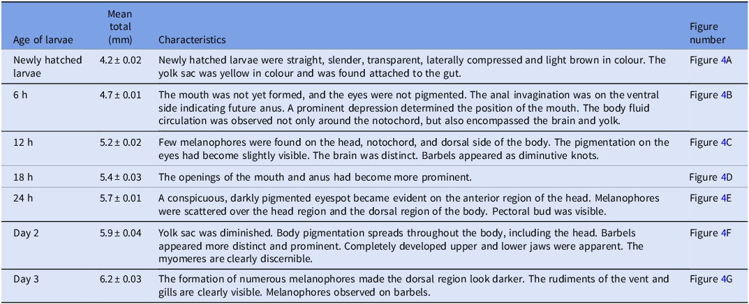
Data collected on various parameters were presented as mean ± SEM (Standard error of mean).
Yolk sac larva: Newly hatched larvae display transparent, slender bodies with a laterally compressed shape and a large, oval-shaped yellow yolk sac attached to the body, measuring 1.83 ± 0.16 mm in length. The hatchlings, which were light brown in colour, were approximately 4.2 ± 0.02 mm long. The head region is comprised of unpigmented eyes and lacks clearly defined mouths and fins. The caudal region was undeveloped and, appeared as a transparent fin fold surrounding the posterior end, with no segmentation or fin rays (D, A P1, P2: absent, C: fin fold only). The notochord remained straight with no upward bending (Figure 5h). The myomeres were prominent, numbering between 30 and 35 (Figure 5A).
(A) Newly hatched larvae. (B) 6 hours post-hatching. (C) 12 hours post-hatching. (D) 18 hours post-hatching. (E) 24 hours post-hatching. (F) 2 days post-hatching. (G) 3 days post-hatching. (H) 4 days post-hatching. (I) 5 days post-hatching. (J) 6 days post-hatching. (K) 7 days post-hatching. (L) 8 days post-hatching.

Early development of Clarias dussumieri; (A) yolk sac larvae, (B) pre-flexion, (C) early flexion, (D) late flexion, (E) early post-flexion, (F) late post-flexion, (G) juvenile, (H) yolk sac larvae – 2.5x, (I) pre-flexion – 2.5x, (J) early flexion – 2.5x, (K) late flexion – 2.5x, (L) early post-flexion – 2.5x, (M) late post-flexion – 2.5x, and (N) juvenile – 2.5x.

Day 3: The larvae had an average length of 6.2 ± 0.03 mm. The head initially bent, was subsequently distinguishable and the eye musculature was fully formed. The operculum was fully developed and fluttering was observed over the branchial arch. The yolk reserve drastically decreased, resulting in enhanced swimming capability of the larvae. The alimentary canal was clearly defined and had become more visible. The blood circulation in the larvae was highly vascularized and visible, due to their translucent nature (Figure 4G). The yolk sac was fully absorbed by 72 ± 2.50 hph, marking the transition to exogenous feeding. Fin rays remained unsegmented (fin fold persists; D, A, P1, P2: absent, C: fin fold)
Pre-Flexion Stage: The pre-flexion stage begins on day 4, with larvae measuring 7.1 ± 0.21 mm. The melanophores disperse throughout the body cephalocaudally, but are significantly concentrated around the head region. The yolk sac exhibited a significant reduction, with vestiges still visible at this stage, marking the initiation of exogenous feeding. The urogenital cleft begins to function, indicating development of the digestive system. Barbels grow longer, consisting of a pair of premaxillary and nasal barbels, and two pairs of mandibular barbels. The caudal fin fold broadened slightly, but remained continuous (C: fin fold; rays unsegmented). D, A, P1, P2: no segmented rays (Figure 5I). Pigmentation began to appear in the caudal region. Melanophores were more concentrated in the dorsal region of the body and fin folds (Figure 5B).
Flexion stage: The flexion stage initiated from day 7 onwards. The total length of the larva at this stage varies between 7.6–10.70 mm. The anal fin became distinct from the body, and the eyes appeared prominent, measuring 0.25 ± 0.01 mm in diameter. The melanophores increase significantly throughout the body. The yolk sac was fully absorbed, and the caudal fin began its transformation, marked by the first rudimentary fin rays in the caudal region (C: rud.) and the initial dorsal flexion of the notochord (Figure 5J and 5K). Concomitantly, the dorsal and anal fin anlagen showed rudimentary raylets (D, A: rud.), the pectoral fin buds were visible (P1: buds), whereas pelvic fins were not yet evident (P2: absent) The four pairs of barbels are clearly visible, and the mouth widens, enhancing prey capture efficiency. By the end of this stage, the dorsal and anal fins are more defined; however, ray counts are not yet reliable because segmentation is incomplete. The stomach and alimentary vent become clearly visible owing to the transparency of the larvae. The number of myomeres varies from 52 to 58 (Figure 5C and 5D).
Post-Flexion Stage: The post-flexion stage is observed from day 10 (average TL 11.34 ± 1.50 mm) and extends to day 17 (average TL 13.7 ± 2.10 mm). The notochord completes dorsal flexion, giving the caudal fin a more structured appearance. The fin rays are segmented and readily visible in the caudal fin, forming a fan like structure with 13–15 rays (C: segmented, Figure 5L and 5M). The dorsal and anal rays are segmented and much over their final extent (D, A: segmented; counts stabilizing). The pectoral fins develop 6–8 rays and are segmented (P1: segmented), with the pectoral spine evident by late post-flexion; pelvic fin anlagen appear and begin segmenting (P2: anlagen/segmenting). The dorsal, anal, and pectoral fins are now well-formed, aiding stability and manoeuvrability. The number of myomeres varied between 55 and 62 (Figure 5E and 5F, respectively).
Juvenile Stage: At this stage, beginning from day 25 onwards, the larvae closely resemble adult fish, with pigmentation patterns similar to those of mature individuals. The average length varies between 15.27 ± 0.02 mm and 17.8 ± 0.05 mm. The mouth is terminal, with the maxilla longer than the mandible, which enhances feeding efficiency. The caudal fin was fully developed, segmented, and had an adult-like structure, supporting rapid swimming and manoeuvrability (Figure 5N). The total number of rays varied between 61–65 in the dorsal fin and 41–45 in the anal fin (D 61–65 and A 41–45) and Pelvic fins have six rays (P2 1,5), whereas pectoral fins have 6–8 including the pectoral spine (P1 1, 5–7). The caudal fin is segmented and adult-like (C 13–15) The number of myomeres varied from 52 to 60 (Fig. 5G). Adults exhibited D 64–68, A 46–56; P1 1, 7; P2 1,5, which the juvenile counts closely approach, consistent with completion of fin ray segmentation at the larva-to-juvenile transition.
Critical period in larval development
The critical period in larval development of C. dussumieri coincided with complete yolk absorption (∼72 h post-hatching), marking the transition to active exogenous feeding and rapid growth. Larvae grew from 4.2 mm at hatch to 6.2 mm by day 3, with enhanced motor skills, gut maturation, and mouth enlargement enabling efficient feeding on prey such as Artemia nauplii. Atmospheric air breathing emerged between 10–13 days post-hatch, indicated by surface gulping behaviours and labyrinth organ maturation, crucial adaptations for survival in oxygen-limited environments.
Behavioural aspects of C. dussumieri
After hatching, larvae measured 1.68–6.20 mm, with transparent bodies and yolk sacs until day three. Larvae initially settled on tank surfaces. By day three, they showed negative phototaxis and fed on Artemia nauplii. During pre-flexion (days 4–5), lengths reached 7.10–7.40 mm, changing from transparent to dark brown. All larvae fed actively by day four. During flexion (days 6–8), larvae measured 7.60–10.70 mm with darker coloration. Post-flexion larvae (day 10) measured 11.34–13.70 mm. While cannibalism remained low, dominant larvae attacked smaller ones’ tails after transitioning to external feeding. Regular feeding and isolation of smaller larvae reduced cannibalism. Juvenile larvae emerged after day 25, measuring 15.27–17.80 mm, showing schooling behaviour.
Morphometry
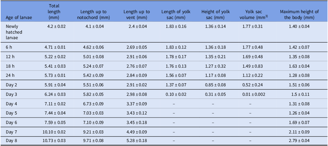
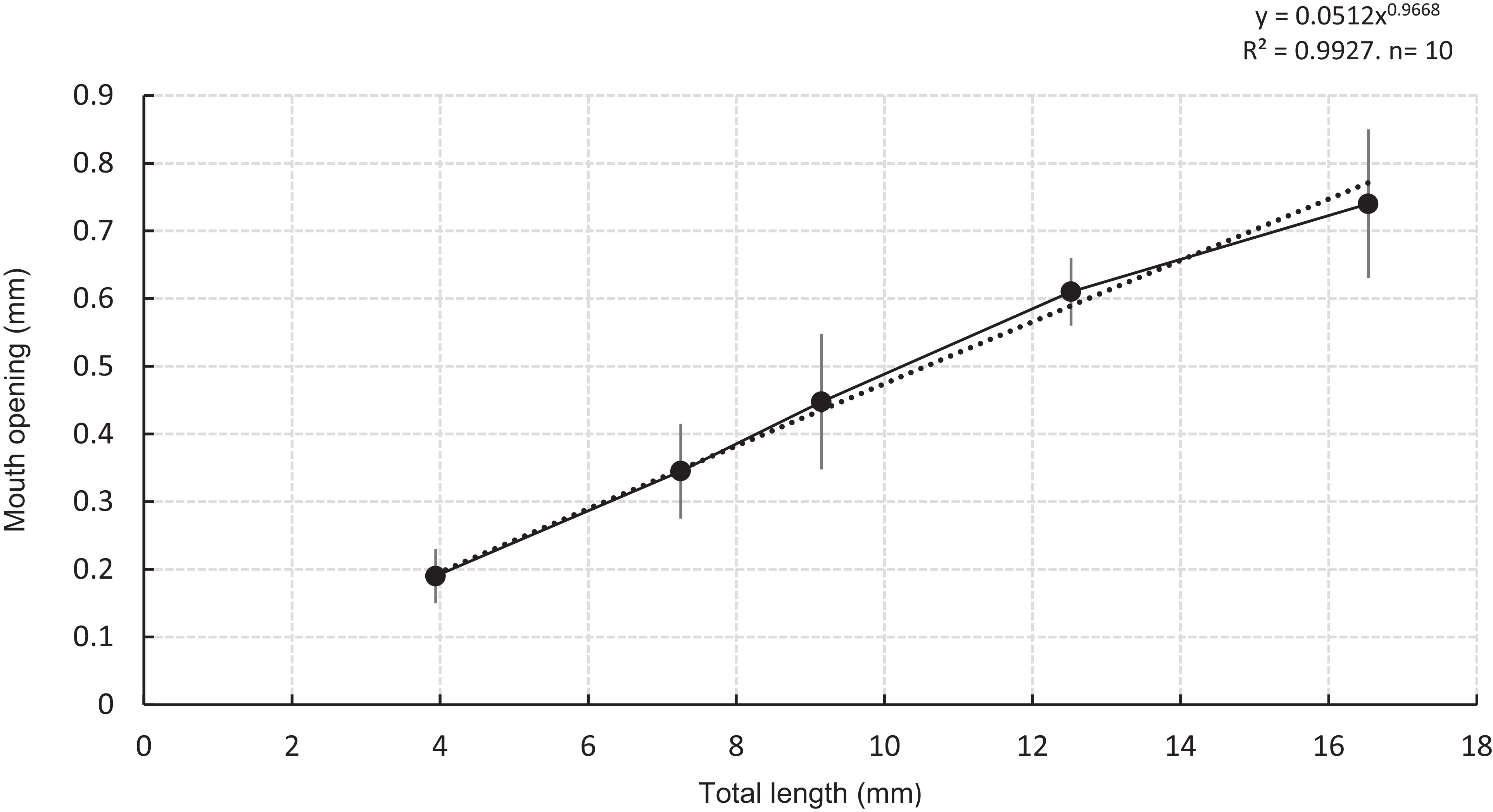
Day-wise variation in TL, MO, and ED across various flexion stages are summarised in Table 3 and visualised in Figure 6. All three metrics increase as development proceeded, growth was relatively slow in the early stages (yolk sac and pre-flexion) and accelerated markedly during the flexion and post-flexion stages. Figure 7 illustrates the allometric relationship between mouth opening and total length, fitted with a power-law trendline (Y = aX b). This power function provided an excellent fit (R 2 = 0.9927, n = 10), and the resulting exponent b was slightly < 1 (b = 0.9668), indicating that mouth opening increases slightly less than proportionally to body length (i.e. slightly negative allometric growth of the gape)
Biometric measurements of Clarias dussumieri larvae at different developmental stages
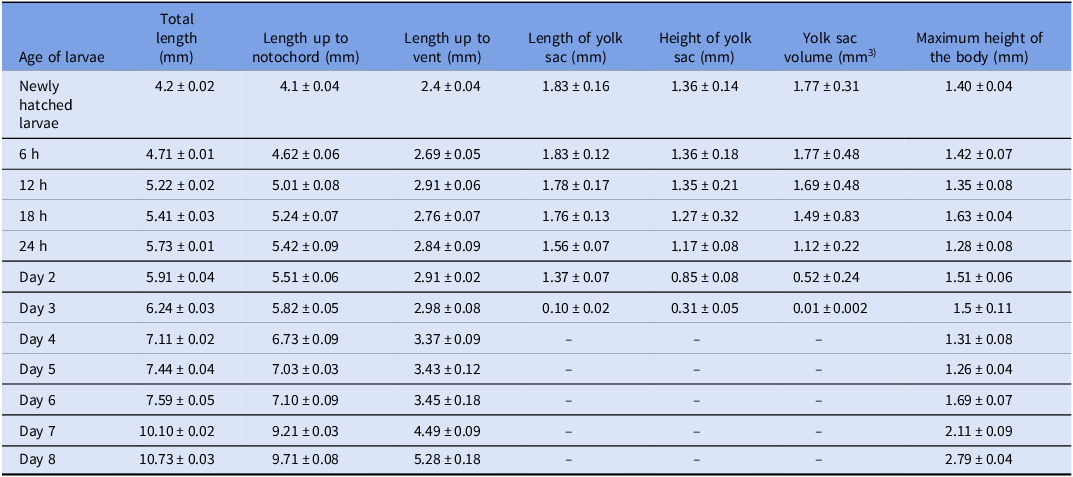
Data collected on various parameters were presented as mean ± SEM (Standard error of mean). YSV was calculated from yolk sac length (L) and height (H) using an ellipsoid approximation, YSV = π/6 LH2 (mm3).
Morphometric development of Clarias dussumieri larvae across days post-hatching (dph).
Notes: Dark bars: mouth opening; light bars: eye diameter; blue line: total length. Data are mean ± SEM (red error bars). The x-axis is labelled in dph with consecutive flexion stages indicated.

Allometric relationship between mouth opening and total length fitted with a power function (Y = aXᵇ).
Notes: Points show mean mouth opening (mm) for each length class with SEM error bars; the dotted curve is the power-law fit y = 0.0512x^0.9668, R 2 = 0.9927 and n = 10. Axes are in millimetres.

Discussion
C. dussumieri is classified as Near-Threatened by the IUCN due to rapid population decline(Abraham, Reference Abraham2011). Captive breeding can help conserve this species and enhance local aquaculture (Fraser, Reference Fraser2008). Detailed embryonic and larval developmental knowledge is essential for effective breeding and culture protocol establishment, expanding on earlier preliminary descriptions (Padmakumar et al., Reference Padmakumar, Bindu, Sreerekha, Joseph, Krishnan, Manu and Basheer2017; Basheer and Lal, Reference Basheer, Lal, Raizada, Pravin, Kumar and Jena2019).
Unlike closely related species (C. gariepinus, C. batrachus, H. fossilis, and P. sutchi) with highly adhesive eggs (Islam, Reference Islam2005; Puvaneswari et al., Reference Puvaneswari, Marimuthu, Karuppasamy and Haniffa2009; Olaniyi and Omitogun, Reference Olaniyi and Omitogun2014a; Roy et al., Reference Roy, Mitra, Biswas, Chakraborty, Pal and Homechaudhuri2019), C. dussumieri eggs were observed slightly adhesive, but easily removable by gentle water flow (Padmakumar et al., Reference Padmakumar, Bindu, Sreerekha, Joseph, Krishnan, Manu and Basheer2017). Egg colouration (brownish-orange) matches C. batrachus (Roy et al., Reference Roy, Mitra, Biswas, Chakraborty, Pal and Homechaudhuri2019), differing from the greenish-brown of C. gariepinus and brownish-green of H. fossilis (Puvaneswari et al., Reference Puvaneswari, Marimuthu, Karuppasamy and Haniffa2009; Olaniyi and Omitogun, Reference Olaniyi and Omitogun2014b). Egg size (1.50 mm) was larger compared to other clariids (Olaniyi and Omitogun, Reference Olaniyi and Omitogun2014a; Roy et al., Reference Roy, Mitra, Biswas, Chakraborty, Pal and Homechaudhuri2019), with fertilized egg diameter (1.70 mm) comparable to earlier reports (Aneesh et al., Reference Aneesh, Salin and Nair2013; Padmakumar et al., Reference Padmakumar, Bindu, Sreerekha, Joseph, Krishnan, Manu and Basheer2017), yet distinct from those reported for C. gariepinus (Osman et al., Reference Osman, Wuertz, Mekkawy, Verreth and Kirschbaum2008; Olaniyi and Omitogun, Reference Olaniyi and Omitogun2014a). These differences may reflect brood size and species-specific variations (Puvaneswari et al., Reference Puvaneswari, Marimuthu, Karuppasamy and Haniffa2009).
The classification of C. dussumieri embryonic development was based on Kimmel et al. (Reference Kimmel, Ballard, Kimmel, Ullmann and Schilling1995). Cleavage follows a meroblastic pattern at the animal pole, common in teleosts (Ninhaus-Silveira et al., Reference Ninhaus-Silveira, Foresti and De Azevedo2006; da Rocha Perini et al., Reference da Rocha Perini, Sato, Rizzo and Bazzoli2010; Rodrigues-Galdino et al., Reference Rodrigues-Galdino, Maiolino, Forgati, Donatti, Mikos, Carneiro and Rios2010). The first mitotic cleavage occurred at 00:36 ± 00:02 h, similar to C. gariepinus but different from C. batrachus, H. bidorsalis, and H. fossilis (Olaniyi and Omitogun, Reference Olaniyi and Omitogun2014a; Roy et al., Reference Roy, Mitra, Biswas, Chakraborty, Pal and Homechaudhuri2019). Until the sixth cleavage, the pattern remained synchronous and regular. Subsequently, cleavage became irregular and asynchronous, producing blastomeres with a ‘mulberry’ form, termed blastula (Olaniyi and Omitogun, Reference Olaniyi and Omitogun2014b). The YSL appears as a darker zone between yolk and blastoderm under microscope (Rodrigues-Galdino et al., Reference Rodrigues-Galdino, Maiolino, Forgati, Donatti, Mikos, Carneiro and Rios2010). Gastrulation completed with blastopore closure at 05:45 h (Kimmel et al., Reference Kimmel, Ballard, Kimmel, Ullmann and Schilling1995). The tail bud and polster marked cranial and caudal parts after gastrulation (Olaniyi and Omitogun, Reference Olaniyi and Omitogun2014a, Reference Olaniyi and Omitogun2014b). Body segmentation formed myotome blocks cephalocaudally. Hatching occurred through muscular contraction and embryo twisting, similar to other catfish species (Islam, Reference Islam2005; Puvaneswari et al., Reference Puvaneswari, Marimuthu, Karuppasamy and Haniffa2009; Olaniyi and Omitogun, Reference Olaniyi and Omitogun2014a). The hatching time of eggs was inversely correlated with the incubation as higher temperatures speed up embryonic metabolism and development rates, facilitating quicker progression through developmental stages to hatching (Mollah et al., Reference Mollah, Taslima, Rashid, Hossain, Sarowar and Khan2011). The embryonic development of C. dussumieri lasted 25 h and 30 min at 26°C, when compared with the other species of Siluriformes: 26 h at 26 ± 0.50°C in C. batrachus (Roy et al., Reference Roy, Mitra, Biswas, Chakraborty, Pal and Homechaudhuri2019) and 17.3 h at 28.5 ± 0.50°C in C. gariepinus (Olaniyi and Omitogun, Reference Olaniyi and Omitogun2014a). This was contrary to the findings of Padmakumar et al. (Reference Padmakumar, Bindu, Sreerekha, Joseph, Krishnan, Manu and Basheer2017), where incubation was completed at 16:30 h at 27 ± 1°C.
Newly hatched larvae of C. dussumieri measured 4.2 ± 0.02 mm, shorter than previously reported (Olaniyi and Omitogun, Reference Olaniyi and Omitogun2014a for C. gariepinus: 5.0 ± 0.50 mm; Padmakumar et al., Reference Padmakumar, Bindu, Sreerekha, Joseph, Krishnan, Manu and Basheer2017: 4.9–5.10 mm), potentially due to broodstock size influences (Roy et al., Reference Roy, Mitra, Biswas, Chakraborty, Pal and Homechaudhuri2019). Initial morphological developments, including mouth formation, eye pigmentation, anal invagination, barbels, and fin fold changes coincided with yolk absorption and exogenous feeding initiation (Kipper et al., Reference Kipper, Taguti, Bialetzki, Makrakis, Baumgartner and Sanches2013). Larvae at this yolk sac stage exhibited transparency, large yolk reserves, and prominent myomeres (30–35), essential for swimming and survival (Behmene et al., Reference Behmene, Bachir Bouiadjra, Attou and Homrani2022). By day three, decreased yolk reserves accompanied eye musculature and operculum formation, further signalling readiness for external feeding (Mir et al., Reference Mir, Srivastava, Bhat, Dar, Sushila, Varghese, Muralidhar and Jain2019; Mahalder et al., Reference Mahalder, Haque, Siddique, Hasan, Alam, Talukdar, Shohan, Ahasan, Hasan and Ahammad2023).
The pre-flexion stage (day 4 onward) was characterized by melanophore dispersion and barbel development, enhancing camouflage and sensory function, similar to other Clariidae species (Olaniyi and Akinbola, Reference Olaniyi and Akinbola2013). Flexion-stage larvae (7.6–10.70 mm) underwent morphological changes, including yolk sac absorption, fin development (Yin and Blaxter, Reference Yin and Blaxter1986), and increased myomere counts (52–58), reflecting improved swimming capability (Olaniyi and Omitogun, Reference Olaniyi and Omitogun2014b; Mahalder et al., Reference Mahalder, Haque, Siddique, Hasan, Alam, Talukdar, Shohan, Ahasan, Hasan and Ahammad2023). Post-flexion larvae (11.34–13.70 mm) displayed segmented fin rays, developed caudal fins, and increased myomeres (55–62), indicating transition toward adult-like morphology (Babu et al., Reference Babu, Anirudhan, Madathumpady, Kurva, Dube, Kodi, Mhaddolkar, Pal, Ignatius and Achamveetil2024). Juveniles (15.27–17.80 mm) resembled adults, exhibiting distinct pigmentation and fin ray structures, critical for optimizing rearing and feeding strategies (Conceição et al., Reference Conceição, Yúfera, Makridis, Morais and Dinis2010).
Sifa and Mathias (Reference Sifa and Mathias1987) described the critical period as the phase of profound physiological and ecological changes in larvae during transition from endogenous to exogenous nutrition. During this period, first feeding timing, food availability, and ability of the larvae to ingest are key survival determinants (Ing & Chew, Reference Ing and Chew2015). Similar to other catfish species (Hag et.al., Reference Hag, Kamarudin, Saad and Daud2012; Adebiyi et.al., Reference Adebiyi, Siraj, Harmin and Christianus2013; Ing and Chew, Reference Ing and Chew2015), C. dussumieri larvae begin exogenous feeding when yolk sac absorption is nearly complete and mouth is fully developed. Fish larvae start feeding once they develop pigmented eyes, open anus, functional jaw and intestine, and horizontal swimming behaviour (Ing and Chew, Reference Ing and Chew2015). Therefore, first feeding for C. dussumieri larvae should occur 72 h post-hatching when they develop essential morphological features for foraging. Atmospheric air utilization and aerial breathing were observed between the 10th and 13th day after hatching, similar to H. fossilis and C. striatus (Marimuthu and Haniffa, Reference Marimuthu and Haniffa2007; Puvaneswari et al., Reference Puvaneswari, Marimuthu, Karuppasamy and Haniffa2009). The sequential feeding strategy from Artemia nauplii to Moina to commercial micro feeds aligns with larval digestive system development and mouth size in clariid catfish, optimizing growth and survival rates (Ayele, Reference Ayele2015; Sales, Reference Sales2011).
Morphometry of fish larvae is essential for studying morphological traits during early development. According to Babu et al. (Reference Babu, Anirudhan, Madathumpady, Kurva, Dube, Kodi, Mhaddolkar, Pal, Ignatius and Achamveetil2024), larval morphometry has applications in studying growth, feeding strategies, and comparing wild and captive fish larvae. TL, MO, and ED increased across pre-flexion, flexion, and post-flexion stages, with slower growth during pre-flexion and faster during post-flexion. Morphometric insights help optimize feeding protocols and stocking densities for larval survival (Gisbert et al., Reference Gisbert, Conklin and Piedrahita2004; Yúfera and Darias, Reference Yúfera and Darias2007). Fish larvae consume prey 25–50% of their mouth gape, making mouth opening to body length ratio crucial for feeding (Shirota, Reference Shirota1970; Bremigan and Stein, Reference Bremigan and Stein1994; Krebs and Turingan, Reference Krebs and Turingan2003). Power regression analysis between MO and TL in C. dussumieri showed negative allometric growth (b = 0.9668), indicating slower MO increase compared to body length. Similar patterns exist in C. macrocephalus and Channa striata (Morioka et al., Reference Morioka, Vongvichith, Phommachan and Ueno2013; Untung and Alfian, Reference Untung and Alfian2021). These align with other freshwater species like Leuciscus, which shows negative allometric growth in eye diameter (Kupren et al., Reference Kupren, Mamcarz, Kucharczyk and Prusińska2008). Power regression models revealed strong scaling relationships between TL and functional traits in C. dussumieri. In Pangasianodon hypophthalmus and Channa striata, relative gape size decreases with growth (Baras et al., Reference Baras, Slembrouck, Cochet, Caruso and Legendre2010; Untung and Alfian, Reference Untung and Alfian2021), demonstrating power regression analysis effectively characterizes ontogenetic scaling patterns.
Conclusion
The present study is the first investigation and documentation of sequential and chronological photomicrographs of the embryonic and larval developmental stages of C. dussumieri for successful larval rearing and juvenile production. The findings of this study provide information that can be used to improve feeding protocols and establish specific conservation management for near-threatened catfish through scaled up production. The present documentation along with previous findings (Padmakumar et al., Reference Padmakumar, Bindu, Sreerekha, Joseph, Krishnan, Manu and Basheer2017), contributes to the retrieval of this threatened species by providing biological tools for conservation and mass scale production for sustainable aquaculture.
Supplementary material
The supplementary material for this article can be found at https://doi.org/10.1017/S0967199426100288.
Acknowledgements
The authors acknowledge the Deputy Director General (Fisheries Science) and, ICAR for their encouragement and support for this work. We are deeply grateful to the Director of the ICAR-NBFGR, Lucknow, for his constant moral support and technical guidance. Special thanks to Kerala University of Fisheries and Ocean Studies (KUFOS), Panangad, Kerala, for providing the pond facility, and to the ICAR-CMFRI, Kochi for facilitating the documentation of embryonic and larval development. The first author acknowledges KUFOS, Panangad, Kerala, India for pursuing PhD. This study was funded by the Indian Council of Agricultural Research (ICAR) Institute Plan Fund, under the CRP-Agrobiodiversity consortium program aimed at conserving indigenous species, for which we are sincerely thankful.
Author contributions
The study was conceptualized and designed by Charan Ravi, V.S. Basheer, and Sarath Varghese, and the methodology was devised and implemented by Sarath Varghese, Abhilash C.P., Saikrishnan K.R., and Charan Ravi. Formal analysis and investigation were carried out by Sarath Varghese, Abhilash, Saikrishnan, Neethu Z., Chandana B.L., and Charan Ravi. The initial draft was prepared by Sarath Varghese, Neethu Z., and Charan Ravi, while V.S. Basheer, Uttam Kumar Sarkar, and T.T. Ajithkumar reviewed and refined the manuscript for clarity and accuracy. Overall supervision and guidance for the project were provided by Charan Ravi V.S. Basheer, T.T. Ajithkumar, and Uttam Kumar Sarkar. The final manuscript was approved by all the authors.
Funding information
This study was funded by the Indian Council of Agricultural Research (ICAR) Institute plan fund, under the CRP-Agrobiodiversity consortium program aimed at conserving indigenous species.
Competing interests
The authors have no conflict of interest to disclose.
Ethical standards
The authors assert that all procedures contributing to this work complied with the ethical standards of the relevant national and institutional guides on the care and use of laboratory animals. This study was approved by the Institutional Animal Ethics Committee of the ICAR – National Bureau of Fish Genetic Resources, Lucknow, India, under approval number G/IAEC/2022/8 dated 25.05.2022. All methods used in this study are reported in accordance with the ARRIVE guidelines for animal research.