Fibrotic myocardial remodelling in repaired tetralogy of Fallot is associated with adverse clinical outcomes.Reference Chen, Dusenbery, Valente, Powell and Geva1–Reference Yim, Riesenkampff, Yoo, Seed and Grosse-Wortmann4 Overlap of T1 between health and disease, including in repaired tetralogy of Fallot, is significant, limiting the clinical usefulness of cross-sectional T1 relaxometry. It has been postulated that the evolution of imaging markers of fibrosis awards a better understanding of disease trajectory versus cross-sectional results alone. We aimed to determine the progression of native myocardial T1 by cardiac magnetic resonance.
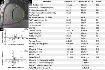
The hospital database was screened for children and adolescents after repaired tetralogy of Fallot who had undergone ≥ one cardiac magnetic resonance examination (n = 37, 12.7 ± 2.6 years, 61% male) at 1.5 T including T1 relaxometry using a modified look-locker inversion recovery approach between November 2013 and February 2018. These had to be ≥ 1 year apart (2.1 ± 0.8 years). Our institutional policy is to routinely refer all tetralogy of Fallot patients >9 years old for surveillance cardiac magnetic resonance evaluation. Global left ventricular, septal, right ventricular diaphragmatic wall, and right ventricular free wall T1 were measured excluding areas of late gadolinium enhancement (Fig 1A).
(a) One of eight T1 relaxometry source images at a mid-ventricular short-axis slice, with regions of interest for global left ventricular T1 (between red and green LV contours), septal T1 (black), right ventricular diaphragmatic wall (pink) and free wall T1 (orange). (b) Baseline and follow-up ECG, exercise test, and magnetic resonance parameters. (c) Relative right ventricular free wall T1 change correlates with right ventricular mass Z-score change and with left ventricular T1 change. RV = right ventricular; LV = left ventricular; RVOT = right ventricular outflow tract; EDV = end diastolic volume; ESV = end systolic volume; EF = ejection fraction; PR = pulmonary regurgitation; RVDW = right ventricular diaphragmatic wall; and RVFW = right ventricular free wall.

Figure 1B shows no significant progression of ECG, exercise capacity, or imaging parameters from baseline. While only 2 patients had predominant right ventricular outflow tract obstruction (right ventricular outflow tract gradient > 30 mmHg, pulmonary regurgitation < 20%), 11 had predominant pulmonary regurgitation (right ventricular outflow tract gradient < 30 mmHg, pulmonary regurgitation > 20%); the rest (24) had a combination of the two. Late gadolinium enhancement imaging was performed in 29 patients (78%) during their first cardiac magnetic resonance and in 7(19%) on follow-up. Six patients (21%) had late gadolinium enhancement extending from the right ventricular outflow tract to adjacent right ventricular free wall beyond the expected surgical site; no new or progressive late gadolinium enhancement was identified. While left ventricular T1 and right ventricular diaphragmatic wall T1 remained similar, right ventricular free wall T1 increased (913 ± 208 ms to 1023 ± 220 ms, p = 0.02). Baseline cardiac magnetic resonance parameters did not predict a change in imaging markers or exercise tolerance. We found no significant relation of either baseline QRS duration or its progression with T1 values for either the right or left ventricle or their progressions. The right ventricular free wall per cent change correlated with left ventricular T1% change (r = 0.51, p = 0.001) and right ventricular mass Z-score change (r = 0.51, p = 0.001, Fig 1C). T1 in patients with late gadolinium enhancement did not differ from the rest.
Ventricular volumes, function, pulmonary regurgitation, right ventricular outflow tract gradient, and exercise capacity remained stable and did not correlate with absolute or relative native right ventricular or left ventricular T1 times in this cohort with repaired tetralogy of Fallot. Right ventricular free wall T1 increased, indicating possible progressive fibrotic remodelling in the right ventricular outflow tract, which has been associated with adverse outcomes in repaired tetralogy of Fallot.Reference Babu-Narayan, Kilner and Li3 We did not find a significant change in left ventricular T1 over time. Nonetheless, T1 increase in the right ventricular free wall correlated with that in the left ventricle. These findings and histological studies suggest cross-talk between the ventricles on a molecular level. Furthermore, the association between progression of right ventricular T1 and that of right ventricular hypertrophy may indicate remodelling consisting of both fibrosis and compensatory hypertrophy. Limitations of our study include the small sample size, prohibiting, for example, a comparison between patients with predominant right ventricular outflow tract obstruction and those with isolated pulmonary regurgitation, a relatively short follow-up interval as well as technical challenges related to partial volume effects in the right ventricular myocardium and possible inter and intra-observer variability.
Increasing right ventricular free wall T1 indicates possible progressive fibrotic remodelling in the right ventricular outflow tract in this pilot study in children and adolescents with repaired tetralogy of Fallot. The value of T1 mapping both at baseline and during serial assessments will need to be investigated in larger cohorts with longer follow-up.
Conflicts of interest
None.
Funding
This research did not receive any specific grant from funding agencies in the public, commercial, or not-for-profit sectors.