Introduction
Orthostatic hypotension (OH), or postural hypotension, describes an excessive fall in blood pressure (BP) on standing. It is a common finding in older people, with prevalence rising from 6% in middle ageReference Fedorowski, Stavenow, Hedblad, Berglund, Nilsson and Melander1 to 12–30% of older people in the communityReference Alli, Avanzini and Bettelli2–Reference Luukinen, Koski, Laippala and Kivela4 and up to 68% in hospital.Reference Vloet, Pel-Little, Jansen and Jansen5–Reference Gorelik, Fishlev, Litvinov, Almoznino-Sarafian, Alon, Shteinshnaider, Chachashvily, Modai and Cohen7 It can present with a wide variety of clinical problems including falls, seizures, dizziness and chest pain.Reference Luukinen, Koski, Laippala and Kivela4, Reference Belmin, Abderrhamane, Medjahed, Sibony-Prat, Bruhat, Bojic and Marquet8–Reference Masaki, Schatz, Burchfiel, Sharp, Chiu, Foley and Curb12 Furthermore, presence of OH is a poor prognostic marker in diabetes,Reference Luukinen and Airaksinen13 heart failure,Reference Tilvis, Hakala, Valvanne and Erkinjuntti14 kidney failureReference Sasaki, Nakahama, Nakamura, Yoshihara, Inenaga, Yoshii, Kohno and Kawano15 and elderly people in general.Reference Räihä, Luutonen, Piha, Seppänen, Toikka and Sourander3, Reference Masaki, Schatz, Burchfiel, Sharp, Chiu, Foley and Curb12
The assessment of patients for OH is therefore a frequently performed clinical measurement and is undertaken for a wide range of conditions; for example in the evaluation of falls, hydration status and blood loss, therapeutic and side-effect monitoring (e.g. in hypertension, Parkinson's disease) and in diagnosis of various conditions, e.g. autonomic dysfunction, Addison's disease, phaechromocytoma and syncope.16–Reference Ewing and Clarke20
In older people the finding is both common but also inconsistentReference Weiss, Grossman, Beloosesky and Grinblat6, Reference Ooi, Barrett, Hossain, Kelley-Gagnon and Lipsitz21–Reference Ward and Kenny23 and there is evidence that routine measurement techniques are frequently suboptimal.Reference Vloet, Pel-Little, Jansen and Jansen5, Reference Cooke, Carew, O'Connor, Costelloe, Sheehy and Lyons24, Reference McKay, Campbell, Parab, Chockalingam and Fodor25 This risks compromising patient care and leading to incorrect or missed diagnoses, inappropriate management, as well as contributing to delays in patient progress and to ineffective use of staff time. For patients undergoing repeated measures, poor and/or varied methodology contributes to inconsistent findings and hence may misinform diagnostic and therapeutic assessments.
It is also recognized that the cardiovascular response to postural challenge (orthostasis) is a spectrum with clinical consequences evident not only due to orthostatic hypotension, but also in response to abnormal fluxes in heart rate (HR).Reference Moya, Sutton and Ammirati19, Reference Jacob, Costa, Shannon, Robertson, Wathen, Stein, Biaggioni, Ertl, Black and Robertson26 Additionally, there are a variety of patterns of OH described; for example, patients who demonstrate an initial transient hypotension, to those whose condition is of a delayed or gradual onset.Reference Moya, Sutton and Ammirati19, Reference Gibbons and Freeman27, Reference Streeten and Anderson28
This review will address the issues surrounding the routine assessment of postural cardiovascular responses in the evaluation of OH and related conditions. The underlying regulatory mechanisms are outlined and considered in terms of clinical assessment. Factors that affect assessment are discussed and guidance is offered in terms of measurements of postural cardiovascular responses in the clinical setting.
Relevant studies were identified from a PubMed search limited to articles published since 2004 in the English language. These papers were supplemented by review of the references of the identified articles and authoritative texts.
Normal cardiovascular responses to postural challenge
In the upright position gravity exerts a downward force on our bodies. This draws blood downward, creating a gradient in perfusion pressures to organs and tissues.Reference Smit, Halliwill, Low and Wieling29, Reference Low30 To maintain perfusion, a series of compensatory neuronal, humoral and endothelial mechanisms adapt HR, stroke volume, cardiac contractility, vascular resistance and venous capacitance.Reference Mathias and Bannister31
On standing upright after a period of recumbency there is displacement of 300–1000 ml of blood into the veins of the legs, pelvis and abdomen.Reference Smit, Halliwill, Low and Wieling29, Reference Grubb32 This displacement is initially rapid, predominantly occurring within the first 10 seconds, although further accumulation continues over 3–5 minutes, with the majority of the redistribution occurring in the pelvic and upper leg veins.Reference Smit, Halliwill, Low and Wieling29 Subsequently there is increased capillary filtration and a reduction in plasma volumeReference Smit, Halliwill, Low and Wieling29, Reference Grubb32 and continuing transcapillary losses approach a steady state after 20–30 minutes.Reference van Lieshout, Wieling, Karemaker and Secher33
Early after attaining an upright posture there is a reduction in venous return to the heart by 25–30%, leading to a transient reduction in stroke volume of approximately 40%Reference Smit, Halliwill, Low and Wieling29, Reference Grubb32 following an initial brief delay due to the blood volume already within the thorax.Reference Low30 To maintain cardiac output there is an acute compensatory increase in HR, systemic vascular resistance and venous tone.Reference Smit, Halliwill, Low and Wieling29–Reference Mathias and Bannister31, Reference Ewing, Campbell, Murray, Neilson and Clarke34 These rapid changes are primarily mediated by the autonomic nervous system via sympatho-excitatory neurones under the influence of the carotid and aortic (high pressure) and pulmonary and cardiac (low pressure) baroreceptors.Reference Mathias, Bannister, Bannister and Mathias10, Reference Smit, Halliwill, Low and Wieling29
Also, as pressure rises, axonal reflexes lead to prompt increases in local smooth muscle tone acting on arteriolar and venous beds, with subsequent redistribution of blood flow.Reference Smit, Halliwill, Low and Wieling29, Reference Low30 In the steady state, these neuronal systems are in a state of continuous activation and have a periodicity that can also be used to differentially assess the functioning of the sympathetic and parasympathetic systems.Reference Mathias and Bannister31, 35
Whilst neuronal control mechanisms are the key to effective cardiovascular responses to initial posture change, hormonal systems have increasing relevance with longer periods of orthostasis.Reference Low30, Reference Mathias and Bannister31 Notably, in the setting of hypovolaemia, these hormonal fluxes are greater and occur earlier, augmenting the initial phases of postural cardiovascular control.Reference Low30
Following orthostatic challenge, the adrenergic and renin-angiotensin-aldosterone systems are activated, contributing to the maintenance of plasma volume.Reference Mathias and Bannister31, Reference Millet, Custaud, Maillet, Allevard, Duvareille, Gauquelin-Koch, Gharib and Fortrat36 Systemic catecholamine levels have been seen to rise within minutes of postural change, whilst changes in renin-angiotensin-aldosterone are more gradual.Reference Low30, Reference Mathias37 Ordinarily, little change is seen in anti-diuretic hormone (ADH) levels unless hypotension develops, when there is a marked increase.Reference Low30
Natriuretic peptides are released in response to stimulation from stretch receptors; these agents have diverse effects and sites of action, and a moderate influence on the activity of the adrenergic and renin-angiotensin-aldosterone systems.Reference Smit, Halliwill, Low and Wieling29, Reference Akashi, Springer, Lainscak and Anker38
Thus there are three distinct phases of normal postural cardiovascular control mechanisms; the first is the initial response related to early volume redistribution and neuronal control systems (about 0–30 s), followed by early steady state related to stabilization of neuronal mechanisms with early hormonal fluxes (about 1–2 min), and finally, during prolonged orthostasis, there is an ongoing maintenance phase of control and increasing hormonal input (about 5+ min).Reference Low30
These mechanisms therefore combine to maintain tissue perfusion. Where there is failure of homeostatic mechanisms it is the cerebral circulation that is most vulnerable, leading to a variety of symptoms including visual disturbances and syncope.Reference Moya, Sutton and Ammirati19
Physiological measurements following orthostasis
Following orthostasis there is a normal transient increase in HR of 10–15 beats per minute, which is maximal about 15 heartbeats after standing.Reference Ewing, Campbell, Murray, Neilson and Clarke34 This normal, prompt physiological response helps facilitate the maintenance of cerebral perfusion and systemic blood pressure. Its presence is an indicator of a functioning parasympathetic autonomic nervous system, is usually transient and is followed by a period of relative bradycardia (assessed by the 30:15 ratio).Reference Ewing and Clarke20, Reference Ewing, Campbell, Murray, Neilson and Clarke34 Furthermore, the lack of an initial HR rise in the setting of OH may aid in the identification of patients with a likely neurogenic mechanism.Reference Gupta and Nair39
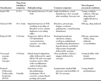
In contrast, in some patients, postural change is associated with excessive increases in HR and with a variety of symptoms including dizziness, visual disturbance, syncope and occasionally falls.Reference Jacob, Costa, Shannon, Robertson, Wathen, Stein, Biaggioni, Ertl, Black and Robertson26, Reference Low30, Reference Grubb32 Where there is a 30 beats per minute increment in HR whilst standing, this is suggestive of the condition Postural Orthostatic Tachycardia Syndrome (POTS).Reference Low30, Reference Grubb32, Reference Mathias40 POTS is associated with a number of medical conditions (e.g. peripheral neuropathy, chronic fatigue syndrome) and muscular deconditioning (Table 1). The underlying mechanisms may include a partial denervation of the autonomic system with the lower limbs specifically involved, an elevation in adrenergic activity on gaining upright postureReference Jacob, Costa, Shannon, Robertson, Wathen, Stein, Biaggioni, Ertl, Black and Robertson26, Reference Low30 and/or disordered central HR control.Reference Grubb32 POTS may be overlooked if HR is not recorded during postural cardiovascular assessment and can be an early finding in the development of more widespread autonomic failure.Reference Low30, Reference Jacob, Shannon, Black, Biaggioni, Mosqueda-Garcia, Robertson and Robertson41
Classification of OH and related conditions, adapted from Moya et al. 2009Reference Moya, Sutton and Ammirati19

CO, cardiac output; SVR, systemic vascular resistance; VR, venous return; CSS, carotid sinus syndrome.
Definition of orthostatic hypotension
The most commonly applied definition is that OH is present where there is a drop in systolic blood pressure of 20 mmHg or more, and/or in diastolic blood pressure of 10 mmHg or more, occurring within the first three minutes of standing (or during head-up tilt-table testing). This consensus definition was published in 1996 by the American Autonomic Society and the American Academy of Neurology (AAS/AAN).42, Reference Kaufmann43 Prior to this there had been inconsistency, with marked variation in definitions used.Reference Ewing and Clarke20, Reference Low30, Reference Atkins, Hanusa, Sefcik and Kapoor44
The consensus statement also highlights that some patients manifest hypotension 10 minutes or more after standing and therefore it is suggested, where clinical suspicion is high and initial testing is negative, that repeated and extended measurements are warranted.42
Although generally accepted, the consensus definition of OH as a BP reduction of 20/10 mmHg is arbitrary.Reference Mathias37, Reference Carey and Potter45, Reference Weiss, Chagnac, Beloosesky, Weinstein, Grinblat and Grossman46 Symptomatic hypotension can occur with blood pressure changes at levels less than this,Reference Mathias37, Reference Weiss, Chagnac, Beloosesky, Weinstein, Grinblat and Grossman46–Reference Carlson48 particularly where there is relative baseline hypotension or obstruction to cerebral blood flow.Reference Mathias and Bannister31 Furthermore, OH was found to be associated with mortality in a linear fashion in the Honolulu heart study, without evidence of a threshold level.Reference Masaki, Schatz, Burchfiel, Sharp, Chiu, Foley and Curb12 Given this and the overall weak association found in many studies between OH and clinical outcomes, some authors have suggested that a review of the AAS/AAN diagnostic criteria would be appropriate.Reference Belmin, Abderrhamane, Medjahed, Sibony-Prat, Bruhat, Bojic and Marquet8, Reference Weiss, Chagnac, Beloosesky, Weinstein, Grinblat and Grossman46, Reference Caine, Alsop and MacMahon49
The 2009 European Society of Cardiology (ESC) syncope guidelines seek to further refine the characterization of OH (Table 1).Reference Moya, Sutton and Ammirati19 In this updated guidance OH is subdivided into initial (occurring within 30 s), classical (30 s to 3 min) and delayed (progressive; which occurs after 3 min). These phases have a logical relationship with the mechanisms involved in the maintenance of blood pressure, and act to reinforce the necessity for the recording of serial measurements of BP in the assessment of postural cardiovascular responses. At present, such subdivisions may suggest differential underlying mechanisms but recommended interventions are not differentiated.Reference Moya, Sutton and Ammirati19
Delayed OH is characterized by a gradual progressive fall in BP, whereas with vasovagal syncope, the typical pattern is of an abrupt change, with or without an associated reduction in heart rate.Reference Moya, Sutton and Ammirati19 It is the gradual nature of the BP fall and the absence of changes in HR that help differentiate delayed OH from vasovagal syncope (Table 1).Reference Moya, Sutton and Ammirati19
In establishing the relevance of a diagnosis of OH it should be noted that the changes in blood pressure seen are often inconsistent and the finding of OH may not be a reliable finding. Where the finding is more consistent and associated with symptoms, it is more likely to be clinically relevant.Reference Ooi, Barrett, Hossain, Kelley-Gagnon and Lipsitz21 Where patients have symptoms associated with postural change and a degree of hypotension develops that does not meet these thresholds, it is relevant for clinicians to consider this. The diagnostic label of symptomatic postural hypotension may be preferable in such circumstances, but it is prudent to ensure the presence or absence of symptoms is recorded during assessment as a matter of routine.
In patients who have a marked HR rise on standing without evidence of intravascular volume depletion, a diagnosis of POTS should be considered. This is often associated with pronounced symptoms including chest pain, dizziness and fatigue.Reference Carlson48
Isolated diastolic orthostatic hypotension
Although the importance of systolic OH is generally accepted and established in relation to falls and symptoms of cerebral hypoperfusion, the significance of isolated diastolic OH, which is less common, is not agreed.Reference Luukinen, Koski, Laippala and Kivela4, Reference Weiss, Grossman, Beloosesky and Grinblat6, Reference Tilvis, Hakala, Valvanne and Erkinjuntti14, Reference Ward and Kenny22, Reference Brignole, Alboni and Benditt47, Reference Eigenbrodt, Rose, Couper, Arnett, Smith and Jones50 The evidence that diastolic hypotension relates to cerebral hypoperfusion or volume depletion is debated, although there are consistent associations with vascular diseases and mortality.Reference Tilvis, Hakala, Valvanne and Erkinjuntti14, Reference Eigenbrodt, Rose, Couper, Arnett, Smith and Jones50
Symptoms in orthostatic hypotension
OH may present with postural dizziness, visual/auditory disturbance, presyncope, syncope or seizures, although symptoms are often milder in older patients than in younger ones and may be absent, even when hypotension is marked.Reference Atkins, Hanusa, Sefcik and Kapoor44, Reference Carlson48, Reference Arbogast, Alshekhlee, Hussain, McNeeley and Chelimsky51 Numerous other less common symptoms have also been reported and include chest pain, the characteristic ischaemic neck/shoulder (‘coat-hanger’) pain, buttock/thigh pain, nausea, palpitations, dyspnoea, episodic confusion, intermittent focal neurological symptoms and non-specific malaise.Reference Mathias40, 42, Reference Arbogast, Alshekhlee, Hussain, McNeeley and Chelimsky51–Reference Gibbons and Freeman53
Whilst in the interpretation of the finding of OH the presence of symptoms (or the development of postural instability or body sway) is helpful, the absence of symptoms does not necessarily indicate a non-significant clinical finding.42, Reference Brignole, Alboni and Benditt47, Reference Carlson48 In older subjects up to 30% with significant OH may be asymptomatic, whilst a further 24% may have atypical symptoms.Reference Arbogast, Alshekhlee, Hussain, McNeeley and Chelimsky51
Causes of orthostatic hypotension
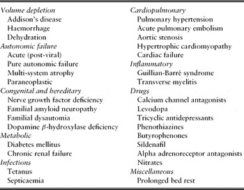
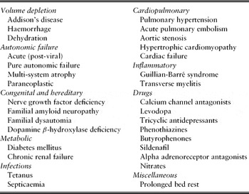
Although OH can be due to a wide variety of conditions (see Table 2), in older people OH is frequently multifactorial; common contributing factors include underlying medical conditions, drug treatments, sarcopenia, blunted hormonal responses, reduced adrenoreceptor and baroreceptor sensitivity and impaired volume regulation.Reference Atkins, Hanusa, Sefcik and Kapoor44, Reference Carlson48, Reference Lahrmann, Cortelli, Hilz, Mathias, Struhal and Tassinari52
Selected causes of orthostatic hypotension
Hypertension is a risk factor for the development of OH.Reference Fedorowski, Stavenow, Hedblad, Berglund, Nilsson and Melander1 When OH occurs in patients with supine hypertension, defining the optimal BP management can be extremely challenging.Reference Mathias and Bannister31, Reference Brignole, Alboni and Benditt47, Reference Shibao, Gamboa, Diedrich and Biaggioni54 There is evidence that OH is a consequence of impaired baroreceptor function, and that by improving control of hypertension there may be an improvement in OH.Reference Naschitz, Slobodin, Elias and Rosner55 This approach must be balanced against the risk of worsening OH and clinical sequelae. The optimal treatment regime in these circumstances remains uncertain and a cautious approach is recommended.
Assessing postural cardiovascular responses
Unfortunately, whilst recommending the routine measurement of postural blood pressure as a technique for identifying OH, many of the key guidelines do not offer specific advice on the active stand technique, the most suitable method for routine clinical assessment.16, 42, Reference Lahrmann, Cortelli, Hilz, Mathias, Struhal and Tassinari52, 56 Where advice is available, it is at times inconsistent and conflicting.Reference Low30, Reference Mathias and Bannister31, Reference Kenny57, Reference Williams, Poulter, Brown, Davis, McInnes, Potter, Sever and McG58 The lack of consistent advice and guidance on the measurement of OH by the active stand method, in contrast with tilt-table testing, is explained by the limited research base upon which evidence can be directly drawn.Reference Lahrmann, Cortelli, Hilz, Mathias, Struhal and Tassinari52
Recommended techniques in the literature
The consensus report of the AAS/AAN42 does not give explicit details in the measurement techniques for the detection of OH, and previous reviewers have noted a continuing concern about how best to assess OH using the active stand technique.Reference Carey and Potter45, Reference Carlson48
The European Federation of Neurological Societies’ guidelines for the ‘Diagnosis and Management of OH’ recommend initially assessment of BP and HR whilst supine and for up to 3 minutes whilst standing. Further recommendations are not given although testing with passive head-up tilt-table testing (HUTT) is advised using a specified protocol.Reference Lahrmann, Cortelli, Hilz, Mathias, Struhal and Tassinari52
A period of rest before BP measurement is recommended for all routine readings.Reference Williams, Poulter, Brown, Davis, McInnes, Potter, Sever and McG58, Reference Chobanian, Bakris and Black59 Most guidance recommends a minimum 5 minute period of rest. Lance et al.Reference Lance, Link, Padua, Clavell, Johnson and Knebel60 studied the measurement of supine BPs in healthy volunteers at 5, 10 and 15 minutes. After 5 minutes supine rest average BP was 113.8/62.6; however, a further reduction occurred over the following 5 minutes with BP at 10 minutes showing no difference to BP at 15 minutes (109.3/60.3 vs. 109.4/59.5). No changes in HR were seen with duration of rest.
As described above, the maintenance of BP is under complex control. Humoral systems are much slower to adapt to postural changes and therefore Mathias & BannisterReference Mathias and Bannister31 advised that an optimal duration of rest before study is at least 20 minutes, whilst acknowledging that this may not be practical in routine clinical work.
Fealey & Robertson (quoted by LowReference Low30) recommend a 2 minute period of measurement with recording made of BP and HR using a cuff sphygmomanometer, stating that this should be preceded by 2 minutes of supine rest. KennyReference Kenny57 also recommends a 2 minute measurement period with a rest period advised of 15 minutes.
Through the use of continuous BP monitors, authors have been able to study closely the patterns of BP response to postural change. The nadir of BP is usually seen at approximately 1–1½ minutes, with a subsequent period of recovery.Reference Ward and Kenny22, Reference Atkins, Hanusa, Sefcik and Kapoor44 However, this is not a universal finding and other patterns are recognized.Reference Moya, Sutton and Ammirati19, Reference Gibbons and Freeman53, Reference Macrae and Bulpitt61
Although some authors suggest a single orthostatic measurement of BP, usually to be performed at 1 or 2 minutes, this approach reduces both the sensitivity and reproducibility of measurements and may fail to capture the nadir.Reference Belmin, Abderrhamane, Medjahed, Sibony-Prat, Bruhat, Bojic and Marquet8, 16, Reference Caine, Alsop and MacMahon49 In contrast, the use of continuous BP monitoring systems has been shown to increase the sensitivity of postural BP responses, but such equipment is not routinely available and requires greater time and expertise to use.Reference Ward and Kenny22, Reference Mathias and Bannister31, Reference Caine, Alsop and MacMahon49
The ESC guidelines suggest that repeated measurements of BP are made over 3 minutes and longer if minor degrees of hypotension are found or if hypotension is progressing.Reference Brignole, Alboni and Benditt47 They recommend also that either a manual sphygmomanometer or continuous beat-to-beat system is used in the performance of BP measurements. The manual technique can be used to monitor BP changes more closely and allows skilled practitioners to track changes in BP.Reference Moya, Sutton and Ammirati19 Using the manual technique, a skilled operator can perform up to three measurements per minute, although venous occlusion can occur.
Fundamental to the accurate recording of postural BP are the generic skills of BP measurement. Adherence to the principles of practice outlined in the British Hypertension Society (BHS) guidance are therefore strongly recommended (e.g. arm position, the selection of the correct cuff size and its location on the arm, loosening of tight clothing, maintenance of equipment).Reference Williams, Poulter, Brown, Davis, McInnes, Potter, Sever and McG58 Nevertheless, physicians’ compliance with such recommendations has been found to be poor.Reference McKay, Campbell, Parab, Chockalingam and Fodor25 Furthermore, where BP readings are made by different individuals using manual sphygmomanometers, there is increased variability in the measured BP.Reference Gorny62, Reference McAlister and Straus63
In contrast, concerns have been raised that many automated sphygmomanometers are not validated for the measurement of BP in patients with significant rhythm disturbances (e.g. atrial fibrillation or with frequent ectopics). Additionally, these systems may fail to record (or delay reading) BPs in situations of low flow (e.g. hypotension) or where there are marked changes or instability in BP; therefore they represent a risk when used for postural BP measurements.Reference Moya, Sutton and Ammirati19, Reference Williams, Poulter, Brown, Davis, McInnes, Potter, Sever and McG58, Reference Gorny62
The use of sitting to standing measures would simplify the assessment of postural cardiovascular responses in many clinical settings. This is recommended by some authorsReference Williams, Poulter, Brown, Davis, McInnes, Potter, Sever and McG58 and is often observed during routine practice.Reference Cooke, Carew, O'Connor, Costelloe, Sheehy and Lyons24 The use of the seated position for recording BP has been investigated and compared with the supine.Reference Cooke, Carew, O'Connor, Costelloe, Sheehy and Lyons24, Reference Netea, Smits, Lenders and Thien64 Whilst resting supine and seated BP measurements are well correlated, particularly in the elderly,Reference Netea, Smits, Lenders and Thien64 the correlation with subsequent measurements after orthostasis is poor. Cooke et al.Reference Cooke, Carew, O'Connor, Costelloe, Sheehy and Lyons24 found only a 16% sensitivity when compared with tilt-table test findings. However, whether OH detected from the seated position is more (or less) sensitive than measurements from supine in terms of clinical outcomes is not established.
Overall, active stand techniques for assessment of orthostatic cardiovascular responses have been shown to be safe, although appropriate manual handling techniques do need to be applied, especially when the procedure is undertaken with a frail, confused patient or when the possibility of resultant syncope is high (e.g. severe autonomic failure).
In some patients a measurement of postural BP may be required immediately at the time of presentation (e.g. in the emergency department or out-patient clinic), whilst in others the measurement can be elective and procedures to ensure optimal recording circumstances can be applied.
Factors influencing postural cardiovascular measurements
BP and HR vary in response to physical activity, emotional and psychological factors, pharmacological agents, dietary and fluid intake and to many chemical stimuli.Reference Vloet, Pel-Little, Jansen and Jansen5, Reference Brignole, Alboni and Benditt47, Reference Williams, Poulter, Brown, Davis, McInnes, Potter, Sever and McG58 Intra-individual findings of OH vary significantly throughout the day by a standard deviation of 12/8 mmHg.Reference McAlister and Straus63 Weiss et al.Reference Belmin, Abderrhamane, Medjahed, Sibony-Prat, Bruhat, Bojic and Marquet8 found OH to be most prevalent in the early afternoon when studied in hospital in-patients. However, other authors have reported prevalence of OH to be greatest in the morning.Reference Gorelik, Fishlev, Litvinov, Almoznino-Sarafian, Alon, Shteinshnaider, Chachashvily, Modai and Cohen7, Reference Belmin, Abderrhamane, Medjahed, Sibony-Prat, Bruhat, Bojic and Marquet8, Reference Ward and Kenny22, Reference Macrae and Bulpitt61 Gorelik et al.Reference Gorelik, Fishlev, Litvinov, Almoznino-Sarafian, Alon, Shteinshnaider, Chachashvily, Modai and Cohen7 studied 121 hospital in-patients and found a particularly high incidence (64.5%) of OH on first rising in the morning, using intermittent measurements and recording BP at 1, 3 and 5 minutes; patients spending longer than 8 hours in bed were at significantly increased risk (P < 0.001).
Whilst some studies have found greater reproducibility in the morning,Reference Vloet, Pel-Little, Jansen and Jansen5, Reference Weiss, Grossman, Beloosesky and Grinblat6, Reference Ward and Kenny22 other authors have not.Reference Belmin, Abderrhamane, Medjahed, Sibony-Prat, Bruhat, Bojic and Marquet8 Post-prandial hypotension is a common finding in frail older people (38–67%) and is considered distinct from OH.Reference Vloet, Pel-Little, Jansen and Jansen5, Reference Weiss, Grossman, Beloosesky and Grinblat6, Reference Fisher, Davis, Srikusalanukul and Budge65 Yet post-prandial orthostatic BP measurements identify a number of patients who do not demonstrate OH at other times.Reference Vloet, Pel-Little, Jansen and Jansen5, Reference Weiss, Grossman, Beloosesky and Grinblat6, Reference Lahrmann, Cortelli, Hilz, Mathias, Struhal and Tassinari52
Patient factors
Not only the time from last meal but also hydration status, hormonal fluxes, medication and anxiety levels all have a significant effect on BP. In addition, muscular tone, use of compression hosiery (e.g. compression stockings), rate of postural change, caffeine/nicotine/salt intake, and postural deconditioning (e.g. prolonged bed rest) have all been shown to affect postural cardiovascular reflexes.Reference Mathias37, Reference Brignole, Alboni and Benditt47, Reference Lahrmann, Cortelli, Hilz, Mathias, Struhal and Tassinari52, Reference Puisieux, Boumbar, Bulckaen, Bonnin, Houssin and Dewailly66
Optimization of the rate of postural change is one of the difficulties in the assessment of postural cardiovascular measurements by the active stand technique; patients with poor mobility vary in the speed they are able to stand, which may take as long as 1 minute, whilst pain and discomfort (e.g. from osteoarthritis) will further influence measurements.Reference Carlson48 Many older people with OH are taking pharmacological preparations that influence BP and HR. The timing of dosing and frequency and properties of the preparations may have differential effects through the day. This is dependent not only on the drug but also on the formulation taken, the absorption, metabolism and elimination of the agents, which will vary between individuals.Reference Weiss, Grossman, Beloosesky and Grinblat6, Reference Luukinen and Airaksinen13, Reference Williams, Poulter, Brown, Davis, McInnes, Potter, Sever and McG58
In addition, BP is affected by anxiety, fear, stress and emotional status. Patients undergoing postural measurements should therefore be given reassurance and have the investigation performed in a calm and relaxed manner. They should be encouraged to stand briskly and receive appropriate support and manual handling techniques to do this.
Environmental factors
BP measurements are influenced by the environment in which they are recorded. Ambient temperature, noise and interruptions all affect cardiovascular measurements.Reference Brignole, Alboni and Benditt47, Reference Irvin and White67 In recording responses to postural change the recommendations are that this should be performed in a quiet, dimly lit room, without interruptions or distractions.Reference Moya, Sutton and Ammirati19, Reference Lahrmann, Cortelli, Hilz, Mathias, Struhal and Tassinari52
Whilst in the research laboratory such variable factors can be controlled, this is much more difficult in the clinic, ward, emergency unit or community setting. Nevertheless, staff should try to ensure that during the recording of OH they are not disturbed and that the environmental noises are kept to a minimum.
Overall reproducibility of orthostatic hypotension in trials
Authors have noted that the finding of OH following an active stand remains inconsistent, with poor reproducibility.Reference Alli, Avanzini and Bettelli2, Reference Weiss, Chagnac, Beloosesky, Weinstein, Grinblat and Grossman46 For authors studying subjects at the same time of day with standardized methodology there still remains a lack of consistency in the finding of OH, with only about one-third of patients showing reproducible findings.Reference Alli, Avanzini and Bettelli2, Reference Belmin, Abderrhamane, Medjahed, Sibony-Prat, Bruhat, Bojic and Marquet8, Reference Ward and Kenny22 Where patients have a consistent finding of OH this has been shown to be more strongly associated with falls.Reference Ooi, Barrett, Hossain, Kelley-Gagnon and Lipsitz21 Thus demonstrating a lack of reproducibility may in itself be clinically relevant.
The reproducibility of postural cardiovascular responses is improved by the use of standardized tilt-table testing using continuous beat-to-beat BP monitoring when compared with intermittent measurements using the active stand technique.Reference Gabbett and Gass68 However, this technique is relatively complex, requiring specialist equipment, and is not widely available for routine clinical measurement. It is, however, considered the reference standard for assessment of OH.Reference Cooke, Carew, O'Connor, Costelloe, Sheehy and Lyons24, Reference Lahrmann, Cortelli, Hilz, Mathias, Struhal and Tassinari52 However, HUTT is a passive process of orthostasis and, as such, the mechanisms involved in maintaining cardiovascular homeostasis are subtly different from a ‘normal’ active stand.Reference Cooke, Carew, O'Connor, Costelloe, Sheehy and Lyons24, Reference Kenny57 Thus although studies using the HUTT approach to assess postural cardiovascular responses are instructive, there is the potential for the finding to be less clinically relevant than measurements by the active stand methodology.Reference Cooke, Carew, O'Connor, Costelloe, Sheehy and Lyons24, Reference Kenny57
In summary, the recommendations on the measurement procedure for investigation of OH by active stand technique in standard reference texts and papers show significant variation. Thus it is not possible to identify a consensus approach or best practice outline solely from such sources.
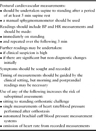
Recommendations (Table 3)
There is a lack of scientific studies comparing various aspects of the active stand methodology in the assessment of orthostatic cardiovascular responses and specific long-term clinical outcomes.Reference Lahrmann, Cortelli, Hilz, Mathias, Struhal and Tassinari52 This has resulted in the development of varied measurement protocols seen in both the clinical setting and published literature. Such varied measurement protocols further contribute to the complexity of study analysis and in comparing the individual studies.Reference Ward and Kenny22, Reference Lahrmann, Cortelli, Hilz, Mathias, Struhal and Tassinari52
Recommendations for assessing orthostatic hypotension

It is notable that the use of automated BP measurement systems is now commonplace in the published literature as well as in clinical practice. It is also noteworthy that not all available systems are validated to the standards of the BHS (a list of validated systems can be accessed on the BHS website: www.bhsoc.org/bp_monitors/automatic_clinic.stm). As automated devices often set a target BP related to previous readings, where there are large changes in BP, the devices take longer, or repeat, measurements. In situations of low flow such devices may be unable to measure the BP; therefore, where there are significant changes in BP such devices may fail or delay readings and thus they are not recommended for the measurement of orthostatic hypotension.Reference Moya, Sutton and Ammirati19
The use of a manual sphygmomanometer can allow the experienced practitioner to undertake repeated assessments quickly. It does, however, require training and development of a skill base. Although the skills of BP measurement are considered a fundamental tool for both doctors and nurses, studies looking at the application of these skills in clinical practice have identified poor procedures and multiple errors.Reference McKay, Campbell, Parab, Chockalingam and Fodor25, Reference McAlister and Straus63, Reference Vloet, Smits, Frederiks, Hoefnagels and Jansen69
Continuous beat-to-beat monitoring systems have been recommended in the measurement of postural cardiovascular measurements.Reference Mathias and Bannister31, Reference Kenny57 These systems do, however, require greater technical skill and are more costly in terms of initial outlay, maintenance and repairs. They have the advantage that continuous monitoring systems allow uninterrupted recordings of the BP and HR. Studies have demonstrated that the use of such systems improves the identification of episodes of transient OH.Reference Ward and Kenny22, Reference Van Der Velde, Van Den Meiracker, Stricker and Van Der Cammen70 However, whether brief episodes of asymptomatic hypotension detected on standing by beat-to-beat systems carry the same implications to those detected by traditional methods is uncertain.Reference Van Der Velde, Van Den Meiracker, Stricker and Van Der Cammen70 The training and investment in equipment that would be required for widespread use of beat-to-beat BP systems is substantial.
The most widely applicable technique for measurement of postural cardiovascular responses is therefore through the use of manual equipment that is appropriately maintained, and is performed by staff who are adequately trained. The use of automated devices, which is now widespread, carries a small risk that the devices will fail to detect significant OH. As long as staff appreciate this and pay close attention to the patient during the measurement period, the attendant risk is low; and this theoretical concern was not highlighted in the studies that used such devices. No matter what approach is used, staff should be performing to the standards of BP measurement as laid down in the BHS guidelines.Reference Williams, Poulter, Brown, Davis, McInnes, Potter, Sever and McG58, Reference McAlister and Straus63
A very high incidence of OH has been demonstrated initially on rising (in the morning) in hospital in-patients, with a high symptom frequency, whilst others have found the peak to be later in the morning.Reference Gorelik, Fishlev, Litvinov, Almoznino-Sarafian, Alon, Shteinshnaider, Chachashvily, Modai and Cohen7, Reference Ward and Kenny22 Where orthostatic BP measurement is to be undertaken first thing in the morning, this should be before there is significant physical activity. Additionally, some patients have symptomatic OH only post-prandially, and thus detection requires clinical suspicion and measurements after meal times. Whilst for some patients (e.g. hospital in-patients) there is flexibility in the timing of postural cardiovascular measurements, this is not the case for many clinical encounters (e.g. emergency unit, out-patient clinics).
The performance of seated to standing BP is not optimal, and this approach should only be considered when there is no alternative. Although it can be useful and does identify some patients with marked OH, it is a significant compromise with a high rate of false negatives.Reference Cooke, Carew, O'Connor, Costelloe, Sheehy and Lyons24, Reference Carlson48, Reference Netea, Smits, Lenders and Thien64
The optimal period of supine rest that should precede a measurement of OH is not defined; it is recommended that all patients have a period of 5 minutes supine rest before measurements are made, in keeping with the JNC and BHS guidelines, although a supine rest period of 10 minutes may offer an advantage.Reference Williams, Poulter, Brown, Davis, McInnes, Potter, Sever and McG58, Reference Chobanian, Bakris and Black59, Reference Lance, Link, Padua, Clavell, Johnson and Knebel60 The use of a single measurement approach has been shown to fail to identify many patients with OH when compared with continuous measurement systemsReference Ward and Kenny22, Reference Caine, Alsop and MacMahon49 or repeated intermittent readings.Reference Luukinen, Koski, Laippala and Kivela4, Reference Belmin, Abderrhamane, Medjahed, Sibony-Prat, Bruhat, Bojic and Marquet8 BP should be recorded immediately on standing and repeated over the following 3 minutes.
Whether the recording period need be as long as 30 minutes or more, as suggested by some authors, is not proven.Reference Cheshire and Phillips71 Given the lack of evidence and resource implications for such a period, it is not recommended that the measurement time is extended beyond 5 minutes in routine clinical settings.
Although uncommon, the finding of orthostasis related sinus tachycardia (HR rise increase 30 beats per minute) is significant and suggestive of POTS.Reference Low30, Reference Grubb32, Reference Mathias40 Furthermore, patients with OH who do not have a HR rise (in the absence of rate-limiting medication) are more likely to have a neurogenic aetiology, and therefore recording the pulse rate can assist in guiding further investigations as to the cause of OH.Reference Gupta and Nair39
As has been discussed, the finding of OH in older people is extremely common. Potentially important features in its analysis are the degree of drop in BP, associated changes (or lack of) in heart rate, and the presence of signs and symptoms and the consistency of the finding. It is important therefore that symptoms are identified and actively sought by directly asking the patient about them.Reference Carlson48 However, as there is a large subpopulation (30%) with marked OH who do not report symptoms, a high index of suspicion should be applied.Reference Arbogast, Alshekhlee, Hussain, McNeeley and Chelimsky51
Conclusion
Bedside assessments of cardiovascular responses to orthostasis are inexpensive, requiring little investment in material costs, and give results immediately. However, poorly performed tests lead to diagnostic errors and inappropriate management.
Unfortunately, there is a lack of research and scientific study comparing methodological approaches in the active stand technique to assess postural cardiovascular responses.Reference Lahrmann, Cortelli, Hilz, Mathias, Struhal and Tassinari52 This is highlighted by the different methodologies given in standard reference publications.Reference Low30, Reference Mathias and Bannister31, Reference Kenny57
Inevitably, this has led to a variety of differing approaches in clinical trials and a notable tendency for researchers to prefer to use the passive technique of HUTT to assess postural cardiovascular responses. This approach, whilst suitable for research purposes, is not applicable as a routine clinical screening or initial assessment tool and has limitations in generalizing findings to day-to-day postural cardiovascular homeostasis.Reference Kenny57
There is consequently a lack of consistency of approach to the measurement of postural cardiovascular responses demonstrated in the contemporary literature and by clinical staff.Reference McKay, Campbell, Parab, Chockalingam and Fodor25, Reference Vloet, Smits, Frederiks, Hoefnagels and Jansen69 In the clinical setting it leads to uncertainty that may contribute to the reluctance of staff to undertake postural cardiovascular assessments and therefore to the under-recognition of the diagnosis.Reference Carey and Potter45, Reference Eigenbrodt, Rose, Couper, Arnett, Smith and Jones50, Reference Lahrmann, Cortelli, Hilz, Mathias, Struhal and Tassinari52, Reference Jones and Huwez72
It is evident that there is a need for greater consistency of approach to postural cardiovascular measurements, informed by the currently available evidence base. This needs to be communicated to staff and support given, as significant concerns about the self-reported approaches to postural cardiovascular measurements have been identified including fundamental procedural errors and poor practice. Reference McKay, Campbell, Parab, Chockalingam and Fodor25, Reference Vloet, Smits, Frederiks, Hoefnagels and Jansen69
By improving staff knowledge through education, supervision and monitoring, the application of the technique should become more consistent and more efficient. The inherent variability of the clinical finding is well established but the need for a robust procedure to identify its presence is essential. Patients who present with falls to hospitals are at a high risk of further falls and injuries and such risks are best prevented by comprehensive assessment and risk factor management.16, 56
OH is a common factor in falls in older people.Reference Kenny57, Reference Rubenstein, Robbins, Josephson, Schulman and Osterweil73 The first step in management is identification through appropriately measured cardiovascular responses to postural stress. Current practice is varied, with inappropriate techniques and suboptimal strategies too often applied. Improvements in methodology will improve case finding and help promote optimal management, potentially reducing falls and their consequences, including fractures, care home admissions and deaths.
Conflicts of interest
The author has no conflicts of interest to declare.