




No CrossRef data available.
Article contents
The proteasome inhibitor MLN-273 blocks exoerythrocytic and erythrocytic development of Plasmodium parasites
Published online by Cambridge University Press: 09 March 2005
Abstract
Protein degradation is regulated during the cell cycle of all eukaryotic cells and is mediated by the ubiquitin-proteasome pathway. Potent and specific peptide-derived inhibitors of the 20S proteasome have been developed recently as anti-cancer agents, based on their ability to induce apoptosis in rapidly dividing cells. Here, we tested a novel small molecule dipeptidyl boronic acid proteasome inhibitor, named MLN-273 on blood and liver stages of Plasmodium species, both of which undergo active replication, probably requiring extensive proteasome activity. The inhibitor blocked Plasmodium falciparum erythrocytic development at an early ring stage as well as P. berghei exoerythrocytic progression to schizonts. Importantly, neither uninfected erythrocytes nor hepatocytes were affected by the drug. MLN-273 caused an overall reduction in protein degradation in P. falciparum, as demonstrated by immunoblots using anti-ubiquitin antibodies to label ubiquitin-tagged protein conjugates. This led us to conclude that the target of the drug was the parasite proteasome. The fact that proteasome inhibitors are presently used as anti-cancer drugs in humans forms a solid basis for further development and makes them potentially attractive drugs also for malaria chemotherapy.
Information
- Type
- Research Article
- Information
- Copyright
- © 2005 Cambridge University Press
INTRODUCTION
Plasmodium falciparum, the causative agent of the most severe forms of malaria, is one of the leading causes of death in tropical Africa, Asia and South America. Despite the fact that current anti-malarial drugs are effective and well tolerated, there is a tendency towards the selection of drug-resistant parasites, the major example being resistance to chloroquine and its derivatives (WHO, 2001). This, together with the lack of an effective vaccine, argues for the need to identify, test and develop new anti-malarial agents.
The 26S proteasome was discovered almost 30 years ago as a multifunctional proteolytic complex present in all eukaryotes (Etlingen & Goldberg, 1977). Studies using synthetic and natural proteasome inhibiting compounds have demonstrated that the ubiquitin-proteasome pathway is responsible for regulating a number of physiological processes, and also has crucial functions in disease development, thus making the proteasome a suitable target for therapeutic drugs. Various groups of proteasome inhibitors have since been developed for this purpose. Lactacystin, a non-peptide proteasome inhibitor, was previously shown to inhibit Plasmodium parasite growth in vitro and was able to decrease parasitaemia in rats, however, none of the animals survived drug treatment for longer than 5 days (Gantt et al. 1998). New peptide boronate inhibitors have been found to be more selective for the protease activity of the 20S proteasome subunit of eukaryotic cells than the non-peptide inhibitor lactacystin (Kozlowski et al. 2001). Moreover, peptide boronates are poor substrates for drug efflux pumps and they do not form part of the common drug resistance mechanism. These inhibitors also notably possess the property of inhibiting apoptosis in some resting or fully differentiated cells but inducing apoptosis in rapidly dividing cells, thus favoring it as an attractive tool to fight fast growing parasites like P. falciparum (Grimm et al. 1996; Grassilli et al. 1998; Stefanelli et al. 1998). In addition to being more potent and stable, the above characteristics make peptide boronates potentially better drug candidates than other proteasome inhibitors. Bortezomib (VELCADE®, formerly PS-341), a dipeptidyl boronic acid proteasome inhibitor (Adams et al. 1999), is effective against a wide variety of cancer cell lines and is capable of reducing tumor growth in animals. It has also demonstrated tolerable toxicities and some clinical responses in Phase I and II clinical trials (Adams et al. 1999; Aghajanian et al. 2002; Orlowski et al. 2002; Voorhees et al. 2003). Consequently, taking advantage of years of development and safety testing of this class of drugs in humans and its known inhibitory activity on the 20S proteasome subunit, we sought to investigate the anti-malarial potential of these compounds.
For this study, we chose a novel small molecule proteasome inhibitor with similar properties to bortezomib, named MLN-273 (Kisselev & Goldberg, 2001). MLN-273 has the advantage of a longer half-life than bortezomib and is hence a better therapeutic agent for infectious disease indications. A less frequent dosing regimen also means fewer side-effects. Recent findings that MLN-273 also acts on mycobacterial proteasomes have lent support for its further development in the treatment of diseases other than cancer. MLN-273 was observed to inhibit Mycobacterium tuberculosis proteasomal protease markedly, and thus sensitize M. tuberculosis to reactive nitrogen intermediates and the host immune system (Darwin et al. 2003). In the case of Plasmodium, the ability of the drug to completely arrest parasite growth makes it a very interesting drug to be exploited as a new therapeutic agent against malarial infections.
MATERIALS AND METHODS
Drugs
MLN-273 was synthesized at Millenium Pharmaceuticals Inc. A stock solution of 100 mM was dissolved in tissue-culture grade dimethyl sulfoxide (Me2SO), aliquoted and stored at −20 °C until use. A stock solution of chloroquine at 100 nM dissolved in 70% ethanol was also prepared. The drugs were diluted with RPMI 1640 medium (PAA, Austria) for P. falciparum cultures and in Dulbecco's modified Eagle medium (DMEM) (PAA) for P. berghei cultures to the appropriate test concentrations for in vitro experiments.
Parasite strains and culture
Two different P. falciparum parasite strains were used for this study, a chloroquine-sensitive strain 3D7 and a chloroquine-resistant strain Dd2. These clonal lines were cultured under standard methods (Trager, 1971) to an initial parasitaemia of 0·5% at a haematocrit level of 1%. Parasite growth was monitored at the times indicated by microscopic analysis of Giemsa-stained thin blood smears, or by HRP2 protein detection (see below).
P. berghei sporozoites were dissected from the salivary glands of infected mosquitoes and resuspended in DMEM supplemented with 10% fetal calf serum (FCS) (PAA), 1× penicillin/streptomycin solution (PAA) and 2 mM glutamine. The sporozoite suspension was added to HepG2 cells plated on 13 mm cover-slips, and infection was allowed to occur for 3 h. After washing, the cells were left to grow for another 24 h before drug treatment. Exoerythrocytic form (EEF) development was followed by labelling of cells with specific antibodies and fluorescence microscopy, as described below.
Detection of levels of HRP2 protein in drug-treated parasites
The effect of compound MLN-273 was examined on parasite growth, based on a 72 h growth inhibition test, as previously described (Noedl et al. 2002; Noedl, Wongsrichanalai & Wernsdorfer, 2003). Briefly, P. falciparum cultures were incubated in 96-well tissue-culture plates in the presence of serial dilutions of the proteasome inhibitor MLN-273, or chloroquine, or a combination of both. Parasite growth during the 72 h incubation period was quantified using a specific commercially available ELISA-based test for P. falciparum (Malaria antigen CELISA™ kit, Cellabs, Australia). This assay is based on the measurement of a histidine- and alanine-rich protein, termed HRP2 produced by P. falciparum parasites in the course of their growth and multiplication. HRP2 protein serves as a good marker, since protein levels have been previously shown to be closely associated with parasite density and development (Desakorn et al. 1997). The HRP2 sandwich ELISA was performed as recommended by the manufacturer. Lysates of red blood cells (RBC) in each well (obtained via repeated freeze-thaw cycles) were determined for their HRP2 protein content, and detected by incubation with specific monoclonal antibodies to HRP2 and horse radish peroxidase-conjugated secondary antibody. The reaction was allowed to develop and the amount of the total HRP2 protein in each well was quantified by measuring the optical density at 450 nm (OD450). Each experiment was carried out at least twice.
Haemoglobin detection
To check for RBC lysis, 1 ml of fresh, spinwashed RBCs was resuspended in 20 ml of RPMI 1640 and incubated for 24 h at 37 °C with or without 100 nM MLN-273. Thereafter the cells were centrifuged at 500 g for 2 min and the resulting supernatant was analysed by spectrophotometry (OD550) for the presence of free haemoglobin.
Flow cytometry
To analyse the RBCs for morphological changes (due to shrinkage or deformation as a result of apoptosis), RBCs were incubated in the absence or presence of the drug under the conditions described above. A 100 μl aliquot was then centrifuged at 500 g for 2 min and resuspended at a haematocrit of 0·5% in phosphate-buffered saline (PBS) and measured on a FACScan Flow cytometer (Becton Dickinson, USA). Cells were analysed using forward and sideward scatter (Lang et al. 2003).
P. falciparum growth in MLN-273 pre-treated RBCs
A volume of 1 ml of uninfected spinwashed RBCs was incubated for 3 h at 37 °C in the presence of 100 nM MLN-273 in RPMI-1640 medium. Following this incubation, cells were spinwashed 3 times by the addition of 10 ml of PBS each time. A P. falciparum culture of mainly ring-stage parasites at a parasitaemia of 5% was diluted 1[ratio ]100 in pre-treated or untreated RBCs and a 72 h growth experiment in 96-well plates was performed, as described above.
Detection of ubiquitination of parasite proteins by Western blot analysis
A highly synchronized 3D7 culture at 10% parasitaemia after 12–18 h of erythrocytic development was treated for 24 h with 100 nM MLN-273. A parallel culture was maintained as a drug-free control. Infected erythrocytes from both treated and untreated cultures were pelleted by centrifugation and lysed in the presence of 0·15% saponin (Sigma, Germany). Following a 30-min incubation on ice, the suspension was centrifuged at 640 g (10 min, 4 °C) and washed extensively with PBS to remove soluble erythrocyte proteins. The resulting pellet contains mainly parasite material. After adding sample buffer and boiling for 5 min, extracted parasite proteins were subjected to SDS-PAGE and were then electrotransfered onto nitrocellulose membrane. The fractions were checked for the presence of protein-ubiquitin conjugates by Western blotting using commercially available mouse anti-ubiquitin antibodies (diluted at 1[ratio ]500) (Santa Cruz Biotechnology, USA) with TBS (20 mM Tris, pH 7·6, 140 mM NaCl) containing 0·1% Tween-20 and 4% skimmed milk. As an internal control of protein quantity, a rabbit antibody raised against P. falciparum 1-Cys peroxiredoxin (Krnajski, Walter & Muller, 2001) recognizing a protein migrating at approximately 31 kDa was used. The secondary antibodies used were horseradish peroxidase-conjugated goat anti-mouse IgG (at 1[ratio ]10000 from Pierce, USA) and goat anti-rabbit IgG (at 1[ratio ]3000 from Dako, Denmark). Blots were developed using the Supersignal West Pico chemiluminescent kit (Pierce), according to the manufacturer's instructions.
In vitro P. berghei development with MLN-273 treatment
HepG2 cells were infected with P. berghei sporozoites and incubated for 24 h before treating with 10, 100 and 1000 nM MLN-273. Control cells were incubated in parallel in medium alone containing the same concentration of Me2SO as in the drug-treated cells. After a further incubation period of 24 h, cells were fixed and parasites were revealed by staining with specific antibodies. In addition to staining patterns, signs of parasite development or degeneration were closely followed by analysing for size and morphological changes in the EEF.
Immunofluorescent analysis
P. berghei-infected HepG2 cells grown on coverslips were fixed with 2% paraformaldehyde (PFA) in PBS for 30 min at room temperature followed by washing with PBS. Cells were permeabilized with ice-cold 100% methanol for 10 min, washed with PBS and blocked in the presence of 10% FCS in PBS for 15 min at RT. Cells were incubated with the primary antibody (diluted to 1[ratio ]100 with 10% FCS in PBS) overnight at 4 °C. Primary antibodies used were mouse anti-P. berghei heat shock protein 90 (HSP 90) antibodies and chicken anti-P. berghei exported protein 1 (Exp 1) antibodies, produced in our laboratory. The former antibody labels parasite-specific HSP90 in the cytosol, regardless of the state of the parasite, and thus serves as a marker to detect both healthy and degenerating parasites. The latter antibody produces an evenly-distributed staining of the entire parasitophorous membrane (PVM), as an indication of parasite viability. A commercially available mouse anti-cleaved caspase-3 (New England Biotech, USA) was also used. This antibody recognizes mammalian caspase-3 in its activated form. After extensive washing with PBS, cells were incubated with the secondary antibody for 1 h at RT. Corresponding secondary antibodies used were anti-mouse antibodies labelled with Alexa Fluor 594 (diluted 1[ratio ]1000 in 10% FCS in PBS) (Molecular Probes, USA) and anti-chicken antibodies conjugated with Cy 2 (diluted 1[ratio ]500 in 10% FCS in PBS) (Dianova, Germany). Cells were washed in PBS and stained with DAPI (1[ratio ]2000 in PBS) (Hoechst, Germany) for 1 min at RT to visualize cell nuclei. After a final rinse with water, mounting medium (Dako) was added and the cells analysed in the microscope (Zeiss Axioskop 2 plus, Zeiss, Germany).
RESULTS
MLN-273 inhibits erythrocytic development of P. falciparum in vitro
In an attempt to examine the effects of MLN-273 on P. falciparum cultures, parasites were first incubated for 72 h with various concentrations of the compound ranging from 1 nM to 1 μM. Results of this preliminary experiment enabled us to estimate growth inhibition occurring between 10 nM and 100 nM of the drug. To determine more precisely the concentration at which parasite growth is inhibited by 50% (IC50 value), serial dilutions starting from 6 nM to 100 nM were carried out and the effects of different drug concentrations tested on the cultures. We observed a strong and very sharp onset of growth inhibition for both chloroquine-sensitive and resistant P. falciparum strains starting at 25 nM. Since there was no difference between both strains, only the results using strain Dd2 are shown (Fig. 1). The IC50 was determined at 35 nM and maximal inhibition, where there was no detectable growth, was reached at a concentration of 70 nM MLN-273.

Fig. 1. Inhibition of Plasmodium falciparum growth by MLN-273. P. falciparum strain Dd2 was incubated with 6, 12, 25, 50, 75 and 100 nM MLN-273 for 72 h and parasitaemia was determined by HRP2-ELISA. Each value on the curve is the average of 2 different experiments.
Further, we analysed the effects of the drug more closely to see if there were phenotypical changes in the P. falciparum cultures. Prior to this analysis, cultures were synchronized to ring stages (12–18 h after invasion of uninfected red blood cells) in the presence of 5% sorbitol, according to standard protocol (Ljungström et al. 2004). The procedure was repeated 3 times to obtain highly synchronized parasites. After incubating the synchronized parasites in the presence of 10 nM or 100 nM MLN-273 for 24 h, 48 h and 72 h, the effects were determined by microscopical analysis of Giemsa-stained thin blood smears (Fig 2). No difference in the morphology or in the parasitaemia was observed when using untreated parasites compared to parasites treated with 10 nM MLN-273. Here, cultures mature normally from ring-stage to late-stage schizonts after 24 h and are seen to re-invade RBCs after 48 h to further develop into the ring stage, both in the absence or presence of 10 nM MLN-273 (Fig. 2A–D).

Fig. 2. Cell cycle arrest of Plasmodium falciparum after treatment with MLN-273. Synchronized P. falciparum cultures (A) were treated with 0, 10 (B–D) or 100 nM (E–G) MLN-273 and grown for 24 h (B, E), 48 h (C, F) and 72 h (D, G). Giemsa-stained thin blood smears were then analysed by light microscopy. The pictures observed in the absence or presence of 10 nM of the drug were very similar to each other and hence only the results using 10 nM of the drug are shown.
In contrast to the above control experiments, addition of 100 nM MLN-273 to the cultures completely stopped parasite development at the initial ring stage after 24 h (Fig. 2E). No further growth was observed after 48 h or 72 h (Fig. 2F and G). Instead, the parasites appear to undergo degeneration. This was evidenced by the disappearance of the parasite cytoplasm in parasite-infected RBCs and the appearance of a dark spot, believed to represent remaining nuclear DNA material.
MLN-273 has no detrimental effects on red blood cells
To ensure that the observed growth inhibition is not due to the effect of MLN-273 on the proteasomes of RBCs, uninfected RBCs were incubated in the presence or absence of 100 nM of the drug for 24 h. The haemoglobin content of the supernatant fraction was determined photometrically, as an indication of the viability of the cells, since RBCs that are affected by the drug are expected to lyse and release significant amounts of haemoglobin. The finding that no difference in the absorbance was observed between treated or untreated cells was taken as evidence that RBCs were not affected by the drug and therefore remained intact (data not shown).
It was recently shown that treatment of RBCs with the Ca2+-ionophore ionomycine induces a number of apoptosis-associated events, such as cell shrinkage and cell membrane blebbing (Lang et al. 2003; Bratosin et al. 2001). To detect any form of cell deformation, MLN-273 treated and untreated RBCs were analysed by flow cytometry. The finding that the morphological behaviour of MLN-273 treated or untreated cells was identical led us to conclude that the drug has no detrimental effects on RBCs and does not induce apoptosis in these cells.
We also investigated the capacity of MLN-273 treated RBCs to support growth of P. falciparum parasites. RBCs were first pre-incubated in 100 nM MLN-273 for 3 h and subsequently used for parasite culture. No difference was detected in parasite development observed over 1·5 cycles of development, regardless of whether treated or untreated RBCs were employed. Apparent normal growth of the parasites in the intact and viable RBCs before or after drug treatment is taken to mean that temporal inhibition of RBC proteasomes by MLN-273 does not interfere with the invasion or the intracellular growth of P. falciparum.
MLN-273 induces accumulation of ubiquitinated parasite proteins
Here, we further investigated the effect of MLN-273 on parasite development in the erythrocyte and asked whether targeting of the drug is selective for the parasite proteasome function rather than the erythrocyte proteasome function. The difficulty in isolating and purifying sufficient amounts of parasite proteasome to carry out enzyme inhibition assays was overcome by indirectly determining the outcome of parasite proteasome inhibition. Based on the knowledge that there is a decrease of overall rate of protein breakdown in the parasite when proteasome activity is inhibited, we checked for the presence of ubiquitin-protein conjugates accumulating abnormally in the cell. Indeed, when synchronized trophozoite fractions were incubated for 24 h in 100 nM of the drug and analysed by Western blotting, ubiquitin-tagged proteins, particularly in the high molecular mass region, were observed to build up. Reaction products as visualized with mouse anti-ubiquitin antibodies were present in significantly higher amounts in treated cells than in untreated cells incubated in medium alone containing Me2SO (Fig. 3). From these results, we concluded that this class of drugs blocked proteosomal degradation of multiply ubiquitin-tagged target proteins, and appeared to affect the proteasome of rapidly growing cells, namely that of P. falciparum parasites in RBCs.

Fig. 3. Immunoblot of parasite proteins using anti-ubiquitin antibodies. (A) Synchronized 3D7 parasite cultures were treated with 100 nM MLN-273 (lane 1) or mock treated (lane 2). Following extraction, parasite proteins were analysed for the presence of ubiquitination. More ubiquitinated proteins were seen to accumulate in the drug-treated cells than in untreated cells. (B) The use of an antibody to a Plasmodium falciparum housekeeping enzyme, peroxiredoxin, showed signals of equal strength in both lanes which confirmed that equal amounts of protein were loaded.
MLN-273 inhibits exoerythrocytic development of P. berghei in vitro
During the hepatic stage, the malaria parasite is known to undergo extensive multiplication resulting in the development of 10–30000 merozoites per cell. To examine the effects of MLN-273 on the exoerythrocytic development of P. berghei in vitro, treated cells were fixed and EEF were visualized by double-staining immunofluorescence using antisera against P. berghei Exp 1 and P. berghei HSP90. Within 48 h, the normal developing Plasmodium EEF was observed to exhibit 3 distinct characteristics. Protein Exp 1 was seen to be expressed on the entire PVM, HSP90 was expressed in the parasite cytosol, while the EEF grew to a large multinucleated body (Fig. 4A, upper panels). Treatment of infected cell cultures with 10 nM MLN-273 did not affect parasite viability to a large extent. Even though the intracellular parasites were slightly smaller than in untreated cultures, Exp 1 expression was still observed to be normal, in being localized to the PVM. Anti-HSP90 antibodies continued to stain the cytosol, and the parasite remained multinucleated (Fig. 4A, panels in second row). In contrast, treatment of infected HepG2 cells with 100 nM MLN-273 affected growth and viability of the EEF in vitro, as demonstrated by the distinct reduction of Exp 1 expression to one area on the PVM. This was also accompanied by a change in the morphology of parasite nuclei, which was seen to condense and to lose its multinucleation (Fig. 4A, panels in third row). At very high concentration (1000 nM), the parasite greatly reduced in size and became almost completely degraded (Fig. 4A, panels in fourth row).

Fig. 4. Effects of MLN-273 on Plasmodium berghei-infected HepG2 cells. (A) P. berghei sporozoite-infected HepG2 cells were treated with different concentrations of MLN-273. The cells were fixed and stained with chicken anti-P. berghei Exp 1 (green) and mouse anti-P. berghei HSP90 antisera (red). Host cell DNA and parasite DNA (white arrow) were visualized with DAPI (blue). The furthermost right panels represent an overlay of the 3 singly stained images. (B) Active mammalian caspase-3 detection was performed in healthy HepG2 (uninfected) controls, and cells treated with 100 nM and 1000 nM MLN-273. Active caspase-3 (red) was detected using a commercially available antiserum. Cell nuclei were visualized with DAPI (blue). An overlay of the anti-active caspase-3 and DAPI is shown in the right panels. Scale bar represents 10 μm.
The effect of the drug on the host cell was investigated by staining with a marker antiserum directed against the active form of caspase-3, which has been shown to be a key mediator of apoptosis in mammalian cells (Kothakota et al. 1997). Whereas no caspase activation was detected in HepG2 control cells and in cells treated with 100 nM MLN-273 (Fig. 4B, upper and middle panels, respectively), the HepG2 cells were positive for caspase-3 activity at 1000 nM MLN-273 (Fig. 4B, lower panels). In being fast replicating cells, the effect of high drug concentration was not surprising. Not only were the HepG2 cells undergoing heavy apoptosis (evidenced by the caspase activation) but the host cell nuclei also showed condensation and fragmentation (visualized from DAPI staining) (Fig. 4B, lower panels). Therefore, the MLN-273 at 100 nM affects only the parasite, but at 1000 nM both the parasite and the host cell are affected cumulatively by the drug.
DISCUSSION
In this study, we have capitalized on the fact that rapidly dividing cells are more sensitive to proteasome-induced apoptosis than non-proliferating cells (Voorhees et al. 2003). The effects of the MLN-273 class inhibitors on the ubiquitin-proteasome pathway were observed to be more pronounced in parasitized cells than normal cells. Our results demonstrate that MLN-273 blocked the development of Plasmodium species. P. falciparum erythrocytic development was blocked at the young ring stages and P. berghei exoerythrocytic forms did not progress further to schizonts. In both Plasmodium species, condensation of the nuclei and shrinkage of the cytoplasm were observed. This led us to speculate that MLN-273 affected cell-cycle progression and thereafter stage transformation through inhibition of the proteasome. Cell-cycle inhibition has also been previously observed for trypanosomes, where development of both procyclic and bloodstream forms stagnated at the G1 and G2 cell-cycle phases (Mutomba et al. 1997). Proteasome inhibition by lactacystin was shown to affect stage transformations of both forms of trypanosomes (Gonzalez et al. 1996) and to prevent encystation of Entamoeba invadens (Gonzalez et al. 1999). Lactacystin concentration required to bring about development inhibition in P. falciparum, trypanosomes and E. invadens are in the lower end of the micromolar ranges (Gonzalez et al. 1996; Mutomba et al. 1997; Gantt et al. 1998), whereas only nanomolar concentrations of MLN-273 were sufficient to retard parasite development. Importantly, MLN-273 at these concentrations affected only Plasmodium development but not the host cells. Both erythrocytes and HepG2 cells retained normal physical morphologies and integrities.
Using anti-ubiquitin antibodies to label ubiquitinated proteins in Western blot experiments, we have shown that there was an apparent accumulation of ubiquitin-protein conjugates within the parasite in drug-treated parasitized erythrocytes compared to those without treatment. The finding that normal cleavage of tagged proteins by enzymes of the proteasome complex did not occur is highly suggestive of the proteasome of P. falciparum being selectively inhibited by the drug. Important future experiments include elucidation of the precise mechanism with which the drug acts on the parasite proteasome, in order to understand the level at which the drug can differentiate between parasite and host proteasomes.
From our data it can also be concluded that growth inhibition is not linked to other markers of drug resistance such as chloroquine resistance, since MLN-273 inhibits growth of resistant and sensitive strains at identical levels. In the event that MLN-273 or derivatives can be successfully developed as an antimalarial drug, it would be most likely used in combination with another chemotherapeutic agent. In preliminary experiments, MLN-273 in combination with chloroquine (at a molar ratio of 1[ratio ]10) on 3D7 parasites showed neither synergistic nor antagonistic effects and growth inhibition was identical to that observed in the presence of MLN-273 alone, despite the fact that chloroquine by itself was also active (data not shown). In view of the fact that proteasome inhibitors act on inflammatory cells and could reduce severe complications associated with the most severe forms of malaria, its development in combination therapy with other known anti-malarial drugs would appear to be a highly desirable goal. It remains to be clarified whether these drugs will also function in proteasome inhibition in vivo at the reported therapeutic concentrations and whether the specificity of these small molecule inhibitors as shown for the in vitro studies will hold true for in vivo studies.
The authors would like to thank Dr J. Clos (Bernhard-Nocht Institute for Tropical Medicine) for helpful discussions and Dr S. Müller (Wellcome Trust Biocenter, University of Dundee) for the kind gift of anti-peroxiredoxin antibody.
References
REFERENCES
ADAMS, J.,
PALOMBELLA, V. J.,
SAUSVILLE, E. A.,
JOHNSON, J.,
DESTREE, A.,
LAZARUS, D. D.,
MAAS, J.,
PIEN, C. S.,
PRAKASH, S. &
ELLIOTT, P. J. (
1999).
Proteasome inhibitors: a novel class of potent and effective antitumor agents.
Cancer Research
59,
2615–2622.Google Scholar
AGHAJANIAN, C.,
SOIGNET, S.,
DIZON, D. S.,
PIEN, C. S.,
ADAMS, J.,
ELLIOTT, P. J.,
SABBATINI, P.,
MILLER, V.,
HENSLEY, M. L.,
PEZZULLI, S.,
CANALES, C.,
DAUD, A. &
SPRIGGS, D. R. (
2002).
A phase I trial of the novel proteasome inhibitor PS341 in advanced solid tumor malignancies.
Clinical Cancer Research
8,
2505–2511.Google Scholar
BRATOSIN, D.,
ESTAQUIER, J.,
PETIT, F.,
ARNOULT, D.,
QUATANNENS, B.,
TISSIER, J. P.,
SLOMIANNY, C.,
SARTIAUX, C.,
ALONSO, C.,
HUART, J. J.,
MONTREUIL, J. &
AMEISEN, J. C. (
2001).
Programmed cell death in mature erythrocytes: a model for investigating death effector pathways operating in the absence of mitochondria.
Cell Death and Differentiation
8,
1143–1156.CrossRefGoogle Scholar
DARWIN, K. H.,
EHRT, S.,
GUTIERREZ-RAMOS, J. C.,
WEICH, N. &
NATHAN, C. F. (
2003).
The proteasome of Mycobacterium tuberculosis is required for resistance to nitric oxide.
Science
302,
1963–1966.CrossRefGoogle Scholar
DESAKORN, V.,
SILAMUT, K.,
ANGUS, B.,
SAHASSANANDA, D.,
CHOTIVANICH, K.,
SUNTHARASAMAI, P.,
SIMPSON, J. &
WHITE, N. J. (
1997).
Semi-quantitative measurement of Plasmodium falciparum antigen PfHRP2 in blood and plasma.
Transactions of the Royal Society for Tropical Medicine and Hygiene
91,
479–483.CrossRefGoogle Scholar
ETLINGEN, J. D. &
GOLDBERG, A. L. (
1977).
A soluble ATP-dependent proteolytic system responsible for the degradation of abnormal proteins in reticulocytes.
Proceedings of the National Academy of Sciences, USA
74,
54–58.CrossRefGoogle Scholar
GANTT, S. M.,
MYUNG, J. M.,
BRIONES, M. R.,
LI, W. D.,
COREY, E. J.,
OMURA, S.,
NUSSENZWEIG, V. &
SINNIS, P. (
1998).
Proteasome inhibitors block development of Plasmodium spp.
Antimicrobial Agents and Chemotherapy
42,
2731–2738.Google Scholar
GONZALEZ, J.,
BAI, G.,
FREVERT, U.,
COREY, E. J. &
EICHINGER, D. (
1999).
Proteasome-dependent cyst formation and stage-specific ubiquitin mRNA accumulation in Entamoeba invadens.
European Journal of Biochemistry
264,
897–904.CrossRefGoogle Scholar
GONZALEZ, J.,
RAMALHO-PINTO, F. J.,
FREVERT, U.,
GHISO, J.,
TOMLINSON, S.,
SCHARFSTEIN, J.,
COREY, E. J. &
NUSSENZWEIG, V. (
1996).
Proteasome activity is required for the stage-specific transformation of a protozoan parasite.
Journal of Experimental Medicine
184,
1909–1918.CrossRefGoogle Scholar
GRASSILLI, E.,
BENATTI, F.,
DANSI, P.,
GIAMMARIOLI, A. M.,
MALORNI, W.,
FRANCESCHI, C. &
DESIDERIO, M. A. (
1998).
Inhibition of proteasome function prevents thymocyte apoptosis: involvement of ornithine decarboxylase.
Biochemical and Biophysical Research Communications
250,
293–297.CrossRefGoogle Scholar
GRIMM, L. M.,
GOLDBERG, A. L.,
POIRIER, G. G.,
SCHWARTZ, L. M. &
OSBORNE, B. A. (
1996).
Proteasomes play an essential role in thymocyte apoptosis.
EMBO Journal
1,
3835–3844.Google Scholar
KISSELEV, A. F. &
GOLDBERG, A. L. (
2001).
Proteasome inhibitors: from research tools to drug candidates.
Chemistry and Biology
8,
739–758.CrossRefGoogle Scholar
KOTHAKOTA, S.,
AZUMA, T.,
REINHARD, C.,
KLIPPEL, A.,
TANG, J.,
CHU, K.,
MCGARRY, T. J.,
KIRSCHNER, M. W.,
KOTHS, K.,
KWIATKOWSKI, D. J. &
WILLIAMS, L. T. (
1997).
Caspase-3-generated fragment of gelsolin: effector of morphological change in apoptosis.
Science
278,
294–298.CrossRefGoogle Scholar
KOZLOWSKI, L.,
STOKLOSA, T.,
OMURA, S.,
WOJCIK, C.,
WOJTUKIEWICZ, M. Z.,
WOROWSKI, K. &
OSTROWSKA, H. (
2001).
Lactacystin inhibits cathepsin A activity in melanoma cell lines.
Tumour Biology
22,
211–215.Google Scholar
KRNAJSKI, Z.,
WALTER, R. D. &
MULLER, S. (
2001).
Isolation and functional analysis of two thioredoxin peroxidases (peroxiredoxins) from Plasmodium falciparum.
Molecular and Biochemical Parasitology
113,
303–308.CrossRefGoogle Scholar
LANG, K. S.,
DURANTON, C.,
POEHLMANN, H.,
MYSSINA, S.,
BAUER, C.,
LANG, F.,
WIEDER, T. &
HUBER, S. M. (
2003).
Cation channels trigger apoptotic death of erythrocytes.
Cell Death and Differentiation
10,
249–256.CrossRefGoogle Scholar
LJUNGSTRÖM, I.,
PERLMANN, H.,
SCHLICHTERLE, M.,
SCHERF, A. &
WAHLGREN, M. (
2004).
Methods in Malaria Research, 4th Edn.
MR4/ATCC,
Manassas,
Virginia.
MUTOMBA, M. C.,
TO, W. Y.,
HYUN, W. C. &
WANG, C. C. (
1997).
Inhibition of proteasome activity blocks cell cycle progression at specific phase boundaries in African trypanosomes.
Molecular and Biochemical Parasitology
90,
491–504.CrossRefGoogle Scholar
NOEDL, H.,
WERNSDORFER, W. H.,
MILLER, R. S. &
WONGSRICHANALAI, C. (
2002).
Histidine-rich protein II: a novel approach to malaria drug sensitivity testing.
Antimicrobial Agents and Chemotherapy
46,
1658–1664.CrossRefGoogle Scholar
NOEDL, H.,
WONGSRICHANALAI, C. &
WERNSDORFER, W. H. (
2003).
Malaria drug-sensitivity testing: new assays, new perspectives.
Trends in Parasitology
19,
175–181.CrossRefGoogle Scholar
ORLOWSKI, R. Z.,
STINCHCOMBE, T. E.,
MITCHELL, B. S.,
SHEA, T. C.,
BALDWIN, A. S.,
STAHL, S.,
ADAMS, J.,
ESSELTINE, D. L.,
ELLIOTT, P. J.,
PIEN, C. S.,
GUERCIOLINI, R.,
ANDERSON, J. K.,
DEPCIK-SMITH, N. D.,
BHAGAT, R.,
LEHMAN, M. J.,
NOVICK, S. C.,
O'CONNOR, O. A. &
SOIGNET, S.-L. (
2002).
Phase I trial of the proteasome inhibitor PS-341 in patients with refractory hematologic malignancies.
Journal of Clinical Oncology
20,
4420–4427.CrossRefGoogle Scholar
STEFANELLI, C.,
BONAVITA, F.,
STANIC, I.,
PIGNATTI, C.,
FARRUGGIA, G.,
MASOTTI, L.,
GUARNIERI, C. &
CALDARERA, C. M. (
1998).
Inhibition of etoposide-induced apoptosis with peptide aldehyde inhibitors of proteasome.
The Biochemical Journal
332,
661–665.CrossRefGoogle Scholar
TRAGER, W. (
1971).
A new method for intraerythrocytic cultivation of malaria parasites (Plasmodium coatneyi and P. falciparum).
Journal of Protozoology
18,
239–242.CrossRefGoogle Scholar
VOORHEES, P. M.,
DEES, E. C.,
O'NEIL, B. &
ORLOWSKI, R. Z. (
2003).
The proteasome as a target for cancer therapy.
Clinical Cancer Res.
9,
6316–6325.Google Scholar
WORLD HEALTH ORGANIZATION (
2001).
Drug Resistance in Malaria.
CSR Resources WHO,
Geneva.

Fig. 1. Inhibition of Plasmodium falciparum growth by MLN-273. P. falciparum strain Dd2 was incubated with 6, 12, 25, 50, 75 and 100 nM MLN-273 for 72 h and parasitaemia was determined by HRP2-ELISA. Each value on the curve is the average of 2 different experiments.

Fig. 2. Cell cycle arrest of Plasmodium falciparum after treatment with MLN-273. Synchronized P. falciparum cultures (A) were treated with 0, 10 (B–D) or 100 nM (E–G) MLN-273 and grown for 24 h (B, E), 48 h (C, F) and 72 h (D, G). Giemsa-stained thin blood smears were then analysed by light microscopy. The pictures observed in the absence or presence of 10 nM of the drug were very similar to each other and hence only the results using 10 nM of the drug are shown.
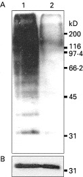
Fig. 3. Immunoblot of parasite proteins using anti-ubiquitin antibodies. (A) Synchronized 3D7 parasite cultures were treated with 100 nM MLN-273 (lane 1) or mock treated (lane 2). Following extraction, parasite proteins were analysed for the presence of ubiquitination. More ubiquitinated proteins were seen to accumulate in the drug-treated cells than in untreated cells. (B) The use of an antibody to a Plasmodium falciparum housekeeping enzyme, peroxiredoxin, showed signals of equal strength in both lanes which confirmed that equal amounts of protein were loaded.
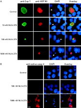
Fig. 4. Effects of MLN-273 on Plasmodium berghei-infected HepG2 cells. (A) P. berghei sporozoite-infected HepG2 cells were treated with different concentrations of MLN-273. The cells were fixed and stained with chicken anti-P. berghei Exp 1 (green) and mouse anti-P. berghei HSP90 antisera (red). Host cell DNA and parasite DNA (white arrow) were visualized with DAPI (blue). The furthermost right panels represent an overlay of the 3 singly stained images. (B) Active mammalian caspase-3 detection was performed in healthy HepG2 (uninfected) controls, and cells treated with 100 nM and 1000 nM MLN-273. Active caspase-3 (red) was detected using a commercially available antiserum. Cell nuclei were visualized with DAPI (blue). An overlay of the anti-active caspase-3 and DAPI is shown in the right panels. Scale bar represents 10 μm.
 You have
Access
- 71
- Cited by