




No CrossRef data available.
Article contents
Diverse experience with the CardioSEAL® and STARFlex® septal occluders
Published online by Cambridge University Press: 21 January 2005
Abstract
Apart from closure of atrial septal defects, there is little information concerning the use of the CardioSEAL family of occluders in congenitally malformed hearts. We review here our initial experience using the CardioSEAL and STARFlex occluders in 12 patients aged 17.3 ± 11.2 years, with a range from 4 to 34 years. Of the patients, 5 had fenestrated extracardiac Fontan procedures, 5 had persistent patency of the arterial duct, 1 had a leak across a Mustard baffle, and the final patient had a huge pulmonary arteriovenous malformation. We implanted successfully 9 CardioSEAL, and 3 STARFlex occluders, with sizes from 17 to 40 mm. In one patient, the occluder embolized to the right pulmonary artery, from where it was retrieved through the catheter. In two patients, there was a trivial residual leak immediately after implantation, but no patient had a residual leak after 6 months of follow-up. We noted improved ventricular dimensions, without any fractures of the arms of the occluders, perforations, or disturbances of flow after 2.4 ± 0.9 years of follow-up. We have demonstrated, therefore, the versatility of the CardioSEAL and STARFlex occluders, which have been used safely and effectively to close a variety of intra and extracardiac communications other than atrial septal defects.
Keywords
Information
- Type
- Original Article
- Information
- Copyright
- © 2004 Cambridge University Press
Transcatheter occlusion of intra or extracardiac communications is increasingly used as an attractive alternative to surgery, attempting to reduce morbidity and operative risks. Several interventional techniques and devices have now been developed, mainly for closure of atrial septal defect, which was occluded as early as 19761 and 1983.2 The devices are designed either to enclose the defect in a sandwich-like fashion,3 or to stent the interatrial communication.4 The CardioSEAL septal occluder, manufactured by NMT Medical, Boston, MA, USA, is a second generation double-umbrella implant constructed of an advanced nonferromagnetic alloy. Its initial use proved satisfactory,5 and from it developed the STARFlex occluder, which had an added flexible self-centring mechanism, along with a pin-pivot mechanism for attachment to the catheter that facilitates pivoting of the occluder along the septum during delivery.6 Both occluders are increasingly used in closure of defects of small to moderate size within the oval fossa.7
In addition to closing defects in the oval fossa, interventional techniques have been used to occlude various intracardiac communications in patients with congenitally malformed hearts, such as closing Fontan fenestrations with coils,8 or umbrella devices.9 Persistent patency of the arterial duct has successfully been treated with coils10 or modified umbrella devices,11 while problematic hepatic veins after a Fontan operation have also been successfully managed with interventional techniques.12 The CardioSEAL occluder has been used for occlusion of postoperative intracardiac communications,13 as well as pulmonary arteriovenous malformations.14
In this report, we show the efficacy and versatility of CardioSEAL and STARFlex occluders in closure of various intra and extracardiac communications other than the atrial septal defect for which they were originally designed, specifically for persistent patency of the arterial duct, fenestrations of an extracardiac Fontan procedure, an unwanted iatrogenic interatrial communication, and a pulmonary arteriovenous malformation.
Methods
Patient population
Between January 2000 and June 2002, we attempted percutaneous transcatheter occlusion using CardioSEAL and STARFlex occluders in 12 patients, 3 male and 9 female, aged from 4 to 34 years, with mean age of 17.3 ± 11.2 years, and a median age 19.7 years. They had various intra and extracardiac communications other than atrial septal defects. In one male and 4 female patients, aged from 5 to 31 years, with mean age of 16.3 ± 12.9 years and a median age of 14.5 years, there was functionally univentricular physiology following completion of the Fontan procedure using a 20 mm extracardiac conduit. A fenestration of 5 mm had been created between the conduit and the pulmonary venous atrium. In another male, and 4 female patients, aged from 4 to 34 years, with mean age of 14.9 ± 12.1 years, and a median age of 9.6 years, there was persistent patency of the arterial duct, the duct itself being greater than 3 or 4 mm in size, permitting a left-to-right shunt of greater than two-to-one, and producing left ventricular volume overload. Of note, the oldest patient with a patent arterial duct had had a previous sternotomy for closure of a ventricular septal defect. A 26-year-old female patient had transposition with ventricular septal defect and pulmonary stenosis with a residual shunt at atrial level >2 to 1 and atrial arrhythmias after an initial Mustard operation and subsequent surgical pulmonary valvotomy. A 25-year-old male patient had hereditary haemorrhagic telangiectasia with cyanosis due to a huge pulmonary arteriovenous malformation. Previous embolization, attempted with a Gianturco-Grifka vascular occlusion device of 9 mm, had been unsuccessful due to the giant size of the feeding artery. The last 2 patients have been previously reported.13, 14
The occluders
The CardioSEAL occluder consists of 2 interconnected umbrellas, each with 4 flexible spring-loaded arms with central and 2 mid-arm hinges, covered with sewn Dacron patches. It is available in 5 nominal sizes corresponding to the diagonal length of each umbrella, giving sizes of 17, 23, 28, 33, and 40 mm. The STARFlex occluder, which comes in sizes 23, 28, 33, and 40 mm, is based on the CardioSEAL occluder, but with the addition of a flexible, self-centring mechanism using microsprings attached in an alternating fashion between the opposing arms of the umbrella, allowing the occluder automatically to adjust to septal morphology without distorting the surrounding structures. The mechanism for attachment is modified to allow the implant to pivot freely against the delivery catheter, thus improving its alignment relative to the septum prior to release.
Procedures and techniques
Informed written consent was obtained from the patient or their guardian prior to the procedure. The protocol was approved by the Institutional Review Committee. The procedure in the patient with atrial shunting after the Mustard operation was performed under general endotracheal anesthesia, using continuous transesophageal echocardiographic monitoring on a System 5, Vingmed Sound, manufactured in Horten, Norway. The remaining procedures were done under conscious sedation.
Closure of persistently patent arterial ducts was attempted if aortography revealed the ducts to be larger than 3 or 4 mm, and hence not suitable for occlusion using single coils. The “stretched” diameter of the duct was assessed by gentle withdrawal of the balloon of a 7 French Swan-Ganz catheter filled with contrast until it passed from the descending aorta to pulmonary arteries, measuring the diameter of the balloon at the time of passage through the patent arterial duct. The size of occluder chosen for closure was at least 2 or 3 times the stretched diameter of the duct, taking care to avoid obstruction of the aorta or left pulmonary artery by using very large occluders. All procedures were performed transvenously following a previously reported technique.15
Patients with fenestrated extracardiac Fontan conduits underwent closure if test occlusion of the fenestration with a balloon produced a systemic venous atrial pressure <15 mmHg, with a rise <4 mmHg, increased aortic saturation, and stable aortic pressures and systemic cardiac output. The size of occluder chosen for closure was approximately twice the “stretched” diameter of the defect, which was assessed using a compliant balloon catheter, manufactured by Meditech, Boston Scientific Corp., Natick, MA, USA. Implantation was performed following the technique described for closure of atrial septal defects.16
The techniques used in the patients with shunting across a Mustard baffle and a giant pulmonary arteriovenous malformation have been previously described.13, 14
The patients with Fontan circulations, and the patient with a Mustard baffle, received heparin at 20 units/kg/h for 24 h, while all patients were placed on cephazolin at 75 mg/kg/day for 24 h, and were discharged to home the day after the procedure. The patients with the Fontan circulation, and the one with a Mustard baffle, were maintained on oral coumadin to keep the International Normalized Ratio twice normal with lifelong endocarditis prophylaxis, while the patient with telangiectasia was also maintained on coumadin due to a history of deep venous thrombosis. Follow-up at 24 h, 3, 6 and 12 months included a physical examination, electrocardiogram and transthoracic echocardiogram, while a chest radiograph was performed 24 h and 6 months after the procedure.
Statistical methods
Descriptive data are expressed as the mean ± standard deviation, with median value and range, as appropriate.
Results
Patent arterial ducts
In four patients, the ducts were deemed to be type A in the Toronto classification,17 with ampullas of significant size, and a narrowest angiographic diameter of 3–4 mm, with a “stretched” diameter from 4 to 7 mm. In these patients, we deployed 2 CardioSEAL occluders of 17 mm, one of 23 mm, and one of 40 mm without complications. A trivial residual shunt was present in 2 patients, but this had disappeared at the 6-month visit, with normalization of the increased left ventricular dimensions. Mild protusion of the occluder for 1–2 mm in the left pulmonary artery was seen in 2 patients without disturbing flow, and with normal velocities measured by echocardiography. No obstruction at the isthmus was observed (Fig. 1). The last patient had left ventricular dilation, with an end-diastolic diameter of 64 mm, due to a left-to-right shunt through the arterial duct of >3 to 1. The arterial duct was type B according to the angiographic classification, without a significant ampulla, and with a narrowest diameter of 5 mm and a “stretched” diameter of 12 mm. Failure to achieve a stable position across the patent arterial duct using a 28 mm CardioSEAL occluder led to its retrieval and deployment of a 40 mm CardioSEAL occluder, which appeared stable and was released with an angiographically satisfactory position and mild residual flow. The occluder embolized to the right pulmonary artery 5 min after implantation, and was retrieved without complications through a 14 French long Mullins sheath.

Figure 1. (a) Lateral projection of contrast injection in the aortic arch shows left-to-right flow through a patent arterial duct with a significant ampulla and narrowing at the pulmonary end (arrows). (b) Injection after deployment of a 17 mm CardioSEAL occluder shows closure of the duct with elimination of left-to-right flow.
Fontan fenestrations
All 5 patients had a surgically created fenestration between the extracardiac Fontan conduit and the pulmonary venous atrium, with significant right-to-left shunt on angiography resulting in an aortic saturation of between 83 and 85%. After test occlusion of the fenestration with a balloon, aortic saturation improved to more than 95% on room air, and systemic venous atrial pressure increased by up to 2 mmHg, while remaining below 15 mmHg. All fenestrations had a “stretched” diameter of 7–8 mm. In 4 fenestrations, we achieved closure using a 17 mm CardioSEAL occluder, and closed the other with a 23 mm STARFlex occluder, with immediate cessation of flow and no complications (Fig. 2).

Figure 2. (a) Lateral projection of contrast injection in the extracardiac Fontan conduit shows right-to-left shunt through the fenestration to the pulmonary venous atrium (arrow). (b) Same injection after deployment of a 17 mm CardioSEAL occluder shows complete closure of the fenestration with cessation of flow.
Other communications
The patient with a residual leak after a Mustard operation had an interatrial communication with a “stretched” diameter of 12 mm, which was occluded with a 23 mm CardioSEAL occluder (Fig. 3).

Figure 3. (a) Contrast injection at the entrance of the inferior caval vein in the heart shows the origin of the baffle leak at the lower portion of the Mustard baffle with shunt from anatomic left-to-right atrium at a certain stage of the cardiac cycle. (b) Contrast injection at the entrance of the caval vein shows the device occluding the site of leakage with no residual shunt.
The patient with hereditary hemorrhagic telangiectasia had a giant pulmonary arteriovenous malformation arising from the pulmonary artery to the left lower lobe, with its feeding vessel measured at 16 mm “stretched” diameter. A 28 mm CardioSEAL occluder deployed in the feeding vessel produced partial obstruction of flow, while deployment of a second 23 mm CardioSEAL occluder proximal to the first achieved obliteration of flow (Fig. 4) and an immediate rise in arterial saturation from 84 to 94%.

Figure 4. (a) Anteroposterior projection of an injection in the artery to the left lower lobe during a previous attempt at occlusion and before the final positioning of the Gianturco-Grifka device shows a very dilated feeding artery (arrow) with rapid filling of a tortuous transverse portion (open arrow) of the pulmonary arteriovenous malformation. (b) Anteroposterior projection of an injection on the left pulmonary artery after placement of both CardioSEAL devices demonstrates absence of flow within the large pulmonary arteriovenous malformation. The contrast enhanced smaller superimposed lesion (arrow) becomes apparent due to absence of contrast in the large pulmonary arteriovenous malformation.
Follow-up
All patients remain stable and free of further intervention over 3.4 ± 0.9 years of follow-up, ranging from 1.6 to 4.5 years. After discharge, no patient had embolization of the occluder, fracture of its arms, myocardial perforation, pericardial effusion, obstruction of pulmonary or systemic veins, impaired atrioventricular valvar function, or thromboembolic events. Left ventricular enlargement resolved completely during the 6 months following the procedure in all the patients with patent arterial duct. The patient with a Mustard operation had sustained improvement of her atrial arrhythmias and exercise tolerance, while the patient with telangiectasia remained with stable pulse oximetry at 94%.
Discussion
Presence of significant left-to-right shunts through communications at the ductal level creating left ventricular volume overload and the possibility for pulmonary hypertension is a recognized indication for closure. Surgical closure is safe in general, albeit that transcatheter occlusion has been increasingly used in order to minimize morbidity, mortality, and hospital stay, and to avoid surgical scarring. Persistent right-to-left shunting through Fontan tunnel fenestrations, if not needed for haemodynamic reasons, carries the risk of right-to-left embolism and creates cyanosis with its potential deleterious effects. Several devices such as coils and/or specifically modified umbrellas have been used for closure of Fontan fenestration and patent arterial ducts.8–11 In this report, we present the acute and medium-term results of our experience using the CardioSEAL and STARFlex occluders in the interventional treatment not of atrial septal defect, for which they were originally designed, but of various other intra and extracardiac lesions, specifically patent arterial ducts, extracardiac Fontan fenestrations, postoperative interatrial communications, and arteriovenous malformations.
In general, the CardioSEAL and STARFlex occluders have proved to be safe, effective and versatile tools for closure of various intra as well as extracardiac communications in congenital heart disease. In our limited series, they were used both in children and adults with few and treatable complications, excellent rates of occlusion, absence of fracture and protrusion of the arms, and without creating obstruction in various intra and extracardiac structures.
In this limited experience, the CardioSEAL occluders proved safer for use in those ducts with significant narrowing towards their pulmonary artery and a sizable ampulla, so-called “Type A”, which can accommodate the left atrial umbrella. One should probably be cautious when dealing with patent arterial ducts with very “stretchable” diameters and short ampullas, factors that may facilitate distortion of the aortic disk and embolization of the occluder, especially given the significant forces exercised on the occluder by the aortic pressure.
Patent arterial ducts with maximal diameters <2.5 mm may safely be occluded, at significantly lower cost compared with the double-umbrella devices, with various types of coils.18 Coils represent the interventional treatment of choice for small patent arterial ducts, but have limited usefulness and high rates of embolization in large and distensible ducts, especially if placement of multiple coils is to be avoided. The Amplatzer duct occluder, a device probably more user-friendly than the CardioSEAL family of occluders, has been extensively used for closure of moderate and large patent arterial ducts with satisfactory results.11 Still, proper implantation of the Amplatzer duct occluder may present technical difficulties in large ducts with specific morphology, such as those that are window-like, where risks for embolization are increased. The CardioSEAL family of occluders, as shown in this study, now offer yet another option for closure of the larger ducts, depending on their morphology.
As the most commonly used technique during a Fontan operation has been creation of an intracardiac baffle through the right atrium connecting the inferior caval vein to the pulmonary arteries, most reports of interventional occlusion of Fontan fenestrations have described closure with coils or devices designed for atrial septal defects.8, 9, 19 The advantages of an extracardiac conduit, namely a decreased amount of atrial surgery and arrhythmias, less leaks and obstructions, improved haemodynamics, and drainage of the coronary sinus to the low-pressure atrium, have led to increasing performance in recent years of the extracardiac Fontan with or without fenestration. Transcatheter occlusion of extracardiac fenestration has been reported before with coils,20 as well as with the Gianturco-Grifka device21 and Amplatzer duct occluders.22 In our small series, the CardioSEAL and STARFlex occluders proved a simple and effective choice for closure of such fenestrations, without producing complications such as induced atrioventricular valvar dysfunction or obstruction of the systemic and pulmonary pathways.
The technical ease of closure, and absence of complications and interactions with other intracardiac structures in our series, is in accordance with previous larger reports in the case of intracardiac Fontan tunnels,9 and is probably due to the smooth and low profile of the CardioSEAL family of occluders after implantation, a fact that makes them one of the devices of choice for occlusion of Fontan fenestrations.
The relatively small number of patients in this study with various types of communications does not permit generalizations. Nevertheless, our experience suggests that the CardioSEAL family of double-umbrella occluders have diverse applications, and may be useful tools in a variety of situations involving attempted occlusion of structures and communications in the interventional catheterization laboratory.
Acknowledgement
We dedicated this study to the memory of Professor Gerd Hausdorf, from the Department of Pediatric Cardiology, Medizinische Hochschule, Hanover, Germany, whose help and inspiration made this work possible.
References
King TD, Thompson SL, Steiner C, Mills NL. Secundum atrial septal defect. Nonoperative closure during cardiac catheterization. JAMA 1976; 235: 2506–2509.Google Scholar
Rashkind WJ. Transcatheter treatment of congenital heart disease. Circulation 1983; 67: 711–716.Google Scholar
Lloyd TR, Rao PS, Beekman III RH, Mendelsohn AM, Sideris EB. Atrial septal defect occlusion with the buttoned device (a multi-institutional U.S. trial). Am J Cardiol 1994; 73: 286–291.Google Scholar
Chan KC, Godman MJ, Walsh K, Wilson N, Redington A, Gibbs JL. Transcatheter closure of atrial septal defect and interatrial communications with a new self expanding nitinol double disc device (Amplatzer septal occluder): multicentre UK experience. Heart 1999; 82: 300–306.Google Scholar
Pedra CA, Pihkala J, Lee KJ, Boutin C, Nykanen DG, McLaughlin PR, Harrison DA, Freedom RM, Benson L. Transcatheter closure of atrial septal defects using the CardioSEAL implant. Heart 2000; 84: 320–326.Google Scholar
Hausdorf G. STARFlex ASD closure: deployment, techniques, equipment. J Interv Cardiol 2001; 14: 69–76.Google Scholar
Carminati M, Giusti S, Hausdorf G, Qureshi S, Tynan M, Witsenburg M, Hess J, Piechaud JF, Bonhoeffer P, Donti A, Ovaert C, Sievert H, Elzenga N, Talsma M, van Oort A, Ernst J, Gewillig M, De Geeter B. A European multicentric experience using the CardioSEAL and STARFlex double umbrella devices to close interatrial communications holes within the oval fossa. Cardiology in the Young 2000; 10: 519–526.Google Scholar
Gamillscheg A, Beitzke A, Stein JI, Rupitz M, Zobel G, Rigler B. Transcatheter coil occlusion of residual interatrial communications after Fontan procedure. Heart 1998; 80: 49–53.Google Scholar
Goff DA, Blume ED, Gauvreau K, Mayer JE, Lock JE, Jenkins KJ. Clinical outcome of fenestrated Fontan patients after closure: the first 10 years. Circulation 2000; 102: 2094–2099.Google Scholar
Patel HT, Cao QL, Rhodes J, Hijazi ZM. Long-term outcome of transcatheter coil closure of small to large patent ductus arteriosus. Catheter Cardiovasc Interv 1999; 47: 457–461.Google Scholar
Bilkis AA, Alwi M, Hasri S, Haifa AL, Geetha K, Rehman MA, Hasanah I. The Amplatzer duct occluder: experience in 209 patients. J Am Coll Cardiol 2001; 37: 258–261.Google Scholar
Moore JW, Murdison KA, Baffa GM, Kashow K, Murphy JD. Transcatheter closure of fenestrations and excluded hepatic veins after fontan: versatility of the Amplatzer device. Am Heart J 2000; 140: 534–540.Google Scholar
Apostolopoulou SC, Papagiannis J, Hausdorf G, Rammos S. Transcatheter occlusion of atrial baffle leak after mustard repair. Catheter Cardiovasc Interv 2000; 51: 305–307.Google Scholar
Apostolopoulou SC, Kelekis NL, Papagiannis J, Hausdorf G, Rammos S. Transcatheter occlusion of a large pulmonary arteriovenous malformation with use of a CardioSEAL device. J Vasc Interv Radiol 2001; 12: 767–769.Google Scholar
Rashkind WJ, Mullins CE, Hellenbrand WE, Tait MA. Nonsurgical closure of patent ductus arteriosus: clinical application of the Rashkind PDA Occluder System. Circulation 1987; 75: 583–592.Google Scholar
Hausdorf G, Kaulitz R, Paul T, Carminati M, Lock J. Transcatheter closure of atrial septal defect with a new flexible, self-centering device (the STARFlex occluder). Am J Cardiol 1999; 84: 1113–1116, A10.Google Scholar
Krichenko A, Benson LN, Burrows P, Moes CA, McLaughlin P, Freedom RM. Angiographic classification of the isolated, persistently patent ductus arteriosus and implications for percutaneous catheter occlusion. Am J Cardiol 1989; 63: 877–880.Google Scholar
Hofbeck M, Bartolomaeus G, Buheitel G, Esser R, Gravinghoff L, Hoffmann W, Kienast W, Michel-Behnke I, Scharabrine EG, Schranz D, Schmaltz AA, Shakhov BE, Singer H, Lindinger A. Safety and efficacy of interventional occlusion of patent ductus arteriosus with detachable coils: a multicentre experience. Eur J Pediatr 2000; 159: 331–337.Google Scholar
Cowley CG, Badran S, Gaffney D, Rocchini AP, Lloyd TR. Transcatheter closure of fontan fenestrations using the Amplatzer septal occluder: initial experience and follow-up. Catheter Cardiovasc Interv 2000; 51: 301–304.Google Scholar
Sommer RJ, Recto M, Golinko RJ, Griepp RB. Transcatheter coil occlusion of surgical fenestration after Fontan operation. Circulation 1996; 94: 249–252.Google Scholar
Zahn EM, Chang AC, Burke RP, Jacobs JP. Transcatheter closure of an extracardiac Fontan fenestration. Ann Thorac Surg 1998; 66: 260–262.Google Scholar
Rueda F, Squitieri C, Ballerini L. Closure of the fenestration in the extracardiac Fontan with the Amplatzer duct occluder device. Catheter Cardiovasc Interv 2001; 54: 88–92.Google Scholar

(a) Lateral projection of contrast injection in the aortic arch shows left-to-right flow through a patent arterial duct with a significant ampulla and narrowing at the pulmonary end (arrows). (b) Injection after deployment of a 17 mm CardioSEAL occluder shows closure of the duct with elimination of left-to-right flow.

(a) Lateral projection of contrast injection in the extracardiac Fontan conduit shows right-to-left shunt through the fenestration to the pulmonary venous atrium (arrow). (b) Same injection after deployment of a 17 mm CardioSEAL occluder shows complete closure of the fenestration with cessation of flow.

(a) Contrast injection at the entrance of the inferior caval vein in the heart shows the origin of the baffle leak at the lower portion of the Mustard baffle with shunt from anatomic left-to-right atrium at a certain stage of the cardiac cycle. (b) Contrast injection at the entrance of the caval vein shows the device occluding the site of leakage with no residual shunt.
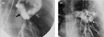
(a) Anteroposterior projection of an injection in the artery to the left lower lobe during a previous attempt at occlusion and before the final positioning of the Gianturco-Grifka device shows a very dilated feeding artery (arrow) with rapid filling of a tortuous transverse portion (open arrow) of the pulmonary arteriovenous malformation. (b) Anteroposterior projection of an injection on the left pulmonary artery after placement of both CardioSEAL devices demonstrates absence of flow within the large pulmonary arteriovenous malformation. The contrast enhanced smaller superimposed lesion (arrow) becomes apparent due to absence of contrast in the large pulmonary arteriovenous malformation.
 You have
Access
- 4
- Cited by