Introduction
Paediatric tracheostomy is relatively uncommon, and has substantial complications.Reference Al-Samri, Mitchell, Drummond and Bjornson1
An extensive selection of paediatric tracheostomy tubes is now available, produced in response to a variety of clinical requirements. Plastic tracheostomy tubes are available, such as like Sims Portex Bivona, Shiley and Tracoe in addition to metal tubes; the former are more commonly used. The Bivona range is becoming increasingly popular, with tubes available in neonatal and paediatric lengths with a range of inner diameters. However, in the relatively uncommon situation of tracheal narrowing in addition to subglottic pathology, a Montgomery T-tube is also an acceptable choice.Reference Tweedie, Skilbeck, Cochrane, Cooke and Wyatt2–Reference Rutter, Yellon, Cotton, Bluestone, Stool, Alper, Arimand, Casselbrant and Dohar4
In an emergency involving a child with multiple airway problems, obtaining an appropriate tracheostomy tube can represent a clinical dilemma, especially in a developing country.
Case report
A 14-year-old boy with subglottic stenosis, who had required a tracheostomy tube for the past two years, underwent partial cricotracheal resection in our hospital.
In the first post-operative week, the boy developed stridor despite having a patent tracheostomy tube in situ.
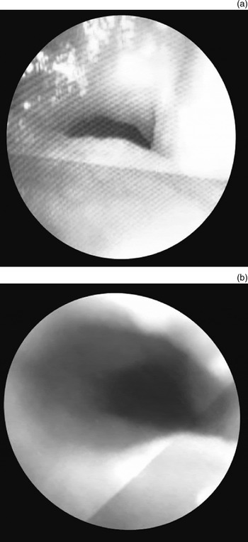
Emergency fibre-optic bronchoscopy showed a collapsing segment extending from the lower end of the tracheostomy tube down to 1 cm above the carina. Balloon dilatation was performed with the aid of the bronchoscope (Figure 1); this improved overall ventilation but did not affect the collapsing segment.
Fibre-optic bronchoscopy showing the patient's collapsing segment: (a) pre-dilatation; (b) post-dilatation.

Since the length of the patient's usual tracheostomy tube was inadequate to prevent stridor, it was evident that a tracheostomy tube with sufficient length to reach and temporarily stent the collapsing segment was urgently needed.
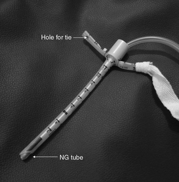
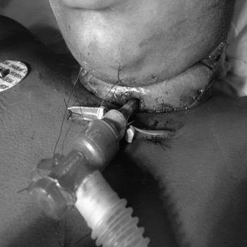
We therefore designed a modified tracheostomy tube for this purpose. After measuring the distance from the carina to the entry point of the tracheostomy stoma, a size five, uncuffed endotracheal tube was divided up to its 12 cm mark. The smooth, blunt tip of a nasogastric tube was then used as an obturator (Figure 2) to facilitate easy insertion. The two halves were used as flanges and sutured to the skin, to prevent accidental disconnection and migration of the endotracheal tube inside the tracheostomy stoma (Figure 3).
The modified, uncuffed endotracheal tube: the smooth, blunt tip of a nasogastric tube is used as an obturator, and a hole is punched into each flange for easy fixation with tape.

The modified, uncuffed endotracheal tube: the tube is divided down to its 12 cm mark, with the two halves used as flanges and sutured to the skin.

The same modified endotracheal tube was maintained as a stent cum tracheostomy tube for a week (Figure 4), following which the patient was successfully decannulated.
Sagittal computed tomography scan showing the lengthy collapsing segment, with the modified endotracheal tube used as both a tracheostomy tube and stent. A = anterior; P = posterior; F = foot

Discussion
Although an extensive selection of paediatric tracheostomy tubes is currently available, a small range of tubes is generally suitable for the majority of children. However, some patients will require other varieties or customised versions of tubes, due to unusual airway pathology. In patients with tracheal pathology, a hyperflexible version is available in paediatric sizes 2.5–5.5 mm (outer diameter), in the Bivona range. This tube's long shaft has an embedded stainless steel coil which safeguards against compression and permits alteration of the intratracheal tube length.
In addition, Montgomery T-tubes (silicone stents) are available in paediatric sizing and are a useful alternative to tracheostomy tubes in complex cases in which a longer period of laryngeal and tracheal stenting is required. The upper limb goes to the larynx, while the lower limb can extend as far as necessary down the trachea towards the carina.Reference Tweedie, Skilbeck, Cochrane, Cooke and Wyatt2, Reference Grillo and Grillo3
The development of tracheomalacia following laryngeal reconstructive surgery is relatively uncommon.Reference Rutter, Yellon, Cotton, Bluestone, Stool, Alper, Arimand, Casselbrant and Dohar4 Our patient developed post-operative tracheomalacia on the seventh post-operative day. Bronchoscopy confirmed that the pathology extended from the lower end of the tracheostomy to just above the carina. This patient urgently required a specialised tracheostomy tube, but this was not readily available in our hospital pharmacy. Hence, we modified the most readily available tube in the pharmacy – an endotracheal tube. Instead of suturing the flanges to the skin, making a hole in them can facilitate fixation with tape (Figure 2).
Conclusion
In emergency cases of tracheomalacia in which an appropriate tracheostomy tube is not available, an endotracheal tube can be modified to act as an emergency tracheostomy tube as well as tracheal stent. This modified endotracheal tube can also act as a simple, temporary alternative to a conventional tracheostomy tube, when a specifically sized paediatric tracheostomy tube is not available.