Introduction
The rat lungworm (Angiostrongylus cantonensis) was first recognized as the cause of human eosinophilic meningitis in the Pacific region after World War II (Alicata, Reference Alicata1991). In the 1950s, Drs. Josephine Mackerras and Dorothea Sandars undertook a series of experiments with parasites from rats that had been caught by dogs used by the rat-gang of the Brisbane City Council in Queensland to elucidate the life cycle of what they recognized as A. cantonensis. Their detailed account of the life cycle of A. cantonensis, transmitted between Rattus norvegicus and the terrestrial slug, Deroceras laeve (as Agriolimax laevis), with its migration through the brain of the rat were seminal works (Mackerras and Sandars, Reference Mackerras and Sandars1954, Reference Mackerras and Sandars1955). Colleagues forwarded additional material to Mackerras and Sandars from wild R. rattus collected from cane fields in Innisfail, north Queensland. Mackerras and Sandars noted that males from these rats possessed long spicules, whereas those from rats from Brisbane had short spicules. Mackerras later reported A. cantonensis in five species of rats, namely the two introduced Rattus species as well as the native Rattus sordidus (as conatus), Rattus fuscipes (as assimilis) and Melomys burtoni (as littoralis) (Mackerras, Reference Mackerras1958). Re-examination of the material from Brisbane, coupled with meticulous experimental infections revealed the presence of a previously undiscovered phenotypically different species of Angiostrongylus in the Australian bush rat, R. fuscipes. This new species was described by Dr Manoon Bhaibulaya and named Angiostrongylus mackerrasae (Bhaibulaya, Reference Bhaibulaya1968, Reference Bhaibulaya1974, Reference Bhaibulaya1975). He reported further that both species occurred in R. norvegicus, but that R. rattus harboured only A. cantonensis and R. fuscipes only A. mackerrasae (Bhaibulaya, Reference Bhaibulaya1968). He later established that the life cycle study of Mackerras and Sandars (Reference Mackerras and Sandars1955) was actually that of the Australian native species, A. mackerrasae, not the invasive A. cantonensis (Bhaibulaya, Reference Bhaibulaya1974). Subsequently, Bhaibulaya (Reference Bhaibulaya and Cross1979) reviewed the morphology and taxonomy of the major species of Angiostrongylus in Eastern Asia and Australia.
The species identity of A. mackerrasae remained unquestioned until relatively recently (Aghazadeh et al., Reference Aghazadeh, Traub, Mohandas, Aland, Reid, McCarthy and Jones2015b; Dusitsittipon et al., Reference Dusitsittipon, Criscione, Morand, Komalamisra and Thaenkham2018; Song et al., Reference Song, Yong and Eamsobhana2018). A series of recent publications have documented co-occurrence of both morphologically defined species A. mackerrasae and A. cantonensis in rats (Rattus spp.) from Brisbane (Aghazadeh et al., Reference Aghazadeh, Reid, Aland, Restrepo, Traub, McCarthy and Jones2015a, Reference Aghazadeh, Harvie, Owen, Verissimo, Aland, Reid, Traub, Mc, Mc and Jones2016). To define the molecular identity of A. mackerrasae, a female specimen from the previous study (Aghazadeh et al., Reference Aghazadeh, Reid, Aland, Restrepo, Traub, McCarthy and Jones2015a) was used for the generation of mtDNA. This mtDNA sequence became the reference for A. mackerrasae. However, compilation of mtDNA data and additional sampling of mtDNA sequences from closely related A. cantonensis and A. malaysiensis revealed that mtDNA A. mackerrasae sensu Aghazadeh et al. (Reference Aghazadeh, Traub, Mohandas, Aland, Reid, McCarthy and Jones2015b) clusters within A. cantonensis using either cox1 or cob genes or the entire mtDNA (Dusitsittipon et al., Reference Dusitsittipon, Criscione, Morand, Komalamisra and Thaenkham2018; Song et al., Reference Song, Yong and Eamsobhana2018). Previous detailed experimental and morphological studies established differences between A. cantonensis and A. mackerrasae (Bhaibulaya, Reference Bhaibulaya1975; Aghazadeh et al., Reference Aghazadeh, Harvie, Owen, Verissimo, Aland, Reid, Traub, Mc, Mc and Jones2016), so it was quite unexpected to see the absence of genetic differences. The specimen used to obtain mtDNA from A. mackerrasae sensu Aghazadeh et al. (Reference Aghazadeh, Traub, Mohandas, Aland, Reid, McCarthy and Jones2015b) is lost. Similarly, the Australian vouchers of A. mackerrasae held at the Australian National Wildlife Collection, CSIRO, Canberra are not preserved in ethanol, thus preventing polymerase chain reaction (PCR) amplification (JŠ and DS, data not shown). To re-evaluate the species identity, we sought material collected from the type host, R. fuscipes, that appeared, based on morphology, to be conspecific with the material used for the description of A. mackerrasae and that could be characterized and compared to sequences from the closely related A. cantonensis.
The aim of the study was to combine morphological and molecular tools to confirm or refute the specific status of A. mackerrasae. Nematodes from previously collected R. fuscipes and R. rattus from Queensland, Australia (Aghazadeh et al., Reference Aghazadeh, Reid, Aland, Restrepo, Traub, McCarthy and Jones2015a, Reference Aghazadeh, Harvie, Owen, Verissimo, Aland, Reid, Traub, Mc, Mc and Jones2016) were used throughout the study. Morphologically identified A. mackerrasae vouchers were characterized genetically using amplification of cox1 followed by the generation of reference complete mtDNA.
Material and methods
Adult specimens of Angiostrongylus spp. from Rattus spp.
The adults of Angiostrongylus spp. from Rattus spp. were retrieved from the collection held at the University of Queensland. All specimens belonged to the previously published field collections from Brisbane and neighbouring areas (Aghazadeh et al., Reference Aghazadeh, Reid, Aland, Restrepo, Traub, McCarthy and Jones2015a). All specimens were dissected from wild Rattus spp. trapped in 2013–2014 and were stored in 70% ethanol at −80°C (Aghazadeh et al., Reference Aghazadeh, Reid, Aland, Restrepo, Traub, McCarthy and Jones2015a).
Morphological identification of Angiostrongylus spp.
All ethanol preserved voucher specimens were cleared in lactophenol and examined under a Zeiss compound microscope by one of us (DMS). The morphology was compared to descriptions of A. cantonensis, A. mackerrasae and A. malaysiensis (Chen, Reference Chen1935; Bhaibulaya, Reference Bhaibulaya1968; Bhaibulaya and Cross, Reference Bhaibulaya and Cross1971) as well as to voucher specimens of the species held at the Australian National Wildlife Collection, CSIRO, Canberra. Voucher specimens for comparison are as follows: A. cantonensis W/L HC# N170 from pulmonary arteries R. norvegicus & R. rattus collected from suburbs of Brisbane, Queensland (1968–1970), A. mackerrasae W/L HC# N171 from pulmonary arteries R. fuscipes collected from Mt. Glorious, Queensland (1969), A. mackerrasae W/L HC# N172 from pulmonary arteries R. fuscipes collected from Mt. Tamborine (1969) and A. malaysiensis W/L HC# N5455 from pulmonary arteries Rattus rattus collected from Luang Probang, Laos. Photographs were made using a Leitz Diaplan compound microscope with a modified telescope adapter connected to a digital camera and processed with Helicon Focus Pro software.
Isolation of genomic DNA
Using a sterile disposable scalpel blade selected ethanol preserved vouchers were bisected at mid-body and ~3 mm were removed, placed in a sterile 1.5 mL Eppendorf tube, dried for 15 min at 55°C in a dry heat block and used for total genomic DNA isolation using an Isolate II Genomic DNA Kit (Bioline, Alexandria, Australia) according to the manufacturers' instructions. Genomic DNA was eluted in 80 μL of Tris buffer (pH = 8.5) and stored at −20°C.
Amplification of cox1 mtDNA
The cytochrome c oxidase subunit 1 gene (cox1) was amplified using primers LCO1490 and HCO2198 (Folmer et al., Reference Folmer, Black, Hoeh, Lutz and Vrijenhoek1994). PCR reactions and amplification conditions were as previously described (Červená et al., Reference Červená, Modrý, Fecková, Hrazdilová, Foronda, Alonso, Lee, Walker, Niebuhr, Malik and Šlapeta2019). The MyTaq™ Red Mix (Bioline, Alexandria, Australia) was used for all PCR reactions. Each PCR was run with a negative control (water). The PCR products (~650 bp) were bidirectionally sequenced using amplification primers at Macrogen Inc. (Amsterdam, Netherlands and Seoul, South Korea). The DNA chromatographs were inspected using CLC Main Workbench 6.9.1 (https://www.qiagenbioinformatics.com/) and compared to reference alignment of Angiostrongylus spp. cox1 (Červená et al., Reference Červená, Modrý, Fecková, Hrazdilová, Foronda, Alonso, Lee, Walker, Niebuhr, Malik and Šlapeta2019).
Whole genome sequencing of Angiostrongylus mackerrasae and assembly of mtDNA
Isolated DNA [N5721 (P43/19-E; JS5347); 110 ng; Table 1] was used for Illumina Nextera XT library construction followed by next generation sequencing (NGS) using 100 bp paired end Illumina HiSeq 2500 sequencing systems at a depth of 1 Gb of raw sequence data (Macrogen, Seoul, Republic of Korea). The complete mitochondrial genome (mtDNA) was assembled from FastQ data using the MITObim pipeline (Hahn et al., Reference Hahn, Bachmann and Chevreux2013) available at https://github.com/chrishah/MITObim with the sequence of A. cantonensis complete mtDNA (GQ398121) as bait as previously described (Červená et al., Reference Červená, Modrý, Fecková, Hrazdilová, Foronda, Alonso, Lee, Walker, Niebuhr, Malik and Šlapeta2019). The assembly was repeated three times using varying percentages of the raw FastQ sequence data (2–10%), keeping mtDNA coverage at 60–100×. The obtained mtDNA sequence was annotated with the aid of MITOS Web Server (Bernt et al., Reference Bernt, Donath, Juhling, Externbrink, Florentz, Fritzsch, Putz, Middendorf and Stadler2013) available at http://mitos.bioinf.uni-leipzig.de/ and aligned to manually checked annotation against all available Angiostrongylus spp. mtDNA in CLC Main Workbench 6.9.1.
Summary of voucher specimens of A. mackerrasae and A. cantonensis analysed in this study

a Australian National Wildlife Collection, CSIRO, Canberra.
b Determinations not possible if only anterior ends present.
Phylogenetic analysis
Available complete mtDNA sequences from Angiostrongylus spp. were retrieved from GenBank, as was that of Aelurostrongylus abstrusus mtDNA as an outgroup. The A. mackerrasae sensu Aghazadeh et al. (Reference Aghazadeh, Traub, Mohandas, Aland, Reid, McCarthy and Jones2015b) nucleotide mtDNA sequence was obtained from a PhD thesis (Aghazadeh, Reference Aghazadeh2016). The complete mtDNA sequences were aligned to identify homologous protein coding regions in CLC Main Workbench 6.9.1. Nucleotide and amino acid sequences were analysed using Mega X (Kumar et al., Reference Kumar, Stecher, Li, Knyaz and Tamura2018) and MrBayes 3.2.6 (Ronquist et al., Reference Ronquist, Teslenko, van der Mark, Ayres, Darling, Hohna, Larget, Liu, Suchard and Huelsenbeck2012). Amino acid substitution model fit was evaluated using a comparison of 56 Maximum Likelihood models in Mega X. A minimum evolution (ME) tree was inferred using amino acid alignment using ME and the JTT substitution model with 500 bootstrap replicates in Mega X. Bayesian analysis in MrBayes was set to mixed model selection (prset aamodelpr = mixed) with rates set to gamma distribution and invariant proportion (lset rates = invgamma). The MCMC was run for 10 000 000 generations sampled every 250 generations and 1000 generations printing frequency. The parameters were checked using Tracer 1.7.1 (Rambaut et al., Reference Rambaut, Drummond, Xie, Baele and Suchard2018). A consensus tree was selected after 25% of initial trees were removed as burnin. The tree was visualized using FigTree 1.4.4 (https://github.com/rambaut/figtree/).
Data and material availability
All voucher specimens were deposited in the helminthological collection of the Australian National Wildlife Collection, CSIRO, Canberra, A. C. T., Australia (Table 1). Sequence data were deposited at SRA NCBI BioProject: PRJNA594091 (https://www.ncbi.nlm.nih.gov/sra/PRJNA594091).
The complete mtDNA genome sequence from A. mackerrasae (N5721) was submitted to GenBank under the accession number MN793157. Individual cox1 sequences from A. mackerrasae and A. cantonensis were submitted to GenBank under the accession numbers MN814821–MN814828. A voucher specimen list and sequence alignments are available at LabArchives (http://dx.doi.org/10.25833/jswk-7e95).
Results
Forty-three voucher specimens removed from the pulmonary arteries and right ventricle of R. rattus and/or R. fuscipes and belonging to either A. cantonensis (n = 19) or A. mackerrasae (n = 24) were analysed in this study (Table 1). All specimens were collected in close vicinity (25 to 75 km) to Brisbane or in Brisbane, Queensland, Australia. The type host of A. mackerrasae is R. fuscipes and type locality is rain forest from Mount Glorious, a locality in close proximity (30 km) to Brisbane.
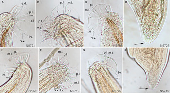
Voucher specimens exhibited typical characteristics of either A. cantonensis or A. mackerrasae and were readily distinguishable (Fig. 1). The prominent difference between males of A. cantonensis and A. mackerrasae was the length of the spicules (Table 2). There was considerable variation in the form of the ventral and lateral rays of the prominent male bursa (A. cantonensis, Fig. 1A–C; A. mackerrasae, Fig. 1E–G). The ventral ray branched at about two-thirds of its length into a small ventro-ventral and a large latero-ventral ray in both species (Fig. 1B and E) but this was not a consistent feature with some branching at half the length and with the ventro-ventral ray nearly as large as the latero-ventral ray in some specimens (Fig. 1A, C, F and G). The postero-lateral rays (p.l.) of A. cantonensis were shorter compared to the medio-lateral (m.l) ray, while in A. mackerrasae p.l. rays were the same or nearly the same length as m.l. rays (Fig. 1A, B and C). Female A. mackerrasae possessed a minute papilla at their tail tips (Fig. 1H, arrow) a feature absent from females of A. cantonensis (Fig. 1D, arrow).
Morphological characteristics of Angiostrongylus cantonensis (A–D) and A. mackerrasae (E–H) from Queensland, Australia. (A) Angiostrongylus cantonensis male bursa with ventral ray branching at about half its length, v.v. and l.v. rays of similar length, lateral rays prominent with m.l. ray typically longest and p.l. ray short (N5723). (B) Angiostrongylus cantonensis male bursa with deeply incised ventral ray branching at about two thirds its length into small v.v. and large l.v. rays, a.l. ray thick, m.l. ray longest and p.l. ray short, stubby (N5729). (C) Angiostrongylus cantonensis male bursa with ventral ray branching at about half its length and v.v. and l.v. rays of equal size (N5716). (D) Angiostrongylus cantonensis female tail tip, note absence of minute terminal papilla (arrow) (N5727). (E) Angiostrongylus mackerrasae male bursa with deeply incised ventral ray branching at about half its length into short narrow v.v. and large l.v. rays, lateral ray arising from common trunk, a.l. ray short, m.l. and p.l. rays of almost equal length (N5720). (F) Angiostrongylus mackerrasae male bursa with slightly incised ventral ray branching at about one-third its length and with v.v. and l.v. rays of similar length, a.l. ray short, m.l. and p.l. rays of almost equal length (N5719). (G) Angiostrongylus mackerrasae male bursa with deeply incised ventral ray branching at half its length into narrow v.v. ray of almost equal length to l.v. ray; lateral rays arising from common trunk with external margin of bursa folded ventrally distorting lengths of m.l. and p.l. rays (N5725). (H) Angiostrongylus mackerrasae female tail tip, note the presence of minute terminal papilla (arrow) (N5715). Abbreviations: v.v. ventro-ventral ray; l.v. latero-ventral ray; a.l. antero-lateral ray; m.l. medio-lateral ray; p.l. postero-lateral ray.

Summary of male Angiostrongylus species spicule length

Eight voucher specimens were selected for initial molecular characterization using the barcoding region cox1. All five specimens morphologically confirmed as A. mackerrasae had 100% identical cox1 regions (655 nt). Three specimens morphologically confirmed as A. cantonensis had 100% identical cox1 regions (655 nt). A pairwise comparison of A. mackerrasae and A. cantonensis demonstrated 61 nt differences (9.3%; 61/655) (Table 3; Supplementary Fig. S1). The cox1 nucleotide sequence translates into 218 amino acids and 3 amino acid (1.4%; 3/218) changes between the two Angiostrongylus species. Compared to the same cox1 region from A. malaysiensis, A. costaricensis and A. vasorum a pairwise nucleotide distance was 9.8–10.5, 12.2–13.7 and 12.7%, respectively. Using the nucleotide sequence corresponding to the amplified and sequenced cox1 region from A. mackerrasae sensu Aghazadeh et al. (Reference Aghazadeh, Traub, Mohandas, Aland, Reid, McCarthy and Jones2015b), it was 100% identical to those cox1 sequences obtained from morphologically confirmed specimens of A. cantonensis, but shared only 90.7% identity with A. mackerrasae.
Comparison of Angiostrongylus nucleotide diversity at cox1 sequences

* – A. mackerrasae sensu Aghazadeh et al. (Reference Aghazadeh, Traub, Mohandas, Aland, Reid, McCarthy and Jones2015b).
Note: Partial cox1 regions amplified using LCO1490 and HCO2198 primers. Percent identity between pairs of sequences expressed (top right) and the number of nucleotide differences between pairs of sequences (bottom left) for the amplified cox1 region. Sequences are identified by their voucher specimen identifier, included is the cox1 sequence for A. mackerrasae sensu Aghazadeh et al. (Reference Aghazadeh, Traub, Mohandas, Aland, Reid, McCarthy and Jones2015b) and A. malaysiensis strain AM6.
A single female specimen of A. mackerrasae (N5721) from R. fuscipes was then subject to whole genome sequencing and mtDNA was assembled (Fig. 2A). Low coverage whole genome NGS of A. mackerrasae DNA (quantity 110 ng) yielded 1.9 G bp with 94% high quality Q30 of the nucleotides. Using MitoBIM, we assembled a complete mtDNA sequences, 13 356 bp (Fig. 2). Three independent MitoBIM runs returned identical mtDNA sequences, each initially baited with A. cantonensis mtDNA (GQ398121) (Lv et al., Reference Lv, Zhang, Zhang, Liu, Liu, Hu, Wei, Steinmann, Graeff-Teixeira, Zhou and Utzinger2012). With the aid of the MITOS Web Server and subsequent manual correction, 12 protein-coding genes representing, 22 tRNA regions and two ribosomal subunits were annotated (Fig. 2A). The partial cox1 sequence obtained by PCR (primers LCO1490-HCO2160, 655 bp) from the same initial DNA following Sanger sequencing was 100% identical to the mtDNA assembled from the NGS data.
Mitochondrial genome of A. mackerrasae: structure and phylogenetic inference. (A) The consensus structure of the mitochondrial genome of A. mackerrasae from Brisbane, Queensland, Australia (N5721). Each tRNA gene was indicated by the amino acid code according to the International Union of Pure and Applied Chemistry. All the genes are located on the same strand and transcribed in the same direction (clockwise). (B) Phylogenetic tree based on 12 mtDNA protein coding genes (3221 amino acids, 1223 variable sites, 730 parsimony-informative sites). The tree was reconstructed using the BI method with mixed model selection (selecting 100% mtREV model). The posterior probability for branch support is depicted next to the nodes as a percentage; ME using JTT amino acid model bootstrap support is also included. The mtDNA accession number, nematode species (colour coded) and known strain/isolate/voucher identity is included. A tree inset (B, inset) shows the branching order of the ME tree with bootstrap support relative to the colour-coded nematode species. Trees were rooted using A. abstrusus.

The Bayesian method of phylogenetic tree inference based on amino acid sequences of 12 protein coding genes of all available Angiostrongylus spp. mtDNA yielded a tree with high (100%) posterior probability for a clade containing A. mackerrasae mtDNA with mtDNAs of A. cantonensis and A. malaysiensis (Fig. 2B). The ME reconstruction tree method identified the same clade with 100% bootstrap support. The amino acid multiple sequence alignment included 3221 amino acids unambiguously aligned residues (1223 variable sites, 730 parsimony-informative sites). The same monophyly of the three species (100% bootstrap support, 500 replicates) was sustained using nucleotide sequence alignment (9,680 nt coding positions) using ME and the evolutionary distances computed using the Maximum Composite Likelihood method with rate variation among sites modelled as a gamma distribution (shape parameter = 0.21). The pairwise nucleotide p-distance between mtDNA (coding region) of A. mackerrasae vs A. cantonensis and A. malaysiensis was 10.9 and 12.2%, respectively. The pairwise amino acid p-distance between mtDNA of A. mackerrasae vs A. cantonensis and A. malaysiensis was 7.1–7.5% and 8.5–8.6%, respectively.
We then focused our attention on the relationship among the three Angiostrongylus species. Using Bayesian inference (BI) A. mackerrasae with A. malaysiensis formed a sister group to A. cantonensis with high posterior probability support (Fig. 2B). Based on a ME tree, A. mackerrasae with A. cantonensis, formed a sister group to A. malaysiensis with moderate 76% bootstrap support (Fig. 2B, inset). To investigate the relationship further we split the mtDNA alignment into individual genes and constructed a ME tree. At cox1 and cox3, A. mackerrasae is a sister group to A. cantonensis and A. malaysiensis at both nucleotide (nt) and amino acid (aa) levels, as is the case for nad3 (aa), cob (nt) nad4 (nt). At cox2 A. mackerrasae is the sister to A. malaysiensis at both the nt and aa levels, as well as for nad5 and nad2 at the nt level, and nad6, nad2 and atp6 at the aa level. At nad5 (aa), nad1 (nt, aa), cob (aa) and nad4 (aa) A. mackerrasae is sister to A. cantonensis.
Discussion
The rat lungworm (A. cantonensis) causes eosinophilic meningitis and is prevalent in Southeast Asia, the Pacific islands and Australia, and continues to spread worldwide (Barratt et al., Reference Barratt, Chan, Sandaradura, Malik, Spielman, Lee, Marriott, Harkness, Ellis and Stark2016; Stockdale Walden et al., Reference Stockdale Walden, Slapcinsky, Roff, Mendieta Calle, Diaz Goodwin, Stern, Corlett, Conway and McIntosh2017; Červená et al., Reference Červená, Modrý, Fecková, Hrazdilová, Foronda, Alonso, Lee, Walker, Niebuhr, Malik and Šlapeta2019). Of the 21 recognized, valid species of Angiostrongylus (Cowie, Reference Cowie2019) only three are neurotropic – A. cantonensis, A. mackerrasae and A. malaysiensis (Spratt, Reference Spratt2015). Whether all three species are able to cause animal and human eosinophilic meningitis remains poorly understood, with a default implication that all disease in animals and humans is caused by A. cantonensis (Prociv et al., Reference Prociv, Spratt and Carlisle2000; Spratt, Reference Spratt2015). Without unequivocal tools to differentiate these three species, A. cantonensis remains the default epithet. Differentiation may rely on morphological characteristics, yet in accidental hosts the presence of only larval stages limits the species differentiation. Molecular tools such as molecular ‘barcoding’ enables rapid and unambiguous identification, provided the marker of choice has sufficient resolution power to differentiate Angiostrongylus species. Both A. cantonensis and A. malaysiensis have been well characterized and the cox1 mtDNA ‘barcoding’ approach has been used to demonstrate sympatry of these parasites in South East Asia (Rodpai et al., Reference Rodpai, Intapan, Thanchomnang, Sanpool, Sadaow, Laymanivong, Aung, Phosuk, Laummaunwai and Maleewong2016; Eamsobhana et al., Reference Eamsobhana, Yong, Song, Prasartvit, Boonyong and Tungtrongchitr2018). Within Australia, molecular evidence for the occurrence of A. cantonensis and A. mackerrasae was addressed using rDNA markers that showed high conservation and limited unequivocal species identification (Chan et al., Reference Chan, Barratt, Roberts, Lee, Shea, Marriott, Harkness, Malik, Jones, Aghazadeh, Ellis and Stark2015). Together with the ambiguity of mtDNA of A. mackerrasae sensu Aghazadeh et al. (Reference Aghazadeh, Traub, Mohandas, Aland, Reid, McCarthy and Jones2015b), it is important to re-establish and provide unambiguous DNA data that would support the existence of the Australian endemic species (Dusitsittipon et al., Reference Dusitsittipon, Criscione, Morand, Komalamisra and Thaenkham2018). We have used specimens of A. mackerrasae to re-assess its morphological identity and complement the original description by Bhaibulaya (Reference Bhaibulaya1968) with molecular data including complete mtDNA. Both A. mackerrasae and A. cantonensis are now present in Australia, with A. mackerrasae being the Australian endemic species principally occurring in native rats, R. fuscipes.
The present study of adult specimens confirms via both morphological and molecular approaches that A. mackerrasae and A. cantonensis are distinct species and readily distinguishable. The short spicule length in male and the presence of a minute papilla on the tail tip of female A. mackerrasae were highlighted as prominent features distinguishing the two species occurring in Queensland, Australia in the original description of A. mackerrasae (Bhaibulaya, Reference Bhaibulaya1968). Our measurement of male spicules are within the ranges of the original description of A. mackerrasae by Bhaibulaya (Reference Bhaibulaya1968) who noted mean spicule length as 490 μm (range 400–560 μm; n = 51) compared to the much longer spicules of A. cantonensis (mean 1240 μm; range 1000–1,460 μm; n = 84). The female terminal papilla is difficult to accurately photograph, and it is noteworthy that the literature contains a number of line drawings of the papilla on the tail tip of A. mackerrasae, but few photographs. For example, note the difficulty of photographing clearly the small projection on the tip of the tail female hybrid A. cantonensis × A. mackerrasae and A. malaysiensis × A. cantonensis in Bhaibulaya (Reference Bhaibulaya1974) and Cross and Bhaibulaya (Reference Cross and Bhaibulaya1974), respectively. Compare these with A. malaysiensis on Fig. 3 in Bhaibulaya and Cross (Reference Bhaibulaya and Cross1971). We confirm that the major characteristic differentiating females of the two species was the presence of a minute terminal papilla at the tip of the tail in A. mackerrasae (Bhaibulaya, Reference Bhaibulaya1968).
Our observations of the male ventral bursal ray branches do not always conform to those described for the two species (Bhaibulaya, Reference Bhaibulaya1968; Bhaibulaya and Cross, Reference Bhaibulaya and Cross1971). For example, in both species the ventral ray was described as branching at a point about two-thirds of its length into a small ventro-ventral (v.v.) and a large latero-ventral ray (l.v.) (Fig. 1B and E). However, we have also noted v.v. and l.v. rays of similar size and length or of smaller size but similar length and originating at a point distal one-half of the common trunk in both species, as occurs in A. malaysiensis (Fig. 1A, C, F and G). Additional bursal rays generally conform to previous descriptions and illustrations (Bhaibulaya, Reference Bhaibulaya1968; Bhaibulaya and Cross, Reference Bhaibulaya and Cross1971), in particular, the mean ratio between length of the m.l. and p.l. was smaller in A. cantonensis than in A. mackerrasae (Bhaibulaya, Reference Bhaibulaya1968).
Re-examination of the nematode vouchers from R. rattus (R297) demonstrated the presence of both species of Angiostrongylus. Previous study from Jervis Bay, New South Wales that compared the ecology and host preferences found A. cantonensis in populations of introduced R. rattus in forests to the north of Jervis Bay, and A. mackerrasae in native R. fuscipes in forests to the south of Jervis Bay (Stokes et al., Reference Stokes, Spratt, Banks, Pech and Williams2007). The apparent geographical separation of these lungworm species may be a consequence of host specificity and negative associations between R. rattus and R. fuscipes that results from interspecific competition (Stokes et al., Reference Stokes, Spratt, Banks, Pech and Williams2007), implying that co-infections will be rare (Aghazadeh, Reference Aghazadeh2016). Availability of the molecular tool to differentiate these two Angiostrongylus species in Australia will enable improved screening of rat faecal samples for the presence of Angiostrongylus co-infections (Bhaibulaya, Reference Bhaibulaya1968; Stokes et al., Reference Stokes, Spratt, Banks, Pech and Williams2007; Aghazadeh et al., Reference Aghazadeh, Reid, Aland, Restrepo, Traub, McCarthy and Jones2015a; Chan et al., Reference Chan, Barratt, Roberts, Lee, Shea, Marriott, Harkness, Malik, Jones, Aghazadeh, Ellis and Stark2015).
Analysis of the mtDNA of A. mackerrasae robustly distinguishes it from A. cantonensis and A. malaysiensis. Phylogenetic studies of Angiostrongylus and other nematode species often rely on the amplification of cox1 (Rodpai et al., Reference Rodpai, Intapan, Thanchomnang, Sanpool, Sadaow, Laymanivong, Aung, Phosuk, Laummaunwai and Maleewong2016; Eamsobhana et al., Reference Eamsobhana, Yong, Song, Prasartvit, Boonyong and Tungtrongchitr2018). The cox1 region is a useful ‘barcoding’ region that may enable distinguishing species or cryptic species, when inter and intra species distance is considered (Yong et al., Reference Yong, Song, Eamsobhana, Goh, Lim, Chow, Chan and Abrahams-Sandi2015b; Červená et al., Reference Červená, Modrý, Fecková, Hrazdilová, Foronda, Alonso, Lee, Walker, Niebuhr, Malik and Šlapeta2019). On the other hand, cox1 may not be the best marker for inferring species phylogeny (Dusitsittipon et al., Reference Dusitsittipon, Criscione, Morand, Komalamisra and Thaenkham2018). Complete mtDNA sequences including the 12 protein coding genes are now frequently considered for phylogenetic reconstructions of nematodes demonstrating superior resolution support compared to single marker gene sequences (Gasser et al., Reference Gasser, Jabbar, Mohandas, Schnyder, Deplazes, Littlewood and Jex2012; Lv et al., Reference Lv, Zhang, Zhang, Liu, Liu, Hu, Wei, Steinmann, Graeff-Teixeira, Zhou and Utzinger2012; Yong et al., Reference Yong, Song, Eamsobhana, Goh and Lim2015a,Reference Yong, Song, Eamsobhana, Goh, Lim, Chow, Chan and Abrahams-Sandib, Reference Yong, Song, Eamsobhana and Lim2016; Červená et al., Reference Červená, Modrý, Fecková, Hrazdilová, Foronda, Alonso, Lee, Walker, Niebuhr, Malik and Šlapeta2019). Previous studies demonstrated a sister relationship between A. cantonensis and A. malaysiensis using either single gene phylogeny or whole mtDNA approaches (Rodpai et al., Reference Rodpai, Intapan, Thanchomnang, Sanpool, Sadaow, Laymanivong, Aung, Phosuk, Laummaunwai and Maleewong2016; Yong et al., Reference Yong, Song, Eamsobhana and Lim2016). The monophyly of A. mackerrasae with A. malaysiensis and A. cantonensis corroborates close morphological identity of these three species (Bhaibulaya, Reference Bhaibulaya and Cross1979; Spratt, Reference Spratt2015). We believe that cox1 revealing A. mackerrasae as a sister to the combined A. cantonensis and A. malaysiensis clade is misleading, because of the superiority of the mtDNA amino acid alignment to resolve nematode phylogeny (Blaxter, Reference Blaxter2003; Hu and Gasser, Reference Hu and Gasser2006).
Conclusions
The morphologically distinct A. cantonensis, A. mackerrasae and A. malaysiensis are genetically distinguishable forming a monophyletic lineage. Our study characterized the molecular identity of morphologically verified A. mackerrasae specimens, including the generation of the complete mtDNA genome. As a result, we conclude that A. mackerrasae sensu Aghazadeh et al. (Reference Aghazadeh, Traub, Mohandas, Aland, Reid, McCarthy and Jones2015b) is a misidentified specimen of A. cantonensis. Hence, the A. mackerrasae species status issue raised by Dusitsittipon et al. (Reference Dusitsittipon, Criscione, Morand, Komalamisra and Thaenkham2018) and Song et al. (Reference Song, Yong and Eamsobhana2018) is now resolved. The availability of the mtDNA genome of A. mackerrasae enables unequivocal genetic identification and differentiation from other Angiostrongylus species for which such data are available.
Supplementary material
The supplementary material for this article can be found at https://doi.org/10.1017/S0031182020000232
Acknowledgements
Mr. You Ning Su and Ms. Debbie Jennings of the Australian National Insect Collection are gratefully acknowledged for their assistance with photography. We thank Richard Malik and David Modrý for discussions.
Financial support
This research received no specific grant from any funding agency, commercial or not-for-profit sectors.
Conflicts of interest
None
Ethical standards
The work with animals was approved by the Animal Ethics Committee of the QIMR Berghofer Medical Research Institute (P1457) and ratified by the University of Queensland Animal Welfare Unit. Specimens of R. fuscipes were collected under permit from the Department of Environment and Heritage Protection of the Queensland Government (WIS12109412).