Introduction
Sarcoidosis has many common head and neck manifestations, including chronic rhinosinusitis, chronic otitis media and laryngeal stenosis. Histopathological analysis shows non-caseating granulomas. Treatment includes steroids, immunosuppressants and occasionally surgery.
Acquired nasopharyngeal stenosis is typically associated with radiotherapyReference Chheda and Postma 1 or nasopharyngeal procedures such as adenoidectomyReference Cheung and Manoukian 2 and uvulopalatopharyngoplasty.Reference Krespi and Kacker 3 Assorted surgical interventions have previously been reported, with varied success.Reference Chheda and Postma 1 – Reference Jones, Guillory and Mair 5
We report a case of a patient with sarcoidosis who developed severe acquired nasopharyngeal stenosis, an association not previously reported. We describe successful management with endoscopic balloon dilatation followed by transoral pharyngoplasty.
Case report
A 40-year-old man with a previous diagnosis of sarcoidosis had been treated for chronic rhinosinusitis and chronic otitis media for six years. He had undergone three functional endoscopic sinus surgery procedures, two tympanostomy tube placements and two glottic dilatations. Some of these had been performed elsewhere prior to the diagnosis of his sarcoidosis, which was established by the presence of non-caseating granulomas on sinonasal and glottic biopsy specimens. He had been commenced on methotrexate and prednisone by a rheumatologist.
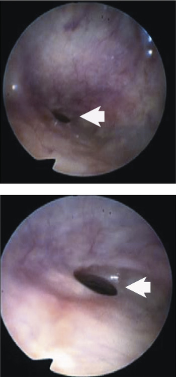
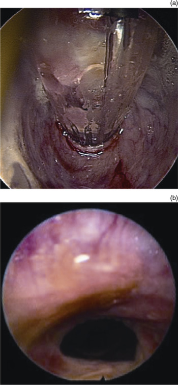
In December 2008, he developed nasal obstruction, snoring and restless sleep. Flexible nasopharyngolaryngoscopy revealed severe stenosis of the nasopharyngeal port, with an estimated diameter of 4 mm, which had not been present on previous endoscopies. He underwent surgery, with transnasal visualisation of the stenotic nasopharyngeal port via a 70° rigid endoscope (Figure 1), serial dilatation to 15 mm with a balloon catheter (Boston Scientific, Natick, Massachusetts, USA) (Figure 2), and injection with 20 mg of topical triamcinalone (Kenalog; Bristol-Myers-Squibb, Princeton, New Jersey, USA). Improvement was seen on follow up, and the patient reported partial resolution of his symptoms; however, some nasal obstruction and sleep difficulty persisted.
Pre-operative endoscopic views of the patient's acquired nasopharyngeal port stenosis (arrows), obtained via transnasal flexible fibre-optic endoscopy.

Endoscopic views showing (a) the balloon catheter expanding the nasopharyngeal port, and (b) the nasopharynx one week after balloon dilatation.

In August 2009, the patient again underwent surgery, during which uvular bifidity and evidence of a submucous cleft palate were observed. A pharyngoplasty was performed by bilateral suturing of the superiorly based posterior tonsillar pillar flaps to the lateral pharyngeal walls, allowing the nasopharyngeal port to be further widened while preserving velopharyngeal competency.
Following this procedure, the patient's symptoms dramatically improved, and he maintained this improvement one year post-operatively. At that time, his only remaining problem was persistent eustachian tube dysfunction.
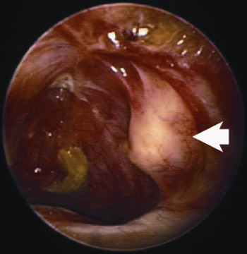
During subsequent surgery for a tympanostomy tube change and rigid nasal endoscopy, we were able to pass a lighted balloon sinuplasty guidewire (Acclarent, Menlo Park, California, USA) through the patient's myringotomies and into the eustachian tubes via the middle-ear cavities, allowing transnasal endoscopic visualisation of a layer of tissue blocking both eustachian tube orifices (Figure 3).
Transnasal, endoscopic view of the obstructed left eustachian tube orifice (arrow) in the nasopharynx, obtained using a 0° rigid endoscope with a lighted guidewire in the eustachian tube (passed through the middle ear via a previously created myringotomy).

Discussion
Sarcoidosis is a multisystem disease primarily affecting adults younger than 50 years of age, with a peak incidence at 20 to 59 years of age.Reference Iannuzzi, Rybicki and Teirstein 6
The aetiology of sarcoidosis is poorly understood. The most accepted theory is that a non-degradable foreign agent, the identity of which is unclear, initiates an inflammatory reaction mediated by macrophages and cluster of differentiation 4 protein positive (CD4+) helper T cells, resulting in the release of cytokines which further propagate the inflammatory response, eventually leading to the formation of non-caseating epithelioid granulomas.Reference Iannuzzi, Rybicki and Teirstein 6 , Reference Lazarus 7
The lungs and intrathoracic lymph nodes are primarily affected, although other areas can also be involved. Approximately 9 per cent of affected patients have head and neck involvement, which can include the middle and inner ears, sinonasal cavities, oral cavity, hypopharynx, and larynx.Reference Braun, Gentine and Pauli 8 Nasopharyngeal involvement has been described but is rare.Reference Braun, Gentine and Pauli 8
The diagnosis of sarcoidosis is made from the combination of a consistent clinical picture, typical radiological findings and a positive biopsy. Biopsies are usually obtained from the most easily accessible affected organ, such as the skin, lacrimal glands, nasal mucosa or oral mucosa. Affected tissues tend to have a nodular appearance with white or yellow discoloration. Transbronchial biopsy may be appropriate in patients without more easily accessible lesions. The serum concentration of angiotensin-converting enzyme is elevated in 30–80 per cent of patients with sarcoidosis, but it is considered a poor diagnostic test due to its highly variable sensitivity and low specificity.Reference Iannuzzi, Rybicki and Teirstein 6 , Reference Lazarus 7 An evaluation for sarcoidosis, with a chest X-ray and biopsy, should be considered in any patient presenting with acquired nasopharyngeal stenosis of unknown aetiology.
-
• Acquired nasopharyngeal stenosis typically occurs due to surgery or irradiation
-
• In spontaneous cases without a clear aetiology, sarcoidosis should be considered
-
• In sarcoidosis with nasal obstruction, nasopharyngeal stenosis should be considered
-
• A patient with sarcoidosis and acquired nasopharyngeal stenosis is reported
-
• Management with endoscopic balloon dilatation and transoral pharyngoplasty was successful
We searched the PubMed database using the open-ended key words ‘nasopharyn*’ and ‘sarcoid*’ along with the closed-ended key words ‘stenotic’ and ‘stenosis’. We did not find any previous reports of acquired nasopharyngeal stenosis occurring in conjunction with sarcoidosis.
There have been many treatments reported in the literature for the management of acquired nasopharyngeal stenosis, including laser excision,Reference Krespi and Kacker 3 , Reference Jones, Guillory and Mair 5 ‘cold knife’ excisionReference Toh, Pearl, Genden, Lawson and Urken 4 and balloon dilatation.Reference Chheda and Postma 1 Modalities for preventing recurrence have included the placement of stents and obturators,Reference Krespi and Kacker 3 , Reference Jones, Guillory and Mair 5 the application of mitomycin C,Reference Jones, Guillory and Mair 5 , Reference Iannuzzi, Rybicki and Teirstein 6 and local flap reconstruction.Reference Toh, Pearl, Genden, Lawson and Urken 4 Our case illustrates the successful initial management of acquired nasopharyngeal stenosis in the setting of sarcoidosis, using balloon dilatation. Local steroid injection was also performed in an attempt to prevent recurrence. Unfortunately, our patient showed evidence of symptomatic, residual, low-grade stenosis post-operatively (Figure 2b). Chheda and PostmaReference Chheda and Postma 1 recently reported the successful use of a similar balloon dilatation technique on three patients with radiation-induced acquired nasopharyngeal stenosis, although one of their patients did require a second dilatation. It is unclear whether a persistent inflammatory process such as sarcoidosis would affect the incidence of long-term recurrence following balloon dilatation, compared with radiation-induced stenosis.
Our patient subsequently underwent transoral pharyngoplasty, which further improved his symptoms to his pre-morbid baseline. The only adverse consequence of this transoral pharyngoplasty was the development of permanent eustachian tube obstruction. It is difficult to determine whether the patient's eustachian tube dysfunction had been worsened by the pharyngoplasty or had simply persisted as a result of the continued inflammatory disease process.
Conclusion
Acquired nasopharyngeal stenosis typically occurs as a result of surgery or irradiation of the nasopharynx. Investigation for sarcoidosis should be considered in patients with spontaneous acquired nasopharyngeal stenosis of unclear aetiology.
Sarcoidosis can present with numerous sinonasal manifestations. Acquired nasopharyngeal stenosis should be considered as a potential cause of nasal obstruction in patients with sarcoidosis.
The presented case demonstrates successful management of acquired nasopharyngeal stenosis in a patient with sarcoidosis, using a combination of endoscopic balloon dilatation and transoral pharyngoplasty. Both these procedures may be considered as first-line treatments for acquired nasopharyngeal stenosis, particularly in patients with rheumatological diseases such as sarcoidosis.