I. INTRODUCTION
Through hundreds of millions of years of evolution, organisms have developed a myriad of ingenious solutions to ensure and optimize survival and success. Many biological (natural) materials are composites of organic (proteins and/or polysaccharides) and inorganic (minerals) components hierarchically assembled into complex structures at ambient temperature and pressure and usually in an aqueous environment.Reference Meyers, Chen, Lin and Seki1, Reference Chen, Lin, Lin, Seki, Stokes, Peyras, Olevsky, Meyers and McKittrick2 These natural composites have exceptional mechanical properties that are far beyond their relative weak constituents and are often multifunctional and possess self-healing ability. Some examples are abalone nacre,Reference Lin and Meyers3, Reference Meyers, Lin, Chen and Muyco4 crab exoskeletons,Reference Chen, Lin, McKittrick and Meyers5 chiton radular teeth,Reference Weaver, Wang, Miserez, Tantuccio, Stromberg, Bozhilov, Maxwell, Nay, Heier, DiMasi and Kisailus6 and squid beaks.Reference Miserez, Schneberk, Sun, Zok and Waite7 In this study, we investigate teeth and scales from four unique fish species, piranha (Serrasalmus manueli), great white shark (GWS, Carcharodon carcharias), arapaima (Arapaima gigas), and alligator gar (Atractosteus spatula), with emphasis on their microstructure and mechanical properties. The uniqueness is derived from the predatory nature of piranha and shark, on one hand, and extraordinary protection proceeded by the scales of arapaima and alligator gar, on the other.
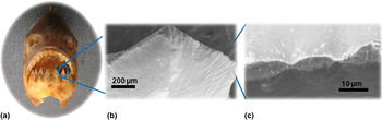
The design principles of sharp biological materials are reviewed by Meyers et al.Reference Meyers, Lin, Lin, Olevsky and Georgalis8 Many sharp edges found in plants, insect proboscises, and teeth have serrations which are known to enhance cutting efficiency.Reference Atkins9Figure 1 shows the structural hierarchy of the cutting mechanisms found in the jaw of a piranha. The jaw is designed with sharp triangular teeth aligned so that as the mouth of the fish closes the initial points of puncture of both the lower and upper jaw are superimposed, as shown in Fig. 1(a). Figure 1(b) is a scanning electron microscopy (SEM) image showing the top view of a piranha tooth. Each tooth exhibits micrometer-sized serrations along its cutting edge, seen in the SEM image of Fig. 1(c). These serrations, approximately 10‑15 μm in wave length, are used to create a highly efficient cutting effect, which converts some of the dragging force into normal force at localized points. There is a superimposed compression and shear, which effectively cuts through skin and muscle. As the jaw further closes, any tissues in the trough of the aligned teeth are severed in a guillotine-like confining action. The piranha tooth has two components: the outer layer is identified as enameloid, an enamel-like material composed of rod-shaped hydroxyapatite nanocrystals, and the inner layer is dentin, which consists of mineralized type-I collagen.
Hierarchical structure of piranha (Serrasalmus manueli) from jaw to tooth to serrations: (a) photograph showing the jaw of piranha; scanning electron microscopy (SEM) micrographs showing (b) top view of the tooth and (c) detailed view of serrations ∼10 μm.

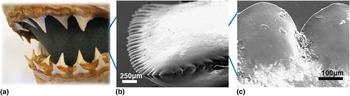
The GWS evolved teeth from the scales of its ascendants. It uses these extremely sharp teeth to perform a very specific killing game. To avoid self-injury, the GWS takes one efficiently large bite out of its prey, then retreats and waits for its victim to undergo shock or hemorrhaging before final consumption. This bite takes only one second to completeReference Diamond10, and the biting stress is measured as high as 600 MPa.Reference Snodgrass, Gilbert, Gilbert, Mathewson and Rall11 Thus, extremely sharp teeth are required. Each tooth is outfitted with a line of large serrations, with up to 300 μm between points. The serrations are perfectly aligned along the cutting edge of the tooth, each creating a mini tooth on the side of its parent tooth. Their shape is close to a hypocycloid. Similar to the piranha tooth, the serrations on this edge maximize the efficiency of the drag force and convert it into points of normal force summed along the side of each serration. Figure 2 shows: (a) an optical image of the overall jaw of a GWS, with multiple rows of teeth, (b) a SEM micrograph of the cutting edge of the tooth with large serrations, and (c) a detailed view of serrations, which have ∼300 μm periodicity. Chen et al.Reference Chen, Lin, Lin, Seki, Stokes, Peyras, Olevsky, Meyers and McKittrick2 compared teeth serration size and body mass of various animals and found that meat eaters, such as the tyrannosaurid dinosaurs, Komodo dragon, and GWS, have serration sizes ranging from 300 to 400 μm, despite the significant difference in body mass and hypothesized that the serration size depends on the mechanical properties of the prey skin and muscle.
Hierarchical structure of great white shark (GWS, Carcharodon carcharias) from jaw to tooth to serrations: (a) photograph of the jaw of GWS showing multiple series of dentition; SEM micrographs showing (b) top view of the tooth and (c) detailed view of serrations ∼300 μm.

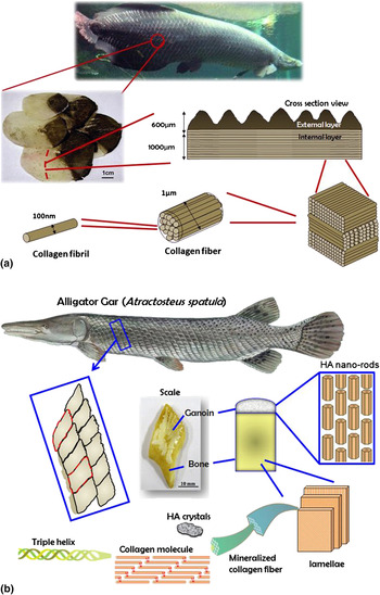
A number of studies on fish scales have been carried out addressing the structural organization, mechanical properties, and design strategies.Reference Onozato and Watabe12–Reference Meyers, Lin, Olevsky and Chen21 Most fish scales have similar material components to other hard tissues such as bones and teeth, composed mainly of type-I collagen fibers and calcium phosphate-based minerals. Arapaima is one of the largest freshwater fish on earth, reaching a length of about 2–2.5 m and a mass over 150 kg. Arapaima inhabits the Amazon River Basin in South America, coexisting in harmony with the piranha, fish known for their voraciousness and sharp teeth. It is believed that the scales of Arapaima serve as armor, providing protection against predators.Reference Meyers, Lin, Olevsky and Chen21, Reference Currey22 Mechanical tests on the scales show that the piranha teeth cannot penetrate the scales.Reference Currey22 The scales consist of an external layer which is highly mineralized and an internal layer, each 50‑60 μm thick and both composed of collagen fibers ∼1 μm in diameter. The mineralized collagen fibers form a cross-lamellar arrangement that produces a laminate composite,Reference Torres, Troncoso, Nakamatsu, Grande and Gomez18, Reference Lin, Wei, Olevsky and Meyers20 as shown in Fig. 3(a). The microhardness of the external layer (550 MPa) is considerably higher than that of the internal layer (200 MPa), consistent with its higher degree of mineralization.Reference Lin, Wei, Olevsky and Meyers20 The scale can be considered as a functionally graded material: the internal layer provides flexibility and toughness while the external layer gives required hardness and wear resistance for protection.
Hierarchical structure of scales from (a) arapaima (Arapaima gigas) and (b) alligator gar (Atractosteus spatula).

The alligator gar is the largest freshwater fish found in North America, measuring up to 3 m and weighing up to 100 kg at maturity. Its size rivals the Arapaima. The scales are diamond-shaped with serrated edges, which can be used to cut potential predators [Fig. 3(b)]. The scales consist of a highly mineralized external layer of ganoin, which is known to be an enamel-like mineral with hydroxyapatite nanorods,Reference Daget, Gayet, Meunier and Sure23 and an internal bony layer made of highly mineralized collagen fibers. Native Americans and swamp inhabitants use alligator gar scales as jewelry.
Probing the mechanical properties of biological materials such as fish teeth and scales is challenging due to their relative small size and complex multilayered structure. Nanoindentation has become a powerful technique that is capable of probing the mechanical properties at micro- and nanometer length scales with high resolution, spatially specific accuracy.Reference Oliver and Pharr24 In recent years, this new technique has been widely applied to studies of bone,Reference Rho, Tsui and Pharr25–Reference Fan and Rho29 cartilage,Reference Ebenstein, Kuo, Rodrigo, Reddi, Ries and Pruitt30–Reference Franke, Göken, Meyers, Durst and Hodge32 and teeth,Reference van Meerbeek, Willems, Celis, Roos, Braerm, Lanbrechrs and Vanherle33–Reference Habelitz, Marshall, Balooch and Marshall37 and extensively reviewed by several groups.Reference Kinney, Marshall and Marshall38–Reference Oyen45 There are many considerations to take into account when determining mechanical properties of biological materials by nanoindentation, such as the effects of viscoelasticity, adhesion, tip selection, storage medium, and sample preparation. Many biological materials exhibit viscoelastic or time-dependent mechanical behavior. The elastic modulus of human cortical bone obtained from the quasi-static nanoindentation tests increased with the indentation loading rate and strain rate.Reference Fan and Rho29 The effects of time-dependent plasticity can be diminished by multiple loading‑unloading cycles followed by a long holding period at maximum load before final unloading.Reference Fan and Rho29 Additionally, the samples can be tested in a dynamic mode, where sinusoidal loads can be applied to measure storage and loss modulus as a function of loading frequency. Most biological materials are naturally hydrated and hydration plays an important role in mechanical properties. Studies on human cortical bone showed that the elastic modulus measured by nanoindentation increased 11–28% after dehydration.Reference Rho, Roy, Tsui and Pharr27, Reference Hengsberger, Kulik and Zysset28 Habelitz et al.Reference Habelitz, Marshall, Balooch and Marshall37 investigated changes in mechanical properties of human dentin and enamel during storage in deionized (DI) water, calcium chloride-buffered (CaCl2) saline solution, and Hank’s Balanced Salt Solution (HBSS). Results showed that the elastic modulus and hardness of dentin and enamel decreased significantly when storing in DI water or CaCl2 solution, while there was no significant change in mechanical properties when storing the specimens in HBSS. A recent study by DickinsonReference Dickinson44 compared the effect of three storage solutions [DI water, HBSS, and phosphate buffered saline(PBS)] on the mechanical properties of human enamel, dentin, and bone. The results showed that storage of mineralized tissues in any of these solutions can significantly (>70%) reduce their mechanical properties possibly due to demineralization. The study indicated the importance of testing biological tissues immediately after extraction.
In this study, we focus on nanomechanical properties of fish teeth and scales using advanced nanoindentation techniques and compare the results with those previously obtained from microindentation and bulk mechanical tests. Comparisons between two types of fish teeth and scales are also made, and the effect of hydration on mechanical properties is discussed.
II. MATERIALS AND METHODS
A. Sample preparation
The piranha teeth and arapaima scales were acquired from the Araguaia River (Amazon Basins) in Brazil. The GWS teeth were obtained from Del Mar, CA, and the alligator gar scales were donated by Dianne Ulery (Dianne Ulery’s Natural Jewelry Art, Alexandria, LA, website: http://www.bonanza.com/Dianne_Ulery). All samples were kept in ambient dry condition before testing. The period of time between the teeth and scales being removed from the animals and tested mechanically was approximately 6 months. For each of the four species, samples were obtained from one animal. Two front teeth were sectioned transversely approximately half way down the cusp from the upper jaws of shark and piranha. The arapaima and alligator gar scales were obtained from the central region of the fish body. Two small pieces (20 mm × 5mm × 2 mm) were cut from the dark (exposed) region from an arapaima scale. For the alligator gar, two specimens were made by cutting a scale in half using a diamond rotating blade through the long axis. All samples were mounted in epoxy with cross-sectional area revealed, followed by grinding and fine polishing.
B. Structural characterization
Optical micrographs of polished samples were taken using a Zeiss Axio imager (Zeiss MicroImaging Inc., Thornwood, NY) equipped with a CCD camera in the reflective light mode. The microstructure was characterized by using a field emission scanning electron microscope (FEI-XL30, FEI Company, Hillsboro, OR). Samples prepared for SEM were mounted on aluminum sample holders and sputter coated with a thin layer of gold.
C. Nanoindentation
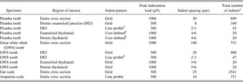
Nanoindentation testing was performed using a TI-950 TriboIndenter (Hysitron Inc., Minneapolis, MN). User-specified parameters for testing each sample are summarized in Table I. Each indent took ∼90 s to complete. Depending on the total number of indents, the tests took from 30 min to 18 h. The testing temperature was 24 °C and the relative humidity was 15% when testing in the ambient dry condition. The nanoindentation instrumentation enclosure ensured that the temperature and humidity were kept fairly constant throughout the testing and no effect was noticed with time.
User-specified parameters for nanoindentation testing on all specimens.

a Outermost (extreme) curves were removed from analysis but are included in the total number of indents performed per specimen location
b User-defined indent locations specified from SPM images
For relatively large areas (on the order of hundreds of microns to several millimeters), regions for testing were identified using an optical microscope integrated into the nanoindentation system. Subsequent grids of indents were performed on specified locations with user-specified peak loads and indent spacing. For smaller areas (less than 100 μm), regions for testing were identified using scanning probe microscopy (SPM), utilizing the nanoindentation tip as the SPM probe to obtain pretest in-situ SPM images. In-situ SPM imaging provided significant information on sample surface morphology on piranha and GWS specimens, which helped to identify desired test regions [dentin‑enameloid junction (DEJ)]. Prior to the measurements, preliminary indents were performed in each region and the size of the residual indent was measured. The peak loads were chosen for different testing conditions to give proper spacing between the indents. For example, the indents in the line profile that were user-defined using SPM imaging were spaced close together, so a lower force (300 μN) was required to not have overlapping elastic and plastic zones. The indents in the DEJ grid were spaced farther apart, so a load of 500 μN was used. A peak force of 1000 μN was applied to probe the mechanical properties throughout the entire cross section of the specimen. In this case, the adjacent indents were spaced 50 or 100 μm apart.
For GWS and piranha specimens, a diamond fluid cell Berkovich probe was utilized to perform tests in both ambient (dry) and hydrated conditions. Samples were immersed in a D-PBS solution during hydrated testing, with a minimum of 2 h of hydration time prior to testing. The measurements were performed in fluid immediately after the samples were fully hydrated to prevent demineralization and degradation of mineralized tissues in solutions, which may affect the mechanical properties. The series of indents were accomplished within 1 h and no physical change of the sample, such as swelling was observed. Tests on GWS and piranha samples were performed in load-controlled feedback mode to various peak forces. A load function consisting of a 5-s loading to peak force segment, followed by a 5-s hold segment, and a 1-s unloading segment was used. For gar and arapaima specimens, a diamond Berkovich probe was utilized to perform load-controlled tests in ambient (dry) conditions. A load function consisting of a 5-s loading to peak force segment, followed by a 2-s hold segment, and a 5-s unloading segment was used. Nanoindentation hardness and reduced modulus (in GPa) were calculated based on the well-established Oliver‑Pharr method for nanoindentation testing.Reference Oliver and Pharr24
III. RESULTS AND DISCUSSION
A. Piranha teeth
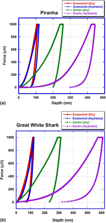
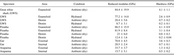
The reduced modulus and hardness (mean ± SD) of the four types of fish samples measured by nanoindentation are summarized in Table II. Figure 4(a) shows representative force‑depth curves of piranha enameloid and dentin tested in ambient dry and hydrated conditions. When tested in ambient dry condition, the enameloid of the piranha tooth has a mean reduced modulus of 86.5 ± 15.9 GPa and a mean hardness of 4.1 ± 0.9 GPa, while dentin has a mean reduced modulus and hardness of 23.0 ± 6.0 and 0.8 ± 0.3 GPa, respectively. The dentin and enameloid were further tested in hydrated condition to understand the effect of hydration on the mechanical properties of dentin and enameloid. The modulus and hardness (81.9 ± 8.4 and 3.1 ± 0.4 GPa) of enameloid in hydrated condition are lower compared with those tested in dry condition and those of hydrated dentin are significantly reduced to 12.4 ± 1.6 and 0.2 ± 0.04 GPa. Enameloid is a highly mineralized tissue with very small amount of organic component and water (<5%) compared with dentin. Thus, the effect of hydration on the mechanical properties of enameloid is not as significant as in dentin. Demineralization of mineralized tissues in solutions is certainly expected, and the first 12‑24 h are crucial. Thus, the samples were tested immediately after full rehydration to minimize the effects of demineralization and degradation. Although it is known that HBSS might be a better storage medium for teeth (particularly for dentin)Reference Habelitz, Marshall, Balooch and Marshall37 compared with PBS which is generally better for bone, the effects of the storage medium on the mechanical properties should not be significant since the samples were kept in the solution for a relatively short period of time (less than 4 h). The hardness values measured from nanoindentation are twice as high as those previously measured by microindentation (enameloid ∼1.5 GPa, dentin ∼0.3 GPa).Reference Chen, Lin, Lin, Seki, Stokes, Peyras, Olevsky, Meyers and McKittrick2, Reference Meyers, Lin, Olevsky and Chen21 This is due to the size effect: the frequency of weak links (cracks, ligaments, voids, interfaces) decreases with decreasing size, leading to higher hardness. This was expressed quantitatively by Yao and GaoReference Yao and Gao46 in their hierarchical structure (Russian doll) theory.
Representative force-depth curves of enameloid and dentin from (a) piranha and (b) GWS tested in ambient dry and hydrated conditions.

Reduced modulus and hardness measured from nanoindentation.

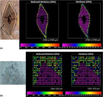
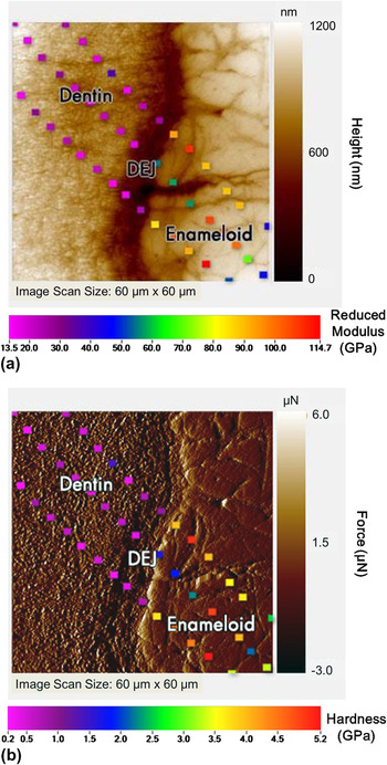
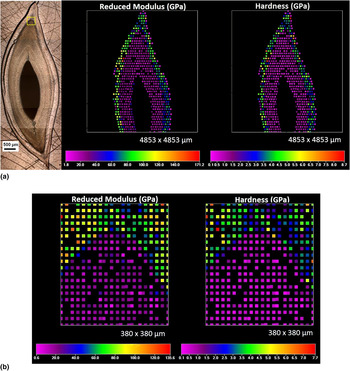
Figure 5(a) shows an optical micrograph of the cross-sectional area in a piranha tooth along with reduced modulus and hardness maps in color scale. It is clearly seen that the external enameloid layer has much higher modulus and hardness compared with internal dentin. A condensed grid of indents (17 × 20, 8 μm apart) was performed at the DEJ of piranha to measure the local mechanical properties at the junction. The zoom-in modulus and hardness maps (128 μm × 152 μm) at the DEJ are shown in Fig. 5(b). Lines of indents (2‑3 μm apart) were performed along the DEJ of the piranha sample in order to take an even closer look at the mechanical properties along the junction. Figures 6(a) and 6(b) show topographical and gradient in-situ SPM images of the DEJ area (60 μm × 60 μm) where the indents were performed. Reduced modulus and hardness values were overlaid on the topographical and gradient in-situ SPM images, respectively, and the difference in mechanical properties between dentin and enameloid can be clearly seen. It should be noted that the peak indentation loads varied from 300 to 1000 μN depending on different testing conditions and locations, as shown in Table I. The loading time was kept the same (5 s) so that the loading rate varied from 60 to 200 μN/s. Fan and RhoReference Fan and Rho29 investigated the effects of viscoelasticity on nanoindentation measurements of human cortical bone and found that the elastic modulus increased with loading rate. However, the effect of loading rate on the mechanical properties of dentin and enameloid should be minor since the magnitude of the loading rate differences in our nanoindentation measurements is only a factor of 3.
(a) Reduced modulus and hardness maps of cross-sectional area in a piranha tooth; (b) zoom-in modulus and hardness maps (128 μm × 152 μm) at the dentin‑enameloid junction (DEJ) region.

(a) Reduced modulus results overlay on the topographical scanning probe microscopy (SPM) image; (b) Hardness values overlay on the gradient SPM image at the DEJ region of piranha (60 μm × 60 μm).

Nanoindentation measurements were performed to map mechanical properties of a piranha tooth at varying length scales, from an entire cross-sectional area, a smaller area enclosing dentin and enameloid, to a more localized region at the DEJ. The space between adjacent indents for nanoindentation ranges from 3 to 50 μm, much smaller compared with that for microindentation, which often reaches ∼200 μm. A limited amount of data points can be obtained using microindentation on small biological samples with a complex microstructure, such as piranha teeth, making nanoindentation a suitable and superior testing method over microindentation.
B. GWS teeth
A cross-sectional optical micrograph of a GWS tooth and corresponding modulus and hardness maps are shown in Fig. 7(a). Higher resolution modulus and hardness maps at the DEJ region are shown in Fig. 7(b). Similar to the piranha, the significant difference in mechanical properties between enameloid and dentin can be clearly seen. The reduced modulus and hardness of shark enameloid tested in ambient dry condition are 84.4 ± 19.9 and 4.1 ± 1.1 GPa, and those of shark dentin are 20.4 ± 5.6 and 0.7 ± 0.2 GPa, respectively. Figure 4(b) shows representative force‑displacement curves of shark enameloid and dentin tested in ambient dry and hydrated conditions. When tested in the hydrated condition, the modulus and hardness of shark enameloid reduce to 77.2 ± 14.8 and 2.6 ± 0.9 GPa, while those of shark dentin drop more drastically to 8.7 ± 3.1 and 0.2 ± 0.1 GPa, respectively. The mechanical properties of dentin and enameloid of shark tooth and the effect of hydration are in good agreement with those of piranha tooth. Whitenack et al.Reference Whitenack, Sinkins, Motta, Hirai and Kumar47 investigated mechanical properties of teeth from two types of sharks, the sand tiger shark (Carcharias taurus) and the bonnethead shark (Sphyrna tiburo), using nanoindentation in dry condition. The reduced modulus and hardness of tiger shark enameloid are 72.61 ± 4.73 and 3.20 ± 0.20 GPa, and those of bonnethead shark enameloid are 68.88 ± 1.50 and 3.53 ± 0.30 GPa, respectively. The tiger shark dentin has reduced modulus and hardness of 28.44 ± 2.21 and 1.21 ± 0.16 GPa, and the bonnethead shark dentin has reduced modulus and hardness of 22.49 ± 1.72 and 0.97 ± 0.07 GPa, respectively. The reported mechanical properties of the tiger and bonnethead sharks are in good agreement with those of the GWS in this study.
(a) Reduced modulus and hardness maps of cross-sectional area in a GWS tooth; (b) zoom-in modulus and hardness maps (380 μm × 380 μm) at the DEJ region.

Despite that the GWS and piranha are significantly different in taxonomy, habitat, body size, and morphology, the microstructure and mechanical properties of their teeth are similar. Both GWS and piranha teeth consist of highly mineralized enameloid and dentin, which is made of mineralized collagen fibrils. Mechanical properties of GWS and piranha measured from nanoindentation are comparable, as shown in Table II. The reduced modulus and hardness of enameloid are 4‑5 times higher than those of dentin when tested in ambient dry condition. The difference in mechanical properties between enameloid and dentin is more significant (7‑15 times) when tested in rehydrated condition. Imbeni et al.Reference Imbeni, Kruzic, Marshall, Marshall and Ritchie48 studied the critical role of dental‑enamel junction in enhancing fracture resistance of human teeth. SEM observation showed that cracks initially formed in enamel tend to penetrate the DEJ and stop when they enter the tougher dentin adjacent to the interface due to extrinsic toughening mechanisms such as uncracked ligament bridging. Such design, i.e., hard and brittle external enameloid enclosing softer and tougher dentin, provides enhanced mechanical properties that are required to fulfill functional purposes.
C. Arapaima gigas scales
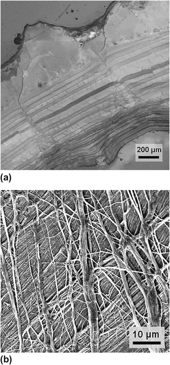
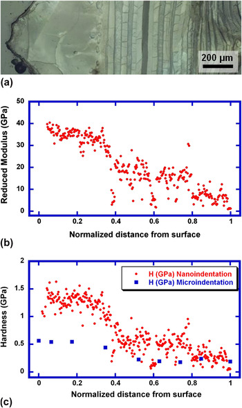
Figure 8(a) is a cross-sectional optical micrograph showing the external and internal layers of an Arapaima scale. The corrugated surface of the external layer corresponds to the ridge structure seen on the surface, whereas the internal layers are characterized by lamellae. Figure 8(b) is a top-view SEM image showing the collagen fiber orientation between two adjacent layers, which has an angle of ∼75°. Figure 9(a) is an optical micrograph showing the cross-sectional hardness mapping through the thickness of an arapaima scale. Figures 9(b) and 9(c) show the reduced modulus and hardness variation through the scale, from the external layer to the internal layer. The reduced modulus measured at the external layer is 33.7 ± 3.7 GPa and gradually decreasing to 15.7 ± 5.1 GPa in the internal layer. The hardness shows the same trend, decreasing with distance from the external layer (1.3 ± 0.2 GPa) to the internal layer (0.5 ± 0.2 GPa). The results are in accord with the calcium and phosphorous mapping through the scale thickness previously reported by Lin et al.Reference Lin, Wei, Olevsky and Meyers20 The external and internal layers are both composed of mineralized collagen fibers, yet the external layer has higher mineral content than the internal one. As a result, the external layer has higher hardness and modulus than the inner layer. Lin et al.Reference Lin, Wei, Olevsky and Meyers20 performed microindentation across the scale. The reported hardness of the outer layer was found to be ∼550 MPa and that of the inner layer was ∼200 MPa [Fig. 9(c)], much lower than the nanoindentation hardness in this study. The significant difference may attribute to that the microhardness tests were performed in rehydrated condition while the nanoindentation measurements were carried out in ambient dry condition. The harder external layer is ideal to serve as protective armor against the sharp teeth of predators and such functional design is widely found in nature, e.g., a hard exocuticle covering endocuticle in crab shells.Reference Chen, Lin, McKittrick and Meyers5 Meyers et al.Reference Meyers, Lin, Olevsky and Chen21 performed penetration tests of piranha teeth into arapaima scales simulating the vigorous piranha bite. Results show that the piranha teeth cannot penetrate the exposed portion of arapaima scales. The piranha teeth fractured and embedded in the scales before further penetration. The highly mineralized external layer of scales plays a key role in protecting arapaima from being attacked by piranha.
(a) Optical micrograph showing the external and internal layers in the scale of Arapaima gigas; (b) SEM image showing the orientation of collagen fibers in adjacent layers.

(a) Cross-sectional optical micrograph showing region where nanoindentation was performed for arapaima scale; (b) reduced modulus and (c) hardness through the thickness of scale. The hardness values measured from nanoindentation are compared with those measured from microindentation.Reference Lin, Wei, Olevsky and Meyers20

D. Atractosteus spatula scales
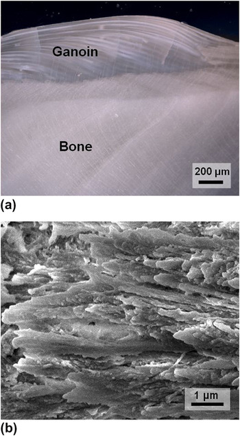
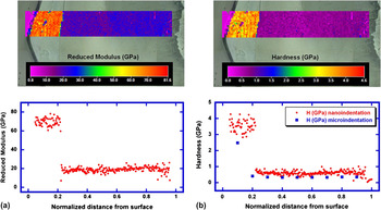
Figure 10(a) is an optical micrograph showing the cross-sectional area of an alligator gar scale, which consists of two regions; an external ganoin layer and an internal bony layer. Figure 10(b) is a SEM image showing the mineralized collagen fibers in the internal bony layer. Figure 11(a) shows the reduced modulus map through the thickness of the gar scale. The modulus measured at the external ganoin layer is 70.8 ± 4.5 GPa and that in the internal bony layer drops to 20.5 ± 2.4 GPa. The modulus of ganoin is comparable to that of enameloid in the fish teeth and that of bony layer is in good agreement with the elastic modulus of bone.Reference Rho, Tsui and Pharr25–Reference Rho, Roy, Tsui and Pharr27 Unlike the gradual change in mechanical properties found in arapaima scales, there is a distinct interface between ganoin and bony layers, which is akin to the DEJ. The hardness of the ganoin layer is 3.6 ± 0.3 GPa and that of bony layer is 0.7 ± 0.1 GPa, as shown in Fig. 11(b). Hardness values measured from microindentation are lower compared with those obtained by nanoindentation. The external ganoin layer has a microhardness of 2.5 GPa, whereas the bony layer has a microhardness of 0.35 GPa. Bruet et al.Reference Bruet, Song, Boyce and Ortiz17 found that the scales from Polypterus senegalus have four layers: ganoin, dentin, isopedine, and bony layers (from external to internal regions). Here, two regions (ganoin and bony layers) are found in alligator gar scales. The modulus (∼62 GPa) and hardness (∼4.5 GPa) of the outer ganoin layer and those of inner bony layer (E ∼ 17 GPa, H ∼ 0.54 GPa) of Polypterus senegalus scales are consistent with our results. The hard, highly mineralized ganoin layer is designed for protection from predator’s attack, and the underlying, more compliant bony layer dissipates energy and provides flexibility of the entire fish body. The multilayered structure and graded mechanical properties seem to be widely adopted in fish scales and biological armor materials, indicating evidence of evolutionary convergence which may inspire the design of novel composites.Reference Munch, Launey, Alsem, Saiz, Tomsia and Ritchie49, Reference Launey, Munch, Alsem, Barth, Saiz, Tomsia and Ritchie50
(a) Optical micrograph showing the cross-sectional area of an alligator gar scale, which consists of two regions, external ganoin layer and internal bony layer; (b) SEM images showing fracture surface of mineralized collagen fibers in the internal bony layer.

Cross-sectional optical micrograph overlapped with (a) reduced modulus and (b) hardness maps through the thickness of an alligator gar scale. The hardness values measured from nanoindentation are compared with those measured from microindentation tests.

IV. CONCLUSIONS
In this study, we investigated the mechanical properties of two types of fish teeth (piranha and GWS) and two types of fish scales (arapaima and alligator gar) using advanced nanoindentation techniques and compared our results with previous microindentation tests. These biological materials provide superior predatory capability (teeth) and protection (scales) in fish and therefore their mechanical properties are essential. Nanoindentation provides position-specific and high-resolution information of mechanical properties, which can probe the nano and microstructure of biological materials. Despite the diversity between fish species, similar design strategies are found in teeth and scales. All samples have a hard, highly mineralized external layer and a tough, less mineralized internal layer. The enameloid in fish teeth is 4‑5 times harder than the dentin. Similar trends are found in the ganoin and bony layers in alligator gar scales as well as the arapaima scales.
The piranha and GWS teeth were further tested in a hydrated condition. The results show that hydration has a strong effect on the mechanical properties of dentin, while no significant difference is observed in the highly mineralized, collagen-deficient enameloid. This study demonstrates that nanoindentation is indeed a powerful technique for probing and elucidating mechanical properties of biological materials, which usually have a complex hierarchical structure and multifunctionality ranging in many length scales.
ACKNOWLEDGMENTS
We thank Ryan Anderson (CalITReference Chen, Lin, Lin, Seki, Stokes, Peyras, Olevsky, Meyers and McKittrick2) for the help on the scanning electron microscopy. Valuable insights and discussion with Ms. Dianne Ulery are greatly appreciated. She kindly donated the alligator gar scales for research purposes. We thank Professor Joanna McKittrick for her enthusiastic support of this project. This research is funded by the National Science Foundation, Division of Materials Research, Biomaterials Program (DMR 0510138) and Ceramics Program (DMR 1006931).