Today's stem cell researchers represent investigators from a broad range of biomedical backgrounds, but at the core lie those whose training is rooted in the field of developmental biology: in this field, the concept of changing one cell type into another is certainly not novel. It was as long ago as 1958 that Sir John Gurdon performed his seminal work in the Xenopus model, wherein he took a nucleus from a mature intestinal tadpole cell and placed it into a recipient enucleated frog egg (Ref. Reference Gurdon, Elsdale and Fischberg1). The egg was found to grow into a tadpole that was genetically identical to the donor, thereby showing that the cytoplasm of the host egg had reprogrammed the transplanted nucleus back to a totipotent cell capable of differentiating into all cell types constituting an adult frog. This experiment showed for the first time that an unknown myriad of factors present in the egg cytoplasm were able to reprogramme the phenotype of fully differentiated cells into totipotent one-cell-stage embryos. This experimental procedure was given the term ‘somatic cell nuclear transfer’ (SCNT) and by the mid 1990s had been extended to several mammalian species, most famously resulting in the cloning of ‘Dolly’ the sheep (Ref. Reference Campbell2). To date no ‘cloning’ of human somatic cells has been reported, but instead nuclear reprogramming of human cells has proved possible by a process of cell fusion between adult and embryonic stem cells (ESCs) (Refs Reference Cowan3, Reference Silva4). These experiments extrapolated Gurdon's concept of the pliability of adult cells secondary to epigenetic (nongenetic) factors in simple organisms such as the frog, to complex mammals, even humans, and later formed the ideological basis for Yamanaka's landmark studies.
Yamanaka reasoned that enforced expression of a limited number of genes critical to the maintenance of pluripotency in ESCs might enable direct reprogramming of somatic cells (Ref. Reference Takahashi and Yamanaka5). To evaluate this hypothesis, he overexpressed a mini-library of 24 candidate reprogramming factors into mouse embryonic fibroblasts (MEFs) carrying a fusion of the genes encoding resistance to β-galactosidase and neomycin, which are expressed from the Fbx15 locus (which is specifically expressed in mouse ESCs and early embryos, but is not crucial for ESC maintenance). In this system, reactivation of the Fbx15 gene (a surrogate marker of the pluripotent state) results in resistance to the antibiotic G418. When MEFs infected with all 24 genes were cultured on feeder cells in ES medium in the presence of G418, drug-resistant colonies emerged, some of which exhibited ES-like hallmarks such as morphology, rate of proliferation and gene expression. In a process of stepwise elimination, four transcription factor genes – Oct3/4 (Pou5f1), Sox2, Klf4 and Myc – were identified as sufficient to give rise to these newly formed pluripotent cells, which were termed induced pluripotent stem cells (iPSCs).
The iPSCs generated by Yamanaka were not initially shown to be capable of achieving germline transmission (the ability of a stem cell transplanted into a recipient blastocyst to give rise to progeny), defined to be a key characteristic of murine ESCs. Subsequently, however, within the space of less than 12 months, he and other groups improved the reprogramming technique sufficiently to produce cells capable of germline transmission, which were epigenetically and developmentally indistinguishable from ES cells (Refs Reference Okita, Ichisaka and Yamanaka6, Reference Wernig7, Reference Takahashi8).
After the reprogramming of mouse fibroblasts, the next challenge was to see whether the technique could be applied to other species – particularly to humans. This landmark was successfully achieved simultaneously by two groups in November 2007 (Refs Reference Takahashi8, Reference Yu9). The fact that the same four factors identified in the murine system were able to confer pluripotency in human cells in the absence of selection indicated that the transcriptional network governing pluripotency is common across these species. Interestingly, however, although the Yamanaka lab used exactly the same four factors from the mouse experiments (Oct4, Sox2, KLF4, Myc), the Thompson group substituted KLF4 and Myc with the homeobox protein NANOG and Lin-28. This cocktail of factors not only had the advantage that the oncogenic protein Myc was omitted, but also suggested that the reprogrammed transcriptional network could be induced by different combinations of factors and initiated the hypothesis that the common pluripotency network could be induced by more than one protocol. Much of the subsequent work since November 2007 has focused on understanding the reprogramming mechanism and improving the reprogramming protocol. This desired improvement is centred around two core objectives: first, to induce pluripotency with no disruption to the genetic content of the cell, and second, to increase the efficiency of the process.
Is an induced pluripotent stem cell similar to an embryonic stem cell?
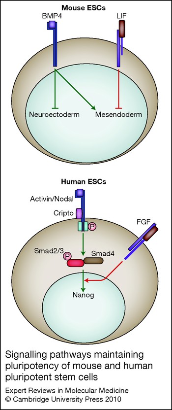
iPSCs and ESCs share the key characteristics of self-renewal and pluripotency. They express the same pluripotency factors (including Oct4, NANOG and Sox2), and can produce cells of all tissues and organs as seen by their ability to form teratomas and generate viable, fertile live-born progeny (Refs Reference Zhao10, Reference Boland11). In addition, fully reprogrammed iPSCs show gene expression and epigenetic states that are highly similar to those in ESCs (Refs Reference Mikkelsen12, Reference Chin13). Therefore iPSCs and ESCs appear to be phenotypically similar. However, recent studies comparing human and mouse ESCs have demonstrated that shared characteristics are not sufficient to demonstrate that two pluripotent cell types are functionally equivalent. Indeed, human and mouse ESCs are phenotypically similar, but they rely on different signalling pathways to control their pluripotent status (see Fig. 1). Mouse ESCs rely on leukaemia inhibitory factor (LIF) and bone morphogenic protein 4 (BMP4), whereas human ESCs rely on activin and FGF2 (Ref. Reference Brons14). This difference in the signalling pathways controlling pluripotency in human and mouse ESCs demonstrates that the cell types are not functionally equivalent, because it implies that different mechanisms control their cell-fate decisions. As a direct consequence, a method of differentiation developed with one pluripotent cell type cannot be used directly with the other pluripotent stem-cell type. Importantly, this divergence between human and mouse ESCs also marks a different embryonic identity. Indeed, our group and others have shown that pluripotent stem cells derived from the epiblast of mouse embryos at post implantation stages (EpiSCs) also rely on activin and FGF2 signalling (Ref. Reference Brons14). Since similar protocols can be used to drive differentiation of human ESCs and mouse EpiSCs into derivatives of the three germ layers, it would suggest that both pluripotent cell types rely on the same signalling pathways to control their differentiation. Therefore, human ESCs and mouse EpiSCs appear to share a common pluripotent state defined by their dependence on activin–nodal signalling. Importantly, this pluripotent state is associated with an embryonic identity corresponding to the late-epiblast stage of postimplantation embryos, from which epiblast stem cells are derived. Therefore, the embryonic identity of mouse ESCs could be closer to the early epiblast of blastocyst embryos blocked in diapause, because genetic studies have shown that LIF signalling is necessary only for this embryonic stage, which is specific to rodents.
Signalling pathways that maintain pluripotency of mouse and human pluripotent cells. Human iPSCs, human ESCs and mouse EpiSCs have been shown to be dependent upon the same cell-signalling pathways for maintenance of their pluripotent state, all critically relying on activin–nodal and FGF signalling. Mouse ESCs however maintain their pluripotent state via a completely different cell-signalling pathway, which is reliant on LIF and BMP4. This suggests that human iPSCs, human ESCs and mouse EpiSCs are all maintained in vitro at a different (later) developmental stage compared with mouse ESCs, thereby helping to explain the different properties observed between these two groups of pluripotent cells. BMP4, bone morphogenetic protein 4; FGF, fibroblast growth factor; LIF, leukaemia inhibitory factor; P, phosphate.

Defining the embryonic identity of iPSCs is thus essential because it will define their reactivity to growth factors and consequently define the efficacy of the methods used to differentiate them. Defining the embryonic identity of mouse iPSCs is rendered more difficult by the possibility of deriving ES-like iPSCs as well as EpiSC-like iPSCs. Indeed, mouse iPSCs derived from cells with a genetic background permissive for ESC derivation usually rely on LIF signalling to maintain their pluripotent status. However, mouse iPSCs derived from cells with a genetic background such as non-obese diabetic (NOD; from which ESCs are impossible to derive with standard culture conditions) appear to rely on activin–nodal signalling to maintain their pluripotency, suggesting that NOD mouse iPSCs resemble EpiSCs rather than ESCs. Interestingly, this phenomenon strictly relies on the culture conditions used during reprogramming, because NOD iPSCs dependant on LIF can be directly generated using defined chemical inhibitors known to improve ESC derivation and avoiding the use of serum and feeders. Therefore, the embryonic identity of mouse IPSCs is defined by their genetic background and the culture conditions used for their derivation. This situation has never been described so far with human iPSCs. On the contrary, recent studies have demonstrated that human iPSCs, similarly to human ESCs and mouse EpiSCs, rely on activin–nodal signalling pathways to maintain the expression of NANOG, which in turn blocks neuroectoderm differentiation induced by FGF2 (Ref. Reference Vallier, Alexander and Pedersen15). Therefore, human iPSCs and human ESCs or EpiSCs rely on the same molecular mechanisms to maintain their pluripotent status. Furthermore, similar signalling pathways drive differentiation of human iPSCs, human ESCs and mouse EpiSCs into neuroectoderm, mesendoderm and extraembryonic progenitors, suggesting that similar mechanisms control the early cell-fate decisions of these pluripotent stem cells (Ref. Reference Vallier16). Together, these results confirm that human iPSCs are functionally equivalent to human ESCs and suggest that the protocols developed over the past 10 years to differentiate human ESCs to diverse cell types could easily be transferable to human iPSCs.
Are iPSCs ‘better’ than ESCs?
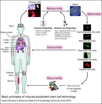
When the first human embryonic stem (ES) cell lines were derived almost 12 years ago, scientists believed new avenues of cell-based regenerative medicine and in vitro disease modelling would be facilitated by differentiating ES cells to given cells of interest. iPSCs, which bear the same core repertoire of useful characteristics, such as unlimited amplification and ability to differentiate into cells of all three germ layers, were therefore hypothesised to possess the same potential applications. However iPSCs are viewed as significant ‘upgrades’ on ES-derived cells for a number of reasons. First, there are probably fewer ethical dilemmas to be overcome with iPSC generation because the starting material (in most cases an adult skin sample) is ubiquitously available and is not dependent upon the use of human embryos. Second, for the purposes of transplantation, iPSCs derived from a patient could be used in an autologous fashion (i.e. transplanted back into the same individual) without the need for harmful immunosuppressants that are a prerequisite to prevent rejection in allogeneic transplants. Third, in vitro disease modelling using iPSCs from individual patients of a given disease or genetic background is far more likely to accurately replicate subtle disease mechanisms than ES-derived models (Fig. 2). Finally, studying the similarities and differences in developmental pathways observed between individuals of diverse genetic backgrounds can only be logistically facilitated using iPSCs.
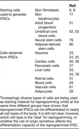
Basic principles of iPSC technology. Reprogramming can be performed starting with a variety of donor somatic cells (depicted here are fibroblasts and blood progenitors; see Table 1 for a complete list). The reprogramming factors can be delivered via either genome integrating (the classical approach as first used by Yamanaka and colleagues) or genome non-integrating methods. The latter techniques have unfortunately so far proven very inefficient. Reprogramming the donor cell back to an induced pluripotent state is followed by differentiation to a target cell(s) of interest. These patient-specific cells should carry the unique genetic signature of the donor and could therefore be used for different research objectives including in vitro disease modelling or drug screening. It is hoped that in the near future such reprogrammed cells may also form part of a cell-based autologous transplantation strategy for treating appropriate diseases. Examples of cells ideally suited to such a strategy include hepatic, neuronal and blood cells.

Examples of cell types used for deriving human iPSCs and cell types differentiated from thema

a Increasingly diverse types of cells are being used as starting material for reprogramming whilst at the same time different groups have shown that reprogrammed iPSCs can be differentiated to nearly all cell types of the body. It is still not clear, however, which cell type is the ‘best’ for reprogramming, or whether the cell of origin somehow affects the differentiation capacity of the reprogrammed iPSC.
Is ‘direct reprogramming’ the mechanism behind iPSC generation?
Understanding of the mechanisms underpinning ‘direct reprogramming’ represents a major challenge for the stem cell field and is crucial to both improving the efficiency of human iPSC generation and for developing new approaches for manipulation of cellular identity, such as ‘forward reprogramming’ or ‘transdifferentiation’ (defined as the transformation of one fully differentiated adult cell to another). The study of these mechanisms remains problematic, however, because the fraction of somatic cells fully reprogrammed by the protocols currently available is extremely low (<5%) and requires an extensive period (at least 2 weeks with human cells). This problem is further exacerbated by the absence of specific markers to select cells undergoing full reprogramming. Performance of informative large-scale experiments, especially in the first few days of reprogramming, therefore remains difficult.
Despite these experimental constraints, important insights into the mechanisms of reprogramming are gradually being uncovered. For a start, it is now commonly acknowledged that the mechanisms involved in direct reprogramming differ considerably from those involved in SCNT. In the latter, a complex mix of factors induce almost immediate epigenetic reprogramming, whereas with direct reprogramming, the overexpressed transcription factors appear to stochastically and therefore very slowly activate their target genes, which eventually recreate the transcriptional network that characterises pluripotent cells. This hypothesis would explain the time required to obtain fully reprogrammed human iPSCs, and also the relative inefficiency of this approach, because key events will only occur randomly. Recent studies have started to reveal some of the mechanisms that could characterise the action of pluripotency factors during human iPSC generation (Ref. Reference Sridharan17). Mechanisms controlling senescence, for example, appear to have a key function in direct reprogramming because Oct4, Sox2 and KLF4 activate the expression of senescence factors such as p16INK4 (CDK4I), which in turn blocks cell proliferation and thus reprogramming (Ref. Reference Banito18). Downregulation of these senescence factors or inhibition of the p53 pathway, the main regulator of senescence, therefore has been shown to significantly improve the efficiency of human iPSC generation (Ref. Reference Hong19). Together, these results provide the first information concerning the mechanisms controlling direct reprogramming (Ref. Reference Krizhanovsky and Lowe20). However, the full understanding of these mechanisms will be more challenging and requires molecular analyses, which are difficult to perform using the current methods of derivation. In addition, recent studies have demonstrated that the identity of the somatic cell strongly influences the efficiency of reprogramming, suggesting that each cell type could express different factors that interfere with the mechanisms leading to production of iPSCs (Ref. Reference Ghosh21). Consequently, understanding the function of each reprogramming factor in ESCs could represent a more immediate solution for uncovering the mechanisms that allow direct reprogramming of somatic cells.
How have iPSCs been used so far?
Disease modelling
Following the first human fibroblast derivation of iPSCs, investigators went on to show that the reprogramming protocol could be used in cells taken from a wide range of mendelian and complex genetic disease backgrounds (Ref. Reference Park22). This observation was then followed by studies proving that human iPSCs could be differentiated into cells similar to adult somatic cells. A range of different human cells with varying relevant functional properties have thus far been generated, including motor and dopaminergic neurons (Refs Reference Dimos23, Reference Soldner24, Reference Karumbayaram25), cardiac cells (Ref. Reference Zhang26), pancreatic cells (Ref. Reference Zhang27), adipocytes (Ref. Reference Taura28), blood cells (Ref. Reference Choi29), vascular cells (Ref. Reference Taura30), retinal progenitors (Ref. Reference Lamba31) and liver cells (Refs Reference Touboul32, Reference Si-Tayeb33, Reference Sullivan34, Reference Gallicano and Mishra35).
However, it was not clear whether the derived cells would be of any ‘clinical’ relevance until publication of two recent landmark studies. In the first study (Ref. Reference Ebert36), investigators described disease modelling of a patient suffering from spinal muscular atrophy (SMA). This study showed that the iPSC-derived neural lineage undergoes selective neuronal death, which could be reversed upon addition of compounds known to increase production of survival motor neuron protein (SMN). However, the authors were not able to produce fully functional neurons displaying the disease phenotype. As a result, they were unable to show that SMN protein was itself reduced in the adult cell type (although a decreased amount of mRNA transcripts was shown), thus making the model an excellent proof-of-concept tool for the production of an in vitro iPSC-based SMA model, but an incomplete disease model in itself. In the second study (Ref. Reference Lee37), the authors describe a patient-specific iPSC-based model of familial dysautonomia. The model impressively showed faithful replication of three key pathogenic features of the disease: (1) defects in gene (IKBKAP) splicing, (2) decreased neurogenesis and (3) defective migration of iPSC-derived neural precursors. However, as with the first study, no evidence was provided to show that decreased protein production was replicated in the fully mature adult cell, nor that lack of protein resulted in adult cell degeneration or paucity of production. Both studies were restricted to the neural lineage, dealt with very small numbers of patients and concerned very rare diseases of developmental abnormality. Furthermore, neither of these models proved that conservation of disease pathology occurring at a protein structural level was possible using human iPSC technology. It is therefore not yet clear whether the myriad of subtle interactions responsible for the conservation of protein translation and trafficking will be preserved following the stressful conditions exerted by ex vivo reprogramming and cellular differentiation upon human iPSCs.
It is significant to recollect at this point that although human ES cells have been studied for over 10 years, we are still unable to reliably achieve fully mature, completely functional cells of interest in vitro. Furthermore, to assume that generation of just one particular cell type will faithfully recapitulate a disease process in vitro is too simplistic. Investigators will need to invest time and resources to establish which cell types are fundamental to the disease of interest and then study how best to aggregate these chosen cells into a novel in vitro platform if new insights into disease processes are to be unearthed. Such efforts will surely be worthwhile because human-iPSC-based platforms offer unique opportunities for patient-specific research that are unachievable with currently available cell lines.
Regenerative medicine
No human trials have thus far been conducted using iPSCs. Yet this is not altogether surprising when we consider that in spite of their much longer history, human ESCs have not yet been approved by the United States Food and Drug Administration (FDA) for use in human clinical trials. However, a Phase I clinical trial based on the use of human-ESC-derived oligodendroglial progenitor cells (GRNOPC1) in subacute thoracic spinal-cord injuries (Ref. Reference Zhang, Denham and Thies38) is being planned and should start shortly. Such advancements therefore suggest it will only be a matter of time before iPSC-based trials are also conducted in regenerative medicine. Indeed, several key studies in the mouse have already established the proof of principal that human iPSCs could be used advantageously in cell-based therapy for degenerative disorders affecting a range of organs, including the eye, the heart and the brain. For example, it has been shown that isolation of a purified human-iPSC-derived photoreceptor population followed by transplantation into a normal mouse retina was capable of anatomical integration and expression of photoreceptor markers (Ref. Reference Lamba31). Whereas in the cardiac field, intramyocardial delivery of human iPSCs within infarcted adult mouse hearts yielded progeny that properly engrafted without disrupting the cytoarchitecture. Moreover, the iPSC treatment restored postischaemic contractile performance, ventricular wall thickness and electric stability, while achieving in situ regeneration of cardiac, smooth muscle and endothelial tissue (Ref. Reference Nelson39). Finally, in the brain, it has recently been shown that neurons derived from reprogrammed mouse fibroblasts could functionally integrate into the foetal brain of rat models of Parkinson disease, which went onto to display improved symptoms 1 month after treatment (Ref. Reference Wernig40).
However, in our opinion, the complex biological interactions that might account for such positive results suggests that this type of study may not be so easily translatable into a human context in the near future. Instead, we believe that cell-based therapies for well-characterised monogenetic disorders that affect a single adult cell type will prove easier, more realisable targets. Such diseases will require expert collaboration between two previously distinct fields of research – stem cells and gene therapy. Encouragingly, some very impressive mouse studies have already set a precedent in this area. The first of these studies used iPSCs derived from a humanised mouse model of sickle-cell anaemia. The sickle haemoglobin allele was corrected by introducing the intact human wild-type β-globin gene (HBB) via homologous recombination. These gene-corrected iPSCs were subsequently used to derive hematopoietic stem and progenitor cells. Transplantation of gene-corrected hematopoietic stem and progenitor cells into three sublethally irradiated autologous recipient mice resulted in robust engraftment and a significant reduction of the sickle-cell haemoglobin as measured by haemoglobin electrophoresis at 4 and 8 weeks after transplantation. Analysis of the peripheral blood of the three transplanted mice revealed increased haemoglobin levels and improvements of red-blood-cell morphology on peripheral blood smears. In addition, an overall improvement of the clinical status of the animals was noted following corrective bone-marrow transplantation (Ref. Reference Hanna41). A similar subsequent report from a different laboratory expanded on this first study by describing how iPSCs derived from mouse-tail-tip fibroblasts could be differentiated into endothelial progenitor cells that express clotting factor VIII, both in vitro and in vivo. The iPSC-derived endothelial progenitor cells were then engrafted into the liver of a haemophilia mouse model and provided phenotypic correction of this bleeding disorder for as long as 3 months (Ref. Reference Xu42).
The most recent work in this field, led by Carlos Belmonte, has however given us the clearest indication that iPSC-based technologies could soon be used within a clinically relevant human context. They showed that following correction of the genetic defect, somatic cells from Fanconi anaemia patients could be reprogrammed to pluripotency to generate patient-specific iPSCs. The corrected Fanconi-anaemia-specific iPSCs could be differentiated into haematopoietic progenitors of the myeloid and erythroid lineages, which were phenotypically normal, and therefore disease free (Ref. Reference Raya43). Unfortunately, the resulting progenitor cells could not engraft into an animal model, which clearly highlights one of the main stumbling blocks of this and the stem-cell field in general: the need to optimise protocols of differentiation to generate homogenous adult-like cells capable of in vivo functionality. Similarly to the ESC field, these early preclinical models will therefore require a great deal of supplementary refinement using a variety of relevant animal models before clinical trials will become feasible. But given that many of these platforms have been set up as a result of the previous work in the ESC field, we could assume that these stumbling blocks will soon be overcome. If they are, then what would be the remaining iPSC-specific barriers to clinical application?
What are the current barriers preventing clinical application of human iPSCs?
Before human iPSCs could safely be used within a clinical context advances in generation and differentiation of cells would have to be achieved. With respect to human iPSC generation, there are three major challenges facing the field. First, the process of generation takes a long time – in our hands it takes at least 100 days from patient consent to production of well-characterised iPSC lines. Such a lag time therefore probably renders the process inappropriate for previously earmarked therapeutic applications such as acute organ failure. Second, the process is inefficient – on average only one iPSC line is generated for every 10,000 cells targeted for reprogramming, making the protocol less reliable than would be required in a clinical setting where human sources of reprogrammable cells are preciously scarce. Finally, the stressful reprogramming process might induce DNA damage to the cell, rendering it unsafe to transplant or genetically nonrepresentative of the disease being modelled. The discussion below considers these problems from a practical perspective.
What starting material should be used for iPSC generation?
Fibroblasts were used as the starting material for reprogramming in the first cohort of iPSC papers (Refs Reference Takahashi and Yamanaka5, Reference Okita, Ichisaka and Yamanaka6, Reference Maherali44). Since then, a wide range of cell types have been found to be capable of generating iPSCs. In the mouse, iPSCs have been generated from bone marrow cells (Ref. Reference Takahashi and Yamanaka5), hepatocytes and gastric epithelial cells (Ref. Reference Aoi45), pancreatic cells (Ref. Reference Stadtfeld46), neural stem cells (Ref. Reference Kim47) and B cells (Ref. Reference Hanna48). Whilst in the human, iPSCs have been generated from skin fibroblasts, keratinocytes (Ref. Reference Aasen49), foetal neural stem cells (Ref. Reference Kim50), blood progenitor cells (Ref. Reference Loh51) and just recently CD133+ (Ref. Reference Giorgetti52) and CD34+ cord blood (Ref. Reference Haase53). Choosing the right starting cell is important because safety concerns have been raised by a report from Yamanaka and co-workers who showed that secondary neurospheres (SNSs) from iPSCs derived from different mouse adult tissues varied substantially in their teratoma-forming propensity (Ref. Reference Miura54). This report might turn out to be functionally related to another recent study, which suggested that different target cells might have intrinsic varied capacities to fully reprogramme to the pluripotent state, although this link has not yet been proved (Ref. Reference Ghosh21).
The cell (tissue) type used as a starting population also appears to have a major impact on the efficiency, as well as safety, of reprogramming. In an elegant set of experiments, Jaenisch and colleagues illustrated the impact of the differentiation state of the cell of origin in the mouse by taking advantage of the highly ordered developmental pathway of sequential DNA rearrangements in heavy- and light-chain immunoglobulin loci during consecutive stages of B-cell maturation (Ref. Reference Hanna48). Although nonterminally differentiated pro- and pre-B cells were readily reprogrammed by stably integrated doxycycline-inducible reprogramming factors under conditions adopted to sustain growth of lymphocytes, terminally differentiated IgM+ IgG+ spleen-derived B cells failed to reprogramme under identical conditions. However, when these differentiated B cells were ‘sensitised’ by altering their mature B-cell identity, reprogramming did occur. Sensitisation was achieved either by introducing the myeloid transcription factor CCAAT/enhancer-binding protein-α or by specific knockdown of the master regulator of mature B-cell development and function, Pax5. These results indicate that nonterminally and terminally differentiated B cells respond differently to the expression of Oct3/4, Sox2, Klf4 and Myc, but provide definite proof for the direct nuclear reprogramming of terminally differentiated adult cells to pluripotency. However, the same team have gone on to show that that all cells (or at least the immune cells used in this elegant mouse model) might be capable of being reprogrammed and that there are two ways for the cells to reprogramme — one that is dependent on cell division, the other that is not (Ref. Reference Hanna55).
What tools should be used for reprogramming?
After identifying the quartet of transcription factors that could induce pluripotency, Yamanaka had to devise a way of getting the factors into the nucleus of the cell of choice and inducing their expression. He chose a retrovirus system that allowed the infection of a high fraction of cells (nearly 80%) and also facilitated a downregulation of the inserted genes after reprogramming because of the inherent ability of ES cells to silence viral promoters. In addition, the use of retroviruses enabled the exogenous factors to be expressed at high levels for long enough to induce the reprogramming transcriptional network. The genetic transduction with exogenous genes, particularly oncogenes such as MYC and KLF4, and the use of integrating retroviral-delivery systems were identified as clear handicaps of iPSC technology with regard to potential future clinical applications. Previous evidence has highlighted the dangers of manipulating the genome (Ref. Reference Hacein-Bey-Abina56): integrated viral vectors have been implicated in gene-expression abnormalities in human iPSCs (Ref. Reference Soldner24) and tumour formation in iPSC-derived mice (Ref. Reference Maherali57). Therefore, investigators began designing novel ways of delivering the factors, such as nonintegrating viruses, piggyBac transposon systems and most recently small molecules and recombinant proteins.
Transgene-free iPSCs can be generated by two basic approaches. The first strategy uses transient factor delivery with vectors that do not integrate into the genome. These experiments stemmed from the question of whether insertional mutagenesis is itself required for iPSC formation and were carried out with mouse cells by two independent groups using either adenoviral vectors (Refs. Reference Stadtfeld46, Reference Zhou and Freed58) or transfected plasmids (Ref. Reference Okita59). In the case of plasmid vectors, repeated rounds of transfection were required to convert foetal fibroblasts into iPSCs, which often contained permanently integrated transgenic DNA. More recently, human fibroblasts have been reprogrammed using self-replicating and selectable episomes, belonging to the Epstein–Barr-virus-derived family of oriP/EBNA1 vectors. Notably, successful iPSC derivation in this system required the addition of three additional factors (Myc, Klf4 and SV40LT) in combination with an alternative reprogramming cocktail comprising Oct4, Sox2, NANOG and Lin-28 (Ref. Reference Yu60). In general, reprogramming with adenoviral or episomal vectors occurs at frequencies 100-times lower than with integrating viral vectors. This is probably due to the dilution of reprogramming factors with cell division, resulting in suboptimal expression of these factors and thus the failure to reprogramme most cells.
The second strategy to produce transgene-free iPSCs entails genomic integration of reprogramming vectors, followed by their excision with highly specific enzymes upon completion of reprogramming. A classical expression–excision system uses vectors flanked by recognition sites for Cre recombinase (34-nucleotide loxP sites). Cre-loxP-based approaches have been used to reprogramme mouse fibroblasts with episomal vectors (Ref. Reference Kaji61) as well as human somatic cells from individuals with Parkinson disease using lentiviral vectors (Ref. Reference Soldner24). Although the efficiencies of iPSC recovery were higher compared with transient delivery approaches, a potential limitation of this technique is that Cre-mediated excision leaves loxP sites at random locations in the genome, which might disrupt the function and expression of endogenous genes (Ref. Reference Vallier, Alexander and Pedersen15). In addition, Cre recombinase might be able to recognise cryptic loxP sites naturally present in the human genome and thus induce genetic abnormalities (Ref. Reference Vallier, Alexander and Pedersen15). In two recent reports (Refs. Reference Woltjen62, Reference Yusa63), an alternative strategy to generate iPSCs at reasonable efficiency and apparently free of genomic alterations was presented. The novel approach is based on the piggyBac transposon, a mobile genetic element from insects that efficiently integrates into the genome preferentially in transcribed regions of mammalian cells. Notably, piggyBac transposons can be entirely removed when re-exposed to transposase. Both integration and excision are catalysed by the piggyBac transposase, which restores the correct genomic sequence in >90% of excision events (Ref. Reference Wang64). However, this approach relies on transfection methods such as nucleofection, whose efficiency is highly variable between cell lines and thus substantial effort is required for optimisation. Consequently, the piggyBac system might be difficult to apply to a broad number of fibroblast lines, especially in a clinical context. A possible way to overcome this might be to use an RNA virus that does not integrate in the host genome. A recent report has shown that the Sendai virus (SeV), which is thought to carry no risk of altering the host genome, is an efficient alternative solution for safely generating iPSCs (Ref. Reference Fusaki65).
Another way to avoid host DNA integration is to completely avoid the use of viruses altogether and generate iPSCs using chemicals and or small molecules. Several groups have already identified chemicals that can improve efficiency or even replace one or two reprogramming factors during iPSC generation (Refs. Reference Huangfu66, Reference Shi67, Reference Shi68, Reference Ichida69, Reference Mali70). Moreover Ding's group have shown that a chemical approach can dramatically improve (200-fold) the efficiency of viral iPSC generation from human fibroblasts, within 7 days of treatment, thus providing an ideal basis for developing safer, more efficient, non-viral methods for reprogramming human somatic cells (Ref. Reference Lin71).
More excitingly, however, two reports have shown generation of protein-induced pluripotent stem cells (piPSCs) from murine (Ref. Reference Zhou72) and human (Ref. Reference Kim73) fibroblasts using recombinant cell-penetrating reprogramming proteins without the requirement of any genetic manipulation. To generate recombinant proteins that can penetrate the plasma membrane of somatic cells, Ding and co-workers designed and fused a poly-arginine (i.e. 11R) protein transduction domain to the C-terminus of four reprogramming factors: Oct4, Sox2, Klf4 and Myc. The proteins were expressed in E. coli in inclusion bodies, which were then solubilised, refolded and further purified before using a calculated repeated protein transduction protocol to reprogramme murine fibroblasts with the aid of valproic acid. The subsequent study by the Kim group was able to generate human iPSCS that did not require addition of further small molecules. They first generated stable mammalian cell lines (HEK293) that could express each of the four human reprogramming factors (Oct4, Sox2, Klf4 and Myc) and fused them with 9R and the Myc tag, then repeatedly applied these recombinant proteins to the human fibroblast culture. Although this protein-induced reprogramming has huge benefits, it is still severely limited by the inefficiency of the process in comparison with viral programming – taking about twice as long (8 weeks) and generating one-tenth of the number of lines (an efficiency of 0.001% versus 0.01%).
Clinical implications
Through its potential applications in regenerative medicine, disease modelling and the study of developmental biology the iPSC field promises to have a huge impact on a range of diseases that have been difficult to understand and even more difficult to treat. This technology could still end up as nothing more than an unfulfilled illusion, a latter-day scientific ‘alchemy’ for clinicians and patients, unless considerable effort is invested into optimising translationally relevant derivation and differentiation technologies. We should also be aware that human iPSCs also present us with many of the same safety issues surrounding the use of ESC therapies, including karyotypic instability and post-transplant tumour formation. Additionally, it is expected that the variability observed between human ESC lines will be exacerbated in human iPSCs, owing to their diverse origins and different modes of derivation (Ref. Reference Hu74). This latter issue probably represents the major challenge in fulfilling the huge expectations surrounding human iPSCs. Variability between human iPSCs could preclude the development of personalised cell-based therapy because it might require the development of specific protocols for each iPSC line generated. Therefore, the important efforts in developing human ESC technologies need to be maintained because they will inevitably reach the clinic more rapidly and also because they represent the ‘bench mark’ of pluripotent stem cells. The experience acquired in this field should act synergistically in development of newer strategies that are not only limited to human iPSCs. Generation of human iPSCs represent a proof of principle that cell identity could easily be changed using a handful of factors. Therefore, an understanding of the mechanisms controlling reprogramming of somatic cells will be essential for the development of better strategies such as transdifferentiation. This has already been suggested to be feasible in two very recent papers demonstrating direct conversion of pancreatic exocrine to endocrine cells and subsequently fibroblasts to neurons, without having to go all the way back to ES-like cells first (Refs Reference Zhou75, Reference Vierbuchen76). Pursuit of this type of directed reprogramming or transdifferentiation would potentially have far quicker and wider practical applications in clinical regenerative medicine than iPSCs would. So the iPSC alchemy story could yet spur a new reality: a new science of cellular reprogramming, which instead of tuning metal into gold, gives us beating hearts from strands of hair.
Acknowledgements
S.T.R. is a Wellcome Trust Clinical Training Fellow and L.V. is an MRC Senior nonclinical Fellow. We thank the reviewers for their helpful comments.