Introduction
Artificial fertilization by intracytoplasmic sperm injection (ICSI) is a technique initiated by microinjecting a sperm into the cytoplasm of an egg. ICSI has been applied not only in assisted reproduction technology to resolve problems related to human male infertility, but also as a potential tool to analyze basic biological issues. However, some technical constraints to this technique are specific to teleosts and therefore only a small number of trials on these fish has been reported (Poleo et al., Reference Poleo, Denniston, Reggio, Godke and Tiersch2001, Reference Poleo, Godke and Tiersch2005; Otani et al., Reference Otani, Iwai, Nakahata, Sakai and Yamashita2009). It is difficult to transfer the protocol established in mammals to teleosts because the fish egg envelope (chorion) is generally too thick, hard and tough for microinjection. In addition, a mammalian egg can accept sperm at any site on the egg, but a teleost sperm can enter the teleost egg only through the micropyle, an entrance located at the animal pole of the egg. Therefore, all previous ICSI experiments on teleosts have adopted the protocol of sperm microinjection through the micropyle with precise control of the injection point. The efficacies of ICSI in teleosts, or the successful rates of fertilization in these studies, were 1.6% in zebrafish (Poleo et al., Reference Poleo, Denniston, Reggio, Godke and Tiersch2001), 8.5% in tilapia (Poleo et al., Reference Poleo, Godke and Tiersch2005) and 13.4% in medaka (Otani et al., Reference Otani, Iwai, Nakahata, Sakai and Yamashita2009), percentages that are lower than those recorded in mammals (e.g. 50% in mice) (Yanagimachi, Reference Yanagimachi2005). Although injecting the sperm into teleosts through the micropyle at the animal pole of the egg is a prerequisite for successful ICSI, the differences in microscope magnifications between eggs and sperm apparently reduce microinjection manoeuvrability. Under the microscope, a single sperm is very small and the egg is comparatively large. Therefore, it is impossible to observe both the egg and sperm simultaneously under the same microscopic view. Even with these challenges, ICSI remains an attractive approach to promote basic and applied studies in various fish and environments, from model and endangered species to aquaculture. If sperm, including immature spermatids and non-motile or poorly motile sperm, can be appropriately cryopreserved for an extended period of time, commercially important and endangered genotypes could be reconstituted using ICSI (Wakayama & Yanagimachi, Reference Wakayama and Yanagimachi1998; Mazur et al., Reference Mazur, Leibo and Seidel2008). However, the major constraint to applying ICSI to teleosts is the technical difficulty of microinjecting a sperm through the micropyle and into a very thin layer of ooplasm at the animal pole of the egg.
When routine ICSI in teleosts is difficult, an alternative method is to inject several sperm into the egg. As far as we know, no previous attempts have been made to fertilize a teleost egg by microinjecting more than a single sperm and observing the subsequent production of viable progeny. Therefore, the purpose of this study was to evaluate precisely the biological effects of injecting multiple sperm into the teleost egg ooplasm. To this end, we induced embryogenesis by microinjecting multiple sperm into the ooplasm of the dojo loach (Misgurnus anguillicaudatus [Teleostei: Cobitidae]) egg and studied the developmental ability, ploidy status, phenotypic expression (albino, recessive trait) and microsatellite DNA genotypes of the resultant progeny.
Materials and methods
Ethics
This study was performed in accordance with the Guide for the Care and Use of Laboratory Animals in Hokkaido University, Japan. All animal experiments were approved by the Animal Studies Ethical Committee of Hokkaido University (approval number 19-2).
Induced maturation and fertilization procedures
Fertilization procedures were similar to those of our previous studies (Fujimoto et al., Reference Fujimoto, Kataoka, Sakao, Saito, Yamaha and Arai2006). Adult diploid males with an albino phenotype (recessive trait) and wild-type diploid females (dominant trait) were used as the parents. Spermiation and ovulation were artificially induced by a single dose of 100 IU human chorionic gonadotropin for males and 500 IU for females (Aska Pharmaceuticals, Tokyo, Japan). After 12 h at 27°C, the gametes were sampled by stripping. Sperm was collected into capillary haematocrit tubes and the sperm content was homogenized in a 1.5-ml microtube containing 200 µl sperm medium (128.4 mM NaCl, 2.7 mM KCl, 1.4 mM CaCl2, 2.4 mM NaHCO3, pH 8.2; Kurokura et al., Reference Kurokura, Hirano, Tomita and Iwahashi1984). The sperm samples were maintained at 4°C for storage. The motility of fresh sperm was always >80%.
Groups of 100–300 eggs were stripped onto plastic 90-mm-diameter Petri dishes covered with polyvinylidene chloride film (Saran wrap, Asahi Kasei, Tokyo, Japan). The eggs were fertilized using the routine dry method. Sperm (50 µl) was added to the egg mass and then activated by a 40-fold dilution with tap water dechlorinated by ageing. After 10 min, the eggs were transferred to another Petri dish without the polyvinylidene chloride film and the water content was increased to 50 ml The eggs were then incubated at 20°C. Any dead eggs, which were identified by their white appearance or delayed developmental stages, were counted and eliminated at 6-h intervals.
Sperm motility in embryo and sperm media
In this experiment, we confirmed whether the loach sperm was immotile in both the 200 mM KCl injection medium and the embryo medium comprising 1.6% egg albumen, 100 IU streptomycin and 100 IU penicillin in Ringer’s solution (7.5 g/l NaCl, 0.2 g/l KCl and 0.2 g/l CaCl2). Both media were diluted with distilled water to provide concentrations ranging from zero (i.e. distilled water) to 100% (undiluted). Sperm from three wild-type diploid males was also diluted with sperm media, as described above. To evaluate sperm motility, we used a glass slide coated with 0.01% bovine serum albumin to prevent the attachment of sperm cells to the glass surface. A small aliquot of sperm was placed on the glass slide and diluted 20-fold by sperm medium (Kurokura et al., Reference Kurokura, Hirano, Tomita and Iwahashi1984). Sperm motility was measured as in previous studies (Yasui et al., Reference Yasui, Arias-Rodriguez, Fujimoto and Arai2009). The percentages of all motile cells (total motility), all cells with straightforward movement along a linear track (progressive motility) and all non-motile cells were evaluated.
Optimization of injection procedure
The eggs were microinjected according to the protocol described in our previous studies (Saito et al., Reference Saito, Fujimoto, Maegawa, Inoue, Arai and Yamaha2006). Aliquots of sperm were diluted using a solution containing 200 mM KCl and 0.5% rhodamine. The dilutions provided sperm concentrations at 105, 106, 107 and 108 sperm ml −1. Each sperm suspension was placed in a borosilicate capillary micropipette, with an open inner diameter of 10 µm. The micropipette was then loaded into an Eppendorf Celltram Vario microinjector with a Narishige M-150 micro-manipulator set (Japan) and the injection procedure was assessed using a Leica M165 FC stereoscopic microscope (Germany).
The dechorionated unfertilized eggs were prepared in a similar way to the procedure described by Tanaka et al. (Reference Tanaka, Takahashi and Ueno2009). Groups of 100–120 eggs were stripped onto a Petri dish, followed by the addition of 50 ml dechlorinated tap water. After 2 min, the perivitelline space was observed and the water was replaced with dechorionation medium (0.12% trypsin and 0.4% urea in Ringer’s solution, modified from Yamaha et al., Reference Yamaha, Usui, Onozato and Hamada1986). After dechorionation (~5 min), the eggs were transferred to an agar-plated Petri dish containing embryo medium.
Approximately 10 nl sperm suspension was injected into the upper central area of the animal pole of each dechorionated egg. Considering the injection volume and sperm dilutions, the number of injected sperm was estimated to be 1, 10, 100 and 1000 cells per injection.
We induced six types of control groups. The first control (Intact control: Co) was induced by fertilization between eggs and sperm before the injection trials. The second control (Intact control: Cf) was induced by fertilization between eggs and sperm after the injection trials. In these two controls, the fertilized eggs with their chorions intact were cultured in dechlorinated tap water. The third control (dechorionated control) was induced by fertilization between eggs and sperm before the injection trials, after which the chorion of the fertilized eggs was removed using the dechorionation medium and the eggs were cultured in the embryo medium. The fourth control (unfertilized control) consisted of eggs activated by only water. The fifth control (non-injected control) was that treated with the sperm suspension at 108 sperm ml −1 poured onto the animal pole of the dechorionated eggs. The sixth control (injected control) was that treated with a solution containing 200 mM KCl and 0.5% rhodamine microinjected into the dechorionated eggs.
Observation of embryonic development
To examine the developmental ability of eggs microinjected with the sperm solution, the eggs were observed at the following stages: 2-cell, blastula, 12-somite and hatching of the control according to the criteria proposed by Fujimoto et al. (Reference Fujimoto, Kataoka, Sakao, Saito, Yamaha and Arai2006). Some eggs (approximately 30 embryos) were fixed at the blastula stage with 2% paraformaldehyde and 2% glutaraldehyde in phosphate buffer for later observation. Normal and abnormal larvae were counted at the hatching stage based on their external appearance. The presence or absence of melanophore pigments was examined on the surface of the larvae.
Ploidy determination and microsatellite DNA genotyping
DNA-content flow cytometry was used to measure the ploidy status of each embryo using the Ploidy Analyzer (Partec). Samples for flow cytometry were prepared according to the method of Fujimoto et al. (Reference Fujimoto, Sakao, Yamaha and Arai2007). DNA samples were extracted and purified from the parent fish and their progeny (20 individuals from the injection group at the highest sperm concentration and 10 individuals from the control) and then amplified by polymerase chain reaction using the primer sets of Mac24, Mac345 and Mac449 according to Morishima et al. (Reference Morishima, Nakayama and Arai2008). Genotyping was conducted using the I3130xdl automated sequencer (Applied Biosystems [ABI]) with GeneMapper v 3.7 (ABI).
Statistical analyses
Data are shown as the mean±standard deviation. All experiments were conducted in triplicate using different egg sources. The obtained data were checked for normality using the Lilliefors test and compared using analysis of variance, followed by Tukey’s multiple range test (P>0.05).
Results
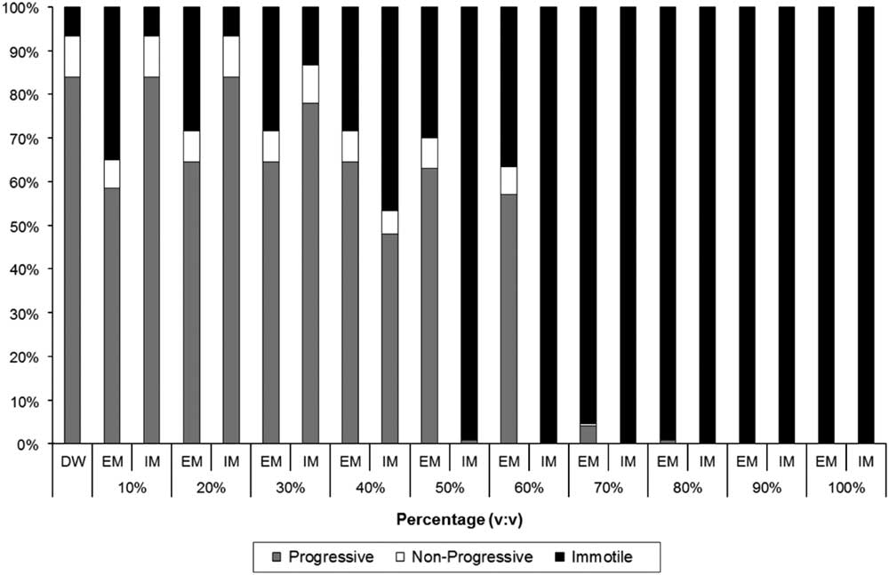
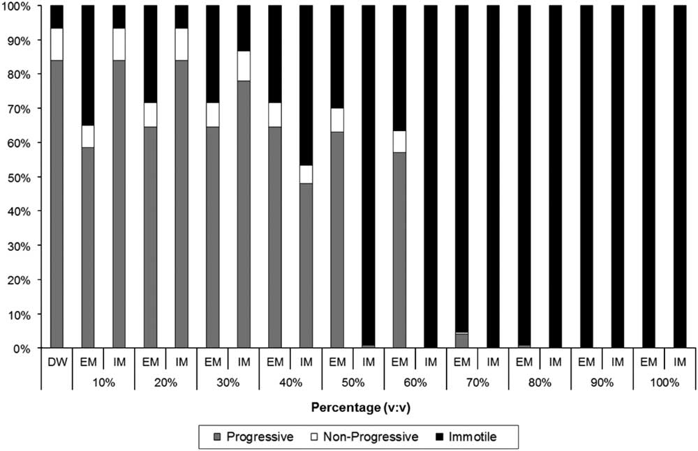
Loach sperm at 80–100% concentration were not motile in either the embryo or injection medium (Fig. 1). The embryo medium at 70% and the injection medium at 40% triggered motility, which was fully activated at lower concentrations (Fig. 1). Percentage of sperm showing progressive motility was 84.0±2.6%, when the sperm was activated by contact with distilled water (Fig. 1). Therefore, the quality of the sperm used here was good.
Motility parameters of loach sperm after mixing with embryo medium (EM) or injection medium (IM) at increasing concentrations ranging from 0 (distilled water, DW) to 100% (undiluted medium). Solid, open and grey rectangles indicate immotility, non-progressive motility and progressive motility, respectively.
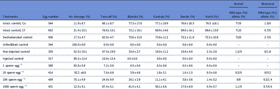
Co, Cf and dechorionated controls showed similar trends in all developmental stages and hatching rates ranged between 68.6% and 76.5% (Table 1). Among the groups that contained eggs injected with sperm, the fertilization success expressed as a percentage of embryos at the two-cell stage increased with an increasing amount of sperm cells injected into the ooplasm. When only one sperm was injected into an egg, the fertilization rate was 7.2±5.9%, with no hatching. Injecting 10 sperm provided similar results, with a fertilization rate of 7.8±6.8% and only two hatched embryos (0.5±0.6%) that comprised one non-pigmented abnormal albino larva and one pigmented normal wild-type larva. Approximately 100 sperm injected per egg increased both the fertilization rate (24.9±9.9%) and hatching (1.4±3.2%), but all were abnormal larvae (six non-pigmented albinos and one pigmented wild-type larva). The highest fertilization rate (87.4±5.1%) was obtained at the highest sperm concentration (1000 sperm per egg), with a hatching rate of 5.9±5.7% (28 larvae) comprising five pigmented normal wild-type, three pigmented abnormal wild-type and 20 non-pigmented abnormal albino larvae.
Egg number; proportion of survived embryos at two-cell, blastula, gastrula, somite and hatched stages; and proportion of normal (wild-type/albino) and abnormal (wild-type/albino) progeny in six types of control groups and sperm-injected (1, 10, 100, 1000 sperm per egg) groups
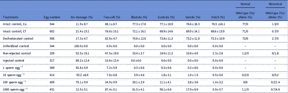
The first control (Intact control: Co) was induced by fertilization between eggs and sperm before the injection trials. The second control (Intact control: Cf) was induced by fertilization between eggs and sperm after the injection trials. In these two controls, the fertilized eggs were cultured with their chorion. The third control (dechorionated control) was induced by fertilization between eggs and sperm before the injection trials, after which the chorion of the fertilized eggs was removed using the dechorionation medium and then cultured in the culture medium. The fourth control (unfertilized control) comprised eggs activated by only water. The fifth control (non-injected control) comprised eggs fertilized by sperm suspension at 108 sperm ml−1 that was poured onto the animal pole of the dechorionated eggs. The sixth control (injected control) comprised dechorionated eggs microinjected with a solution containing 200 mM KCl and 0.5% rhodamine.
The eggs in the unfertilized control did not show any cleavage. Some eggs began to exhibit cleavage-like segmentation in the injected control, but did not develop further. In the non-injected control eggs, some developed and viable larvae hatched.
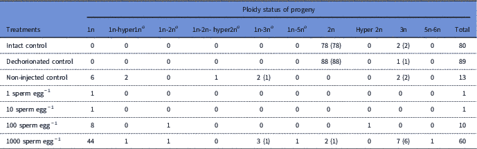
The ploidy status of the progeny sampled from all the treatments before hatching is provided in Table 2. The progeny used for ploidy analysis comprised normal embryos with wild-type pigmentation and abnormal embryos with/without pigmentation. In the controls (i.e. Co, Cf and dechorionated), normal embryos were measured and the majority (98–99%) were diploid, with only a small number of triploid progeny. In groups containing eggs injected with sperm, the progeny died before the beginning of pigmentation, but their ploidy status was estimated. The non-injected control gave six haploid, five mosaic and two triploid progeny, in which normally pigmented wild-type progeny with haploid–triploid mosaicism (n=1) and triploidy (n=2) were observed. Injecting 1 sperm and 10 sperm gave two abnormal haploids (1 fish in each group). Injecting 100 sperm gave haploids (n=8), a haploid–diploid mosaic (n=1) and a hyperdiploid (n=1), but all were abnormal. When approximately 1000 sperm were injected, a predominant appearance of haploids was observed (n=44), together with diploids (n=2), triploids (n=7) and various types of mosaicism (n=7). In this group, the normal progeny were diploid (n=1), triploid (n=6) and haploid–triploid mosaics (n=1).
Ploidy status of progeny in control groups and sperm-injected (1, 10, 100, 1000 sperm per egg) groups
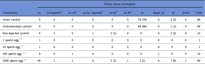
Number in parenthesis denotes the number of normal and pigmented wild-type progeny.
a Dash indicates mosaicism of cell populations with different ploidy status.
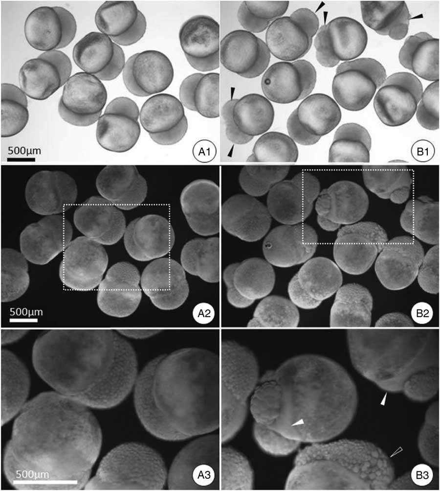
Most injected eggs exhibited abnormal development that was expressed especially during the blastula stage. Among the embryos developed from the injected eggs, unusual development was detected by observing blastomeres of different sizes as well as those containing anuclear cells (Fig. 2).
Blastula embryos from the control (A) and injected groups (B). Top (A1, B1): embryos under a stereomicroscope using normal light. Middle (A2, B2): embryos under epi-illumination and a dark background. Bottom (A3, B3): boxed area with dashed lines from A2 and B2. Note the abnormal embryos with irregular cell proliferation, reduced or divided blastoderms (solid black arrowheads in B1), acellular development (solid white arrowheads in B3) and asynchronous cell development showing blastomeres of different sizes (open white arrowheads).
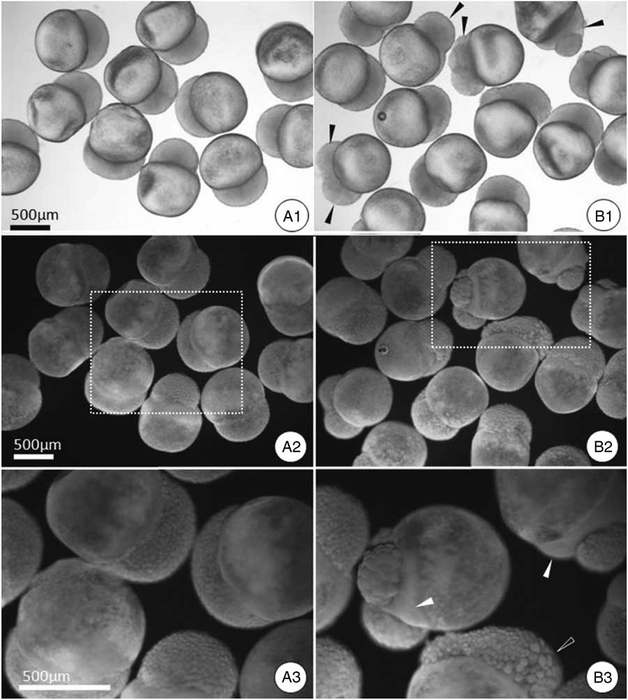
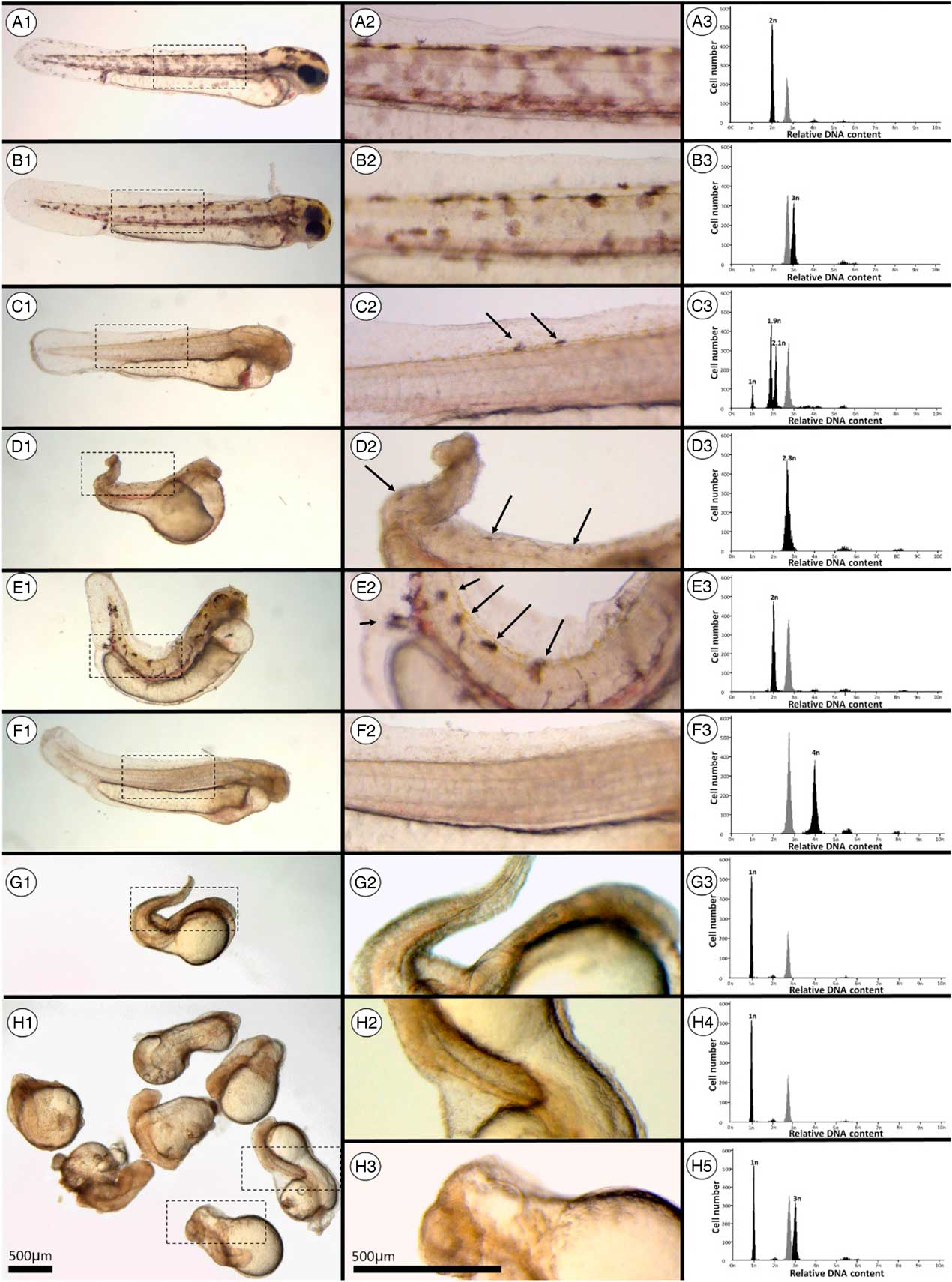
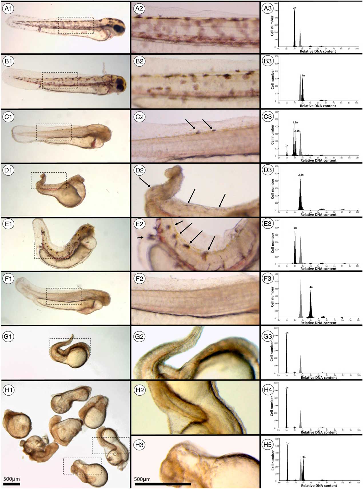
Figure 3 shows the external gross appearance, presence or absence of melanophore pigments (visible marker of paternal genome) and ploidy status based on flow cytometric analyses of progeny developed from fertilized eggs in the control groups and those injected with sperm. Control progeny with normal appearance and wild-type body pigmentation showed diploidy (Table 3 and Fig. 3 A1–A3). Among the progeny of the injected groups (Table 3), haploid (Fig. 3 G1–G3), diploid (Fig. 3 E1–E3), triploid (Fig. 3 B1–B3), tetraploid (Fig. 3 F1–F3), hypotriploid (Fig. 3 D1–D3) and haploid–hypodiploid–hyperdiploid mosaic (Fig. 3 C1–C3) progeny were observed. Abnormal diploid (Fig. 3 E2) and normal triploid progeny (Fig. 3 B2) gave apparent expressions of melanophore pigments on their body surface, but other normal haploid–hypodiploid–hyperdiploid mosaic (Fig. 3 C2) and abnormal hypotriploid (Fig. 3 D2) progeny exhibited few pigments within a limited area on their body surfaces. Abnormal haploid (Fig. 3 G1–G3, H1, H2, H4), abnormal haploid–triploid mosaic (Fig. 3 H1, H3, H5) and normal-looking tetraploid (Fig. 3 F1–F3) progeny without melanophore expression were also observed in the injected group.
External appearance (left, 1), its magnified image in a boxed area (centre, 2) and flow cytometric histogram (right, 3) showing the ploidy status of progeny 4 days after fertilization using wild-type female (dominant phenotype) and albino male (recessive phenotype) (A) and progeny developed from injecting multiple numbers of sperm (B–H). Arrows indicate pigmentation. In each flow cytometric histogram, the grey peak denotes somatic cells of goldfish as a standard reference. Progeny shown in B–G correspond to Injected group, nos. 1–6 in Table 3.
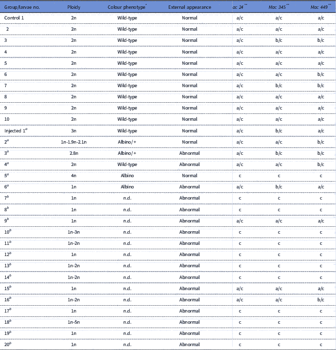
Ploidy status, colour phenotype, external appearance and microsatellite DNA genotypes of progeny from control (normal fertilization) and microinjection of approximately 1000 sperm from albino male into the ooplasm of an egg from wild-type female
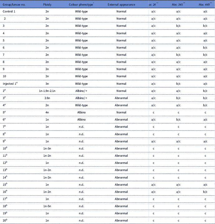
*Wild-type: normal pigmentation, albino: no pigmentation, +: partially pigmented, n.d.: not determined.
**Female genotypes (a/b or a/a): 120/120 at Mac 24 (linkage group 16), 514/546 at Mac 345 (linkage group 6), 228/240 at Mac 449 (linkage group 14). Male genotypes (c/c): 112/112 at Mac 24, 506/506 at Mac 345, 236/236 at Mac449.
a Progeny 1–6 correspond to individuals shown in Fig. 3B–G .
b Progeny 7–20 have the same abnormal morphology patterns shown in Fig. 3H .
Microsatellite DNA genotyping was conducted in the above-mentioned progeny from the injected group of approximately 1000 sperm injected per egg (nos. 1–6 in Table 3) and the haploid, haploid–diploid, haploid–triploid and haploid–pentaploid progeny of the same injected group showed an extremely abnormal appearance and were sampled before hatching (nos. 7–20 in Table 3). All control group (normal wild-type) progeny had heterozygous genotypes consisting of alleles from both the female and male parents in the three independent loci that we examined. In contrast, in the injected group, 12 (nos. 5, 7, 8, 10–14 and 17–20 in Table 3) of 20 progeny demonstrated only paternally derived alleles and therefore were androgenotes with an all-male inheritance (Table 3). Among these androgenetically developed progeny, only the tetraploid progeny (no. 5 in Table 3) exhibited a normal appearance (Fig. 3 F1–F3); the haploid, haploid–diploid, haploid–triploid and haploid–pentaploid mosaics demonstrated inviable abnormalities (Fig. 3 G1–G3, H1–H5, Table 3). From the injected group, three haploid (nos. 6, 9 and 15 in Table 3) and one haploid–diploid mosaic (no. 16 in Table 3) had both maternally and paternally derived alleles and did not develop androgenetically (Table 3). Among these non-androgenotes, one haploid (no. 6 in Table 3) demonstrated the albino phenotype, but also had microsatellite alleles from both parents. Four fully or partially pigmented progeny with triploidy (Fig. 3 B1–B3, no. 1 in Table 3), haploid–hypodiploid–hyperdiploidy (Fig. 3 C1–C3, no. 2 in Table 3), hypotriploidy (Fig. 3 D1–D3, no. 3 in Table 3) and diploidy (Fig. 3 E1–E3, no. 4 in Table 3) were presumably initiated by fertilization with injected sperm because they showed both maternally and paternally derived alleles at three loci (Table 3). Among these progeny, triploid (Fig. 3 B1–B3, no. 1 in Table 3) and haploid–hypodiploid–hyperdiploid mosaic progeny (Fig. 3 C1–C3, no. 2 in Table 3) displayed a normal appearance (Table 3).
Discussion
We confirmed that sperm were not motile in either the embryo or the injection medium. We injected a sperm suspension into the ooplasm of each dojo loach egg that was activated in advance with ambient water, followed by dechorionation. As with zebrafish (Poleo et al., Reference Poleo, Denniston, Reggio, Godke and Tiersch2001), the dojo loach eggs are activated by contact with water, as evidenced in Tanaka et al. (Reference Tanaka, Takahashi and Ueno2009). Embryos developed in the groups that contained eggs injected with sperm. This suggests that fertilization or embryogenesis should be the result of injected or non-injected non-motile cells reaching the oocytes. Increasing the number of injected sperm increased the fertilization success, as indicated by the number of cleaved eggs. Presumably, a larger number of sperm increased the probability of forming the male pronucleus. Because the breakdown of the sperm plasma membrane (Morozumi et al., Reference Morozumi, Shikano, Miyazaki and Yanagimachi2006) and nuclear envelope (Yamashita et al., Reference Yamashita, Onozato, Nakanishi and Nagahama1990) is a prerequisite for the decondensation of the sperm nucleus and the subsequent formation of the male pronucleus for successful fertilization, part of the injected sperm are likely to transform into male pronuclei and then proceed with the process of fertilization and subsequent embryogenesis.
Small but significant numbers of progeny developed and sometimes hatched from the eggs injected with sperm. Most resultant embryos were abnormal and inviable, but a few progeny exhibited normal to near-normal appearance. The highest eupolyploid was a tetraploid and one mosaic that included pentaploid and hexaploid cell populations, although injecting 1000 sperm per egg was conducted in this study. Haploidy was the most frequent ploidy in eggs injected with sperm. These results indicate that activated eggs still retained the ability to continue with the fertilization reaction; however, it is unclear why only one sperm or several sperm contributed to embryogenesis, even after injecting hundreds and thousands of them into the ooplasm.
Microsatellite genotyping revealed that there are two types of haploids. The first was generated by androgenesis resulting from inheriting only paternally derived alleles. Androgenotes were haploid–diploid, haploid–triploid and haploid–pentaploid mosaics and tetraploids. These results suggested that a single sperm or multiple sperm injected into the ooplasm became male pronuclei and contributed to embryogenesis without incorporating the egg nucleus. The second type of haploid exhibited both maternally and paternally derived alleles. These haploids are considered mosaics comprising the following two types of haploid cell populations: androgenetically developed haploid cells from the male nucleus and gynogenetically developed haploid cells from the female nucleus. This suggests that each haploid cell population that independently formed from the sperm or egg pronucleus developed into an embryo without syngamy.
Injecting multiple numbers of sperm into an egg of the dojo loach gave rise to progeny with various ploidy statuses, a large proportion of which were androgenotes developed from the male pronucleus/pronuclei transformed from the injected sperm. Another small proportion of progeny developed from the fusion of both female and male pronuclei or probable mosaics comprising maternally derived cells from the female pronucleus/pronuclei and paternally derived cells from the male pronucleus/pronuclei. No pure gynogenetic progeny were observed. These results suggested that injecting multiple numbers of sperm into the egg ooplasm should have potential as a new biological technique to induce artificial androgenesis.
Financial support
This study was supported in part by KAKENHI Grants-in-Aid for Young Scientist (B) (grant number 18780138) to T.F. and for Challenging Exploratory Research (grant number 25660161) to K.A. from the Japan Society for the Promotion of Science (JSPS).
Conflicts of interest
There are no conflicts of interest.
■