Introduction
Ebstein’s anomaly is a rare congenital heart disease of variable severity, and the prognosis remains poor in cases requiring surgical intervention in the early neonatal period. It is a life-threatening condition, particularly in cases of a circular shunt, so that prompt interruption of this lethal circulation by pulmonary artery ligation immediately after birth is crucial for survival. Reference Barre, Durand, Hazelzet and David1 We present our experience in managing a very critically ill fetus by emergency surgery using the consecutive two-stage Starnes strategy immediately after delivery.
Case report
At 31 weeks of gestation, fetal echocardiography showed severe tricuspid regurgitation, and the fetus was referred to our hospital, where a diagnosis of Ebstein’s anomaly was made. Echocardiography performed every other week showed increasing pulmonary regurgitation, leading to rapid progression of the circular shunt and occurrence of fetal edema, which could have resulted in fetal death, so an urgent caesarean section was planned at 36 weeks of gestation under cardiac surgery standby.
The baby’s first cry was confirmed but soon became bradycardic, so intubation was performed immediately, and chest compression was initiated. Concurrently, a peripheral venous line was secured, from which adrenaline was injected to restore the heart rhythm.
Subsequently, surgery started with one peripheral venous access, an electrocardiogram monitor, and a pulse oximeter to make haemodynamic improvement a top priority. Five minutes into the surgery, that is, 24 min after birth, his oxygen level increased immediately after ligation of the main pulmonary artery, and his heart rate stabilised.
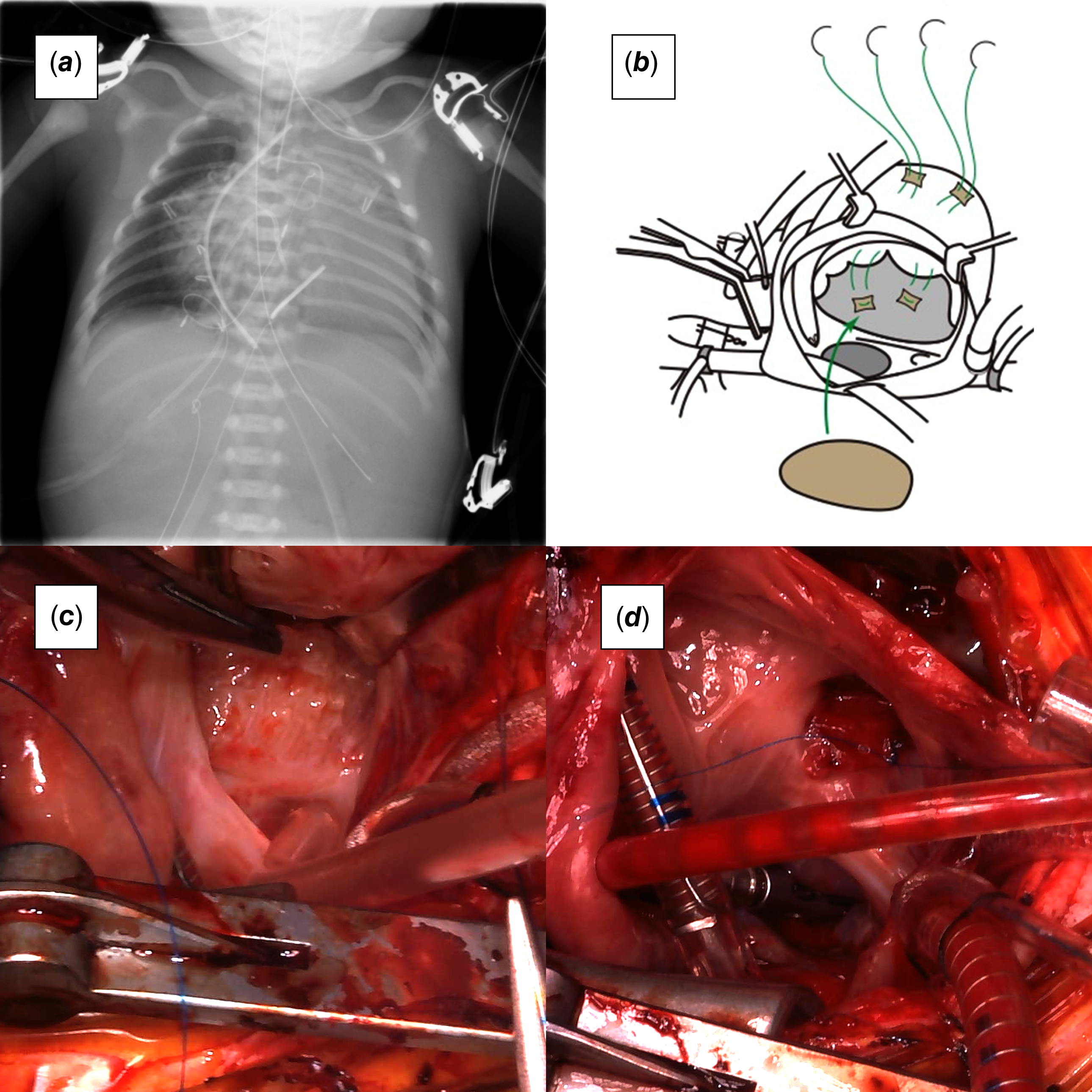
An arterial pressure line was placed directly into the ascending aorta and a central venous catheter into the right atrium. Instead of the ductus, the bilateral pulmonary arteries were clamped during assisted cardiopulmonary bypass. Subsequently, he underwent right ventricle plication, tricuspid valve closure with a pericardial patch, and atrial septal defect creation (Figure 1a–d). To avoid atrioventricular block, the patch was not sutured around the atrioventricular node, which is similar to the effect of fenestration. The patient was weaned off cardiopulmonary bypass with satisfactory haemodynamics. However, his lungs had poor gas exchange capacity, and he could not fully expel carbon dioxide.
( a ) Plain chest X-ray after the initial surgery. ( b ) Surgical schema. Right ventricular plication, tricuspid valve closure with a fenestrated patch. ( c ) Intraoperative photography of the dysplastic and plastered tricuspid valve. ( d ) Intraoperative photography of the tricuspid valve closure with a fenestrated glutaraldehyde-treated autologous pericardium patch.

In the paediatric intensive care unit, high-frequency oscillation support was introduced for 4 days, and he was extubated on postoperative day 36. He underwent a systemic-to-pulmonary shunt surgery at 1 month. Thus, the consecutive two-stage Starnes strategy could save the baby. He had a bidirectional Glenn surgery at 6 months old and is waiting for total cavopulmonary connection in a stable condition.
Discussion
The Starnes procedure, reported in 1991, Reference Starnes, Pitlick, Bernstein, Griffin, Choy and Shumway2 is a useful palliative procedure in the single ventricle strategy for Ebstein’s anomaly, but it is known to have a high operative mortality rate. Because the lungs are hypoplastic due to marked cardiac dilatation, an early neonatal systemic-to-pulmonary shunt may fail due to neonatal pulmonary hypertension or worsen cardiac function due to diastolic coronary blood flow steal. Additionally, compression of the dilated right ventricle restricts left ventricular function, making maintaining circulation difficult. Reference Lang, Oberhoffer and Cook3
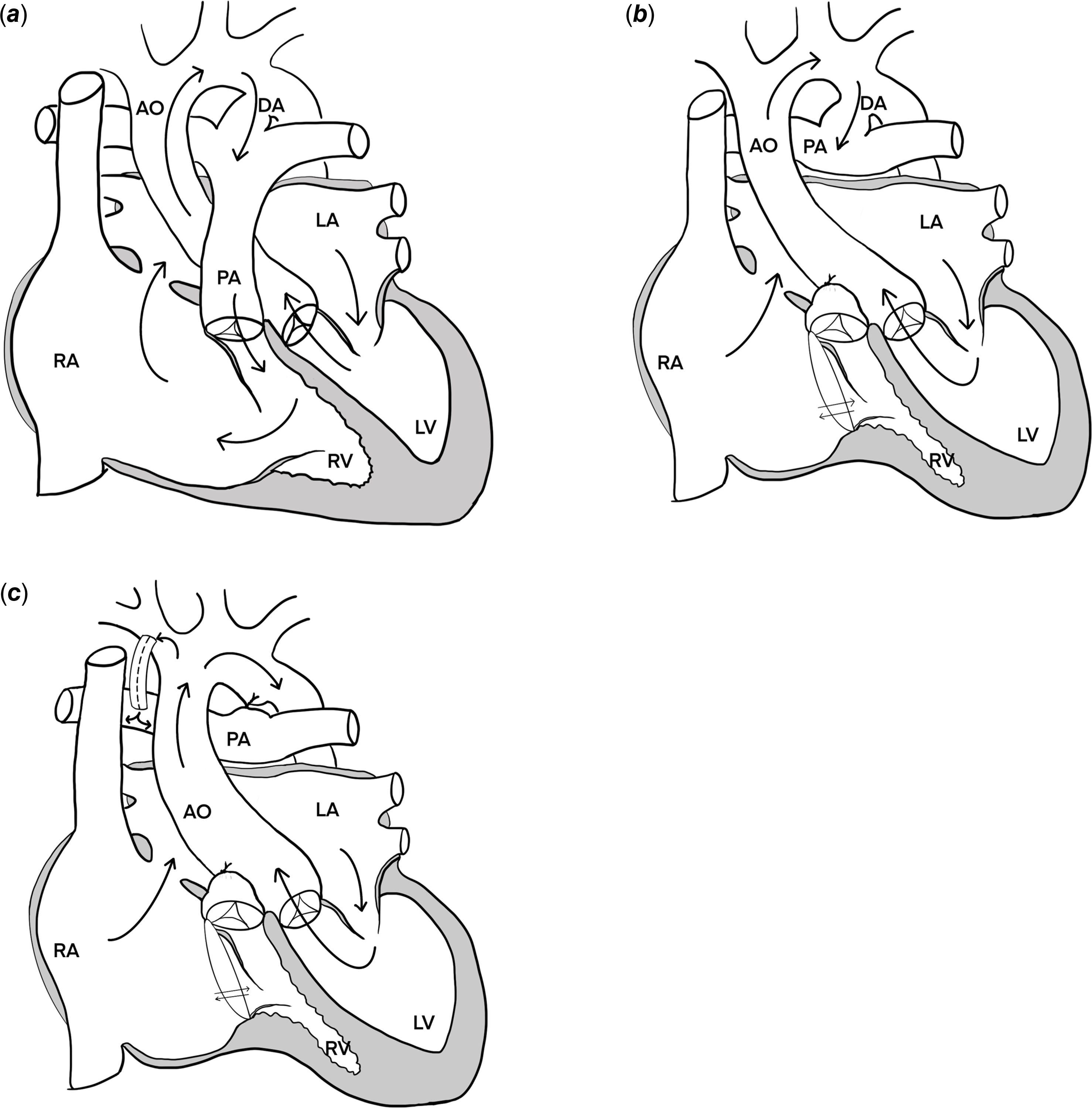
We hypothesised that the three components of the Starnes procedure, including right ventricle exclusion, atrial septal defect creation, and systemic-to-pulmonary shunt, could be organised and reconstructed in a consecutive two-stage strategy to control postoperative circulatory fluctuations (Figure 2a–c). The strategy of performing systemic-to-pulmonary shunt initially, followed by right ventricle exclusion, may avoid the use of cardiopulmonary bypass during the initial surgery. However, the former operation only changes the pulmonary blood flow source from the ductus to a shunt graft and does not improve haemodynamics; therefore, a second-stage operation is required immediately afterwards. In addition to that, early shunt surgery during the neonatal period may lead to diastolic hypotension, impaired coronary perfusion, and acute deterioration of myocardial function. Therefore, we prefer a standby approach to delay shunt placement until after the neonatal phase when pulmonary vascular resistance has stabilised.
Haemodynamic schematic representations. ( a ) At birth, the patient exhibits a circular shunt. ( b ) After the initial surgery, the main pulmonary artery is ligated, and the tricuspid valve is closed using an autologous pericardial patch with fenestration. Pulmonary circulation is maintained via ductus-dependent circulation. ( c ) Following the second stage, pulmonary circulation is maintained via a shunt instead of the ductus. RA = right atrium; RV = right ventricle; PA = pulmonary artery; DA = ductus arteriosus; AO = aorta; LA = left atrium; LV = left ventricle.

Conversely, right ventricle exclusion and atrial septal defect creation can be expected to improve haemodynamics. Although open heart surgery with cardiopulmonary bypass in neonates remains challenging, it is performed to improve haemodynamics with acceptable risk. Therefore, we believe that right ventricle exclusion should be performed first to improve left ventricular function. Subsequently, a systemic-to-pulmonary shunt is performed around 1 month when pulmonary vascular resistance has stabilised. This approach requires precise monitoring of ductal formation and size, along with careful regulation of medication, inspired oxygen concentration, and ventilator settings to balance pulmonary circulation. Although this strategy is not universally applicable to all cases, we believe it provides a safer initial pathway for patients with extreme circulatory fragility.
We have experienced three other cases of neonatal Ebstein’s anomaly managed using a similar strategy in our institution, and all reached total cavopulmonary connection in satisfactory condition.
Financial support
This research received no specific grant from any funding agency, commercial, or not-for-profit sectors.
Competing interests
The authors declare none.