


Introduction
Heart injuries are usually caused through penetrating or blunt traumas. The most common causes of penetrating injuries are firearms or cutting and piercing tool injuries. Although penetrating cardiac injuries are a rare type of injury in frequency, they are critically important, owing to their high mortality rates. Reference Campbell, Thomson, Muckart, Meumann, Van Middelkoop and Botha1 In relation to the localization of penetrating heart injuries, their frequencies from highest to the lowest can be listed as the right ventricle, left ventricle, right atrium, and left atrium, respectively, Reference Karasu, Tokat and Uzun2 a finding consistent with the gross anatomy of the heart in which the right ventricle forms the largest part of the sternocostal surface of the heart facing to anterior chest wall.
A 46-year-old male patient lying unconscious on the ground was found and brought to the emergency room. His initial examination revealed a uniform 1cm x 1cm bullet wound on his left cervical area with relatively stable hemodynamic condition. His further evaluations indicated that the patient suffered from a gunshot and the bullet core stuck in his heart. Consequently, the patient was taken into the operation room for emergent surgical intervention to save his life. During the intervention, the bullet core was removed and the left ventricle was repaired. This study aimed to present this case, focusing on the fact that the apparent injury zone might not always coincide with the actual injury site, along with considering importance of fast transport, rapid preoperative evaluation, preparation, and selection of right operative approach.
Case Presentation
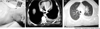
A 46-year-old male patient lying unconscious on the ground was found and referred to the emergency room with bruises around his left eyelid, knees, and nose, in addition to a uniform 1cm x 1cm wound on his left cervical area with no active bleeding (Figure 1A). His close examination revealed that the wound was due to a bullet entry; his general situation was poor with unsteady consciousness, prone to light sleep, with the presence of response to the audible stimulus. His blood pressure was 90/60mmHg and his pulse was 120 per minute. Heart sounds sounded deeply, and on the left, breathing sounds decreased.
(A) Entrance of the Bullet; (B) CT Image of the Bullet Core Lodged in the Left Ventricle; and (C) Left Lung Injury with the CT Image of the Left Hemothorax.
Abbreviation: CT, computed tomography.

Initially, the patient was evaluated with ultrasonography. Since the ultrasonographic examination showed pericardial fluid, along with considering the state of his consciousness, the patient was further assessed with neck computed tomography (CT) and contrast-enhanced chest CT. His CT showed a foreign body (bullet core) inside the left ventricle with unclear demarcation of inner or outer wall of the left ventricle (Figure 1B), hemothorax and pneumothorax on the left side (Figure 1C), suspected left atrium injury, and pericardial effusion. Consequently, the patient was also evaluated with transthoracic echocardiography (ECHO), in which a foreign body that was not clearly distinguished from the posterior surface of the left ventricle and did not form a tamponade. Upon these findings, the patient was taken into the operating room.
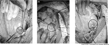
For the surgery, a median sternotomy was performed under general anesthesia, and simultaneously, a femoral artery and vein cannulation were performed from the right femoral region. Since active bleeding was observed when the pericardium was opened, cardiopulmonary bypass was initiated. Hemodynamic stability was achieved without cardiac arrest, accompanied by a heart-lung pump. Subsequently, approximately one liter of blood from the pericardial cavity and 0.8 liters of blood from the left thoracic cavity were drained. The bullet was noted to being stuck in the myocardium near the obtuse marginal branch of the circumflex artery on the posterior surface of the left atrium (Figure 2A). The area of active bleeding in the left atrium auricle was repaired using Prolene sutures and the bleeding was stopped (Figure 2B). Afterward, the area in the left ventricle where the bullet core stuck was exposed with a scalpel and the bullet core was removed from the ventricle. There was no coronary artery damage, and the area was repaired using Teflon felt-supported polypropylene sutures (Figure 2C).
(A) Bullet Core in the Wall of the Left Ventricle; (B) Appearance of the Left Atrial Appendage after its Surgical Repair; and (C) Repaired Left Ventricle after Removing the Bullet Core.

No pathology was detected in the postoperative examinations. Ejection fraction of the patient was assessed as 50% during the ECHO. Likewise, no pathology was also noticed on the electrocardiogram/ECG. In the post-operative period, the patient did not need blood transfusion, as well as did not have any indication of hypoxia, kidney failure, or fluid-electrolyte disorder. Eventually, the patient was discharged on post-operative day nine since the patient was hemodynamically stable and showed no additional pathology.
Prior to the discharge of the patient, verbal and written consent were obtained from the patient for using his photographs taken during the operation, information regarding his examinations, and stages related to the operation without specifying his name in a case presentation in order to contribute to the literature.
Discussion
Mortality in penetrating cardiac injuries varies depending on the area of the injury, duration of transport and intervention, the presence of additional organ injury, the amount of blood loss that occurs, and the presence of tamponade. Reference Carr, Buterakos, Bowling, Janson, Kralovich and Copeland3 At their studies on the penetrating cardiac injuries, Buckman, et al reported that 78% of all deaths occurred in the first hour after cardiac injury. Reference Buckman, Badellino and Mauro4 In another similar study, Hardikar, et al stated that prehospital mortality rate was 70%-80%. Reference Hardikar, Thakur, Karmarkar, Ambike, Kanetkar and Golhar5 Survival rate among the patients arriving at the hospital on time, receiving immediate diagnose, and getting quick surgical intervention if needed is shown to be 74%. Reference Tyburski, Astra and Wilson6
The major finding in heart injuries is usually cardiac tamponade. Although there was no cardiac tamponade in the present case, the patient was immediately taken into surgery room since his radiological evaluations and ECHO indicated heart injury.
One of the most important diagnostic criteria for heart injuries is the site and the direction of entry of the damaging factor into the body. Reference Jebara and Scede7 In the present case, although the patient had an injury in the cervical region where cardiac injury would not have been considered in the first place, the results of brain CT, contrast thorax CT, and ECHO indicated heart injury, thereby facilitating instantaneous decision for the surgery. Present observation showed the importance of considering cardiac injury in differential diagnosis, regardless of hemodynamic stability.
The first assessment of the patient in the emergency room by a cardiovascular surgeon, implementation of radiological and ECHO tests on time, and rapid assessment of their results, in addition to findings showing heart injury and rapidly taking the patient in the emergency operation room, were life-saving for the patient. The time elapsed between the evaluation of the patient in the emergency room and the start of the operation was 27 minutes and the time between ECHO and the operating room was 20 minutes.
Several studies report that while mortality rate varies between 54%-94% in the patients developing cardiac arrest and receiving emergent intervention due to the arrest after penetrating heart injuries, it is shown to be reduced to 13.5%-40.0% in the patients receiving emergent intervention without cardiac arrest after penetrating heart injuries. Reference Attar, Suter, Hankins, Sequeira and McLaughlin8–Reference Peter, Hugh and Christoph10 In the present case, although the initial ECHO of the patient revealed pericardial effusion not causing to cardiac tamponade, a tamponade clinic began developing in the period passed for taking the patient on the operation table. The onset of tamponade clinic pointed out presence of a potential active bleeding and prompted an urgent need for cardiopulmonary bypass. Even though left anterolateral thoracotomy is a preferable approach in the heart injuries, since it requires fewer surgical tools and offers a rapid access, median sternotomy method is often favored in the studies reported in the literature because it not only provides better entree to entire chambers of the heart, aorta, pulmonary arteries, and veins, but it also ensures better exploration and allows convenient switch to cardiopulmonary by-pass if necessary. Reference Topaloğlu, Aras and Çağlı11–Reference Mitchell, Muakkassa, Poole, Rhodes and Griswold16 Accordingly, median sternotomy was preferred for the present case.
Numerous studies in the literature report higher mortality rate among the patients developing cardiac arrest or becoming hemodynamically unstable after penetrating heart injury. Reference Attar, Suter, Hankins, Sequeira and McLaughlin8–Reference Peter, Hugh and Christoph10 The cases where cardiopulmonary bypass is needed for repair include major vascular and/or atrial/ventricular injury, excessive bleeding, or difficult access to the injury site. Reference Asension, Berne and Demetriades9,Reference Mittal, McAleese, Young and Cohen17 In the present case, the sternotomy and pericardial opening procedures, along with simultaneous peripheral cannulation, were initiated prior to development of cardiac arrest and impairment of perfusion. In addition, cardiopulmonary bypass was also introduced when active bleeding was spotted in opened pericardium. This approach precluded the formation of hemodynamic instability and prevented the deterioration of cerebral perfusion in the patient.
Several studies indicate that patients are lost due to profuse bleeding after sternotomy during central cannulation and opening of the pericardium; however, peripheral cannulation compared to the central cannulation can save time and creates a more comfortable surgical field without causing interruption of the cerebral circulation. In the present case, the time elapsed between the beginning of the operation and the commencement of cardiopulmonary bypass was 10 minutes, and the peripheral cannulation allowed for simultaneous initiation of cardiopulmonary bypass once the active bleeding was seen after opening of the pericardium.
Limitations
The most important limitations for the present case include the followings: the present study centers around a single case, lacks information concerning patient series, and the use of peripheral cannulation during surgery cannot be generalized since there is no need for peripheral cannulation in every patient with penetrating heart injury. Overall, patient-based approach is required in the treatment of patients with penetrating heart injury.
Conclusion
Even if the area of injury is not compatible with cardiac injury, the patients who are hemodynamically impaired should be evaluated for heart injury, and the patients with the possibility of heart injury should be taken into emergent surgery. Among the most important factors in reducing mortality for heart injury patients are swift transportation, rapid evaluation, and operation with the accurate surgical technique. Furthermore, the present observation indicated that preparation of peripheral cannulation during the time passed for the opening of the pericardium to maintain hemodynamic stability in patients with high possibility of cardiac injury appeared to reduce mortality and contribute to the protection of the myocardium through permitting the initiation of cardiopulmonary bypass in case of noticing active bleeding after opening of the pericardium.
Conflicts of interest
The authors claim no conflict of interest.